Question 109
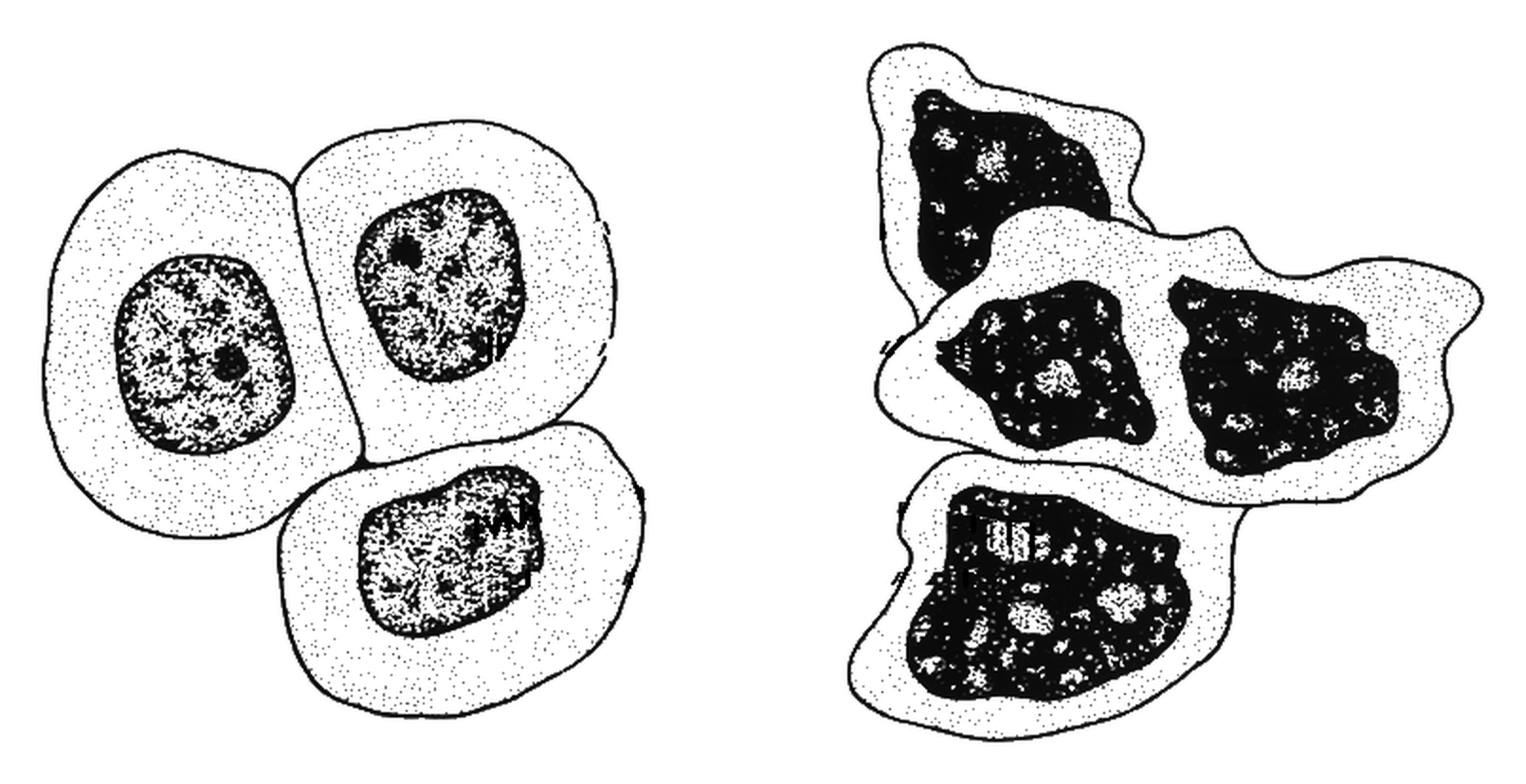
HA doctor takes two samples of cells from the same tissue in a patient. The cells in sample A are normal. The cells in sample B were taken from a tumour. The figure shows a drawing of the two samples seen using a light microscope. Sample A is on the left of the figure and sample B is on the right.
Figure: Drawings of normal cells and of cells taken from a tumour, seen using a light microscope
a.[2]
Give two differences, visible in the figure, between the cells in sample B and the cells in sample A.
b.[2]
In the figure, one of the normal cells in sample A is 30 mm wide. The real width of this cell is 0.02 mm. Calculate the magnification of the figure.
c.[2]
Some of the cells in sample B contain more than one nucleus. Explain what this suggests about cell division in these cells.
d.[2]
The doctor cannot tell from the figure alone whether the tumour is benign or malignant. Explain why.
2.2 Animal tissues, organs and organ systems Questions
179 exam-style questions on AQA GCSE Biology 2.2 Animal tissues, organs and organ systems, covering 2.2.1 The human digestive system, 2.2.2 The heart and blood vessels, 2.2.3 Blood, 2.2.4 Coronary heart disease: a non-communicable disease, 2.2.5 Health issues, 2.2.6 The effect of lifestyle on some non-communicable diseases, and 2.2.7 Cancer. Each one has a worked solution and a mark scheme showing where the marks go.