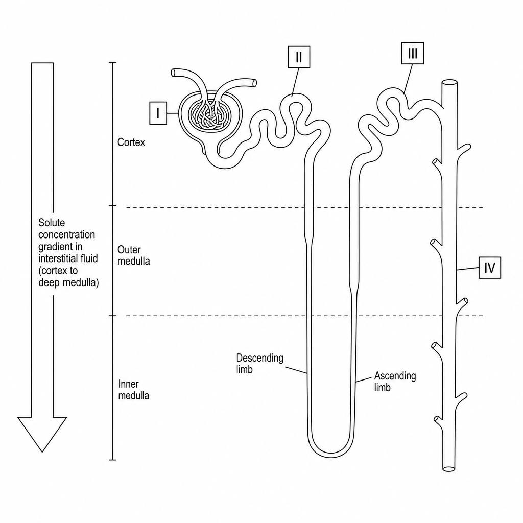
The diagram below shows the anatomical structure of a single mammalian nephron, highlighting different functional regions and the surrounding medullary concentration gradient.
a.
[3]
Complete the table below by matching the correct Roman numeral or numerals (I\text{I}I, II\text{II}II, III\text{III}III, or IV\text{IV}IV) of the nephron to each physiological description.
| Physiological Description | Roman Numeral(s) |
|---|---|
| Contains cells with specific co-transporter proteins that carry out selective, active reabsorption of all filtered amino acids and glucose. | |
| Has walls whose permeability to water is dynamically regulated by the insertion of aquaporins under the influence of antidiuretic hormone (ADH). | |
| Site of ultrafiltration where blood plasma is forced across a three-layered filtration barrier under high hydrostatic pressure. |
b.[3]
Explain the specific physiological processes occurring in the ascending and descending limbs of the loop of Henle that establish the osmotic concentration gradient shown by the downward-pointing arrow in the diagram.