The diagram shows a simplified schematic of a specialized epithelial cell lining the mammalian small intestine.
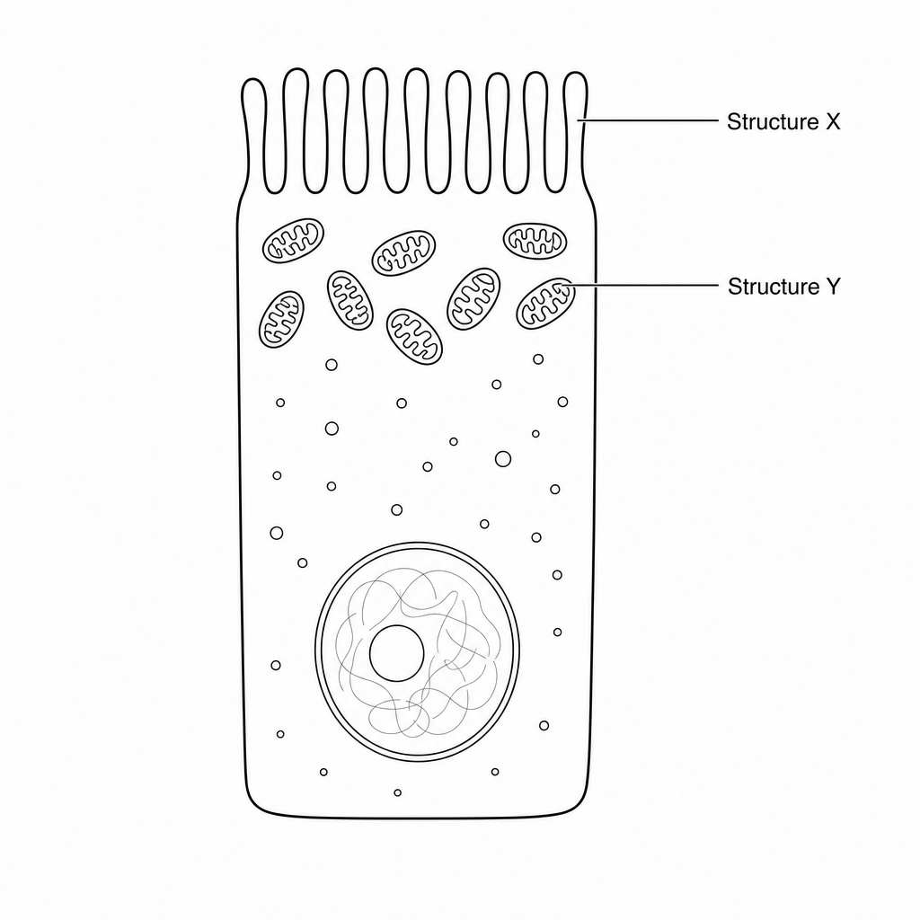
Which of the following correctly identifies Structure X, Structure Y, and the main biological function of this cell?
Structure X: Cilia; Structure Y: Mitochondria; Function: Sweeping mucus along the trachea\text{Structure X: Cilia; Structure Y: Mitochondria; Function: Sweeping mucus along the trachea}Structure X: Cilia; Structure Y: Mitochondria; Function: Sweeping mucus along the trachea
Structure X: Microvilli; Structure Y: Mitochondria; Function: Active transport of digested nutrients\text{Structure X: Microvilli; Structure Y: Mitochondria; Function: Active transport of digested nutrients}Structure X: Microvilli; Structure Y: Mitochondria; Function: Active transport of digested nutrients
Structure X: Microvilli; Structure Y: Lysosomes; Function: Intracellular digestion of macromolecules\text{Structure X: Microvilli; Structure Y: Lysosomes; Function: Intracellular digestion of macromolecules}Structure X: Microvilli; Structure Y: Lysosomes; Function: Intracellular digestion of macromolecules
Structure X: Flagella; Structure Y: Ribosomes; Function: Protein synthesis and secretion\text{Structure X: Flagella; Structure Y: Ribosomes; Function: Protein synthesis and secretion}Structure X: Flagella; Structure Y: Ribosomes; Function: Protein synthesis and secretion