Skip to content
MathsGenie logo
Open app

Course home

  1. A Level
  2. Biology OCR A
  3. Question bank

Transport in animals

EasyMediumHard
123456789101112131415161718192021222324252627282930313233
Question 23

An electrocardiogram (ECG) records the electrical activity of the heart over time. The grid on an ECG trace is standardized: the horizontal axis represents time, where 5 large grid squares correspond to exactly 1.0 s1.0\text{ s}1.0 s (each large square is 0.2 s0.2\text{ s}0.2 s).

The diagram below shows the resting ECG traces of three different individuals:
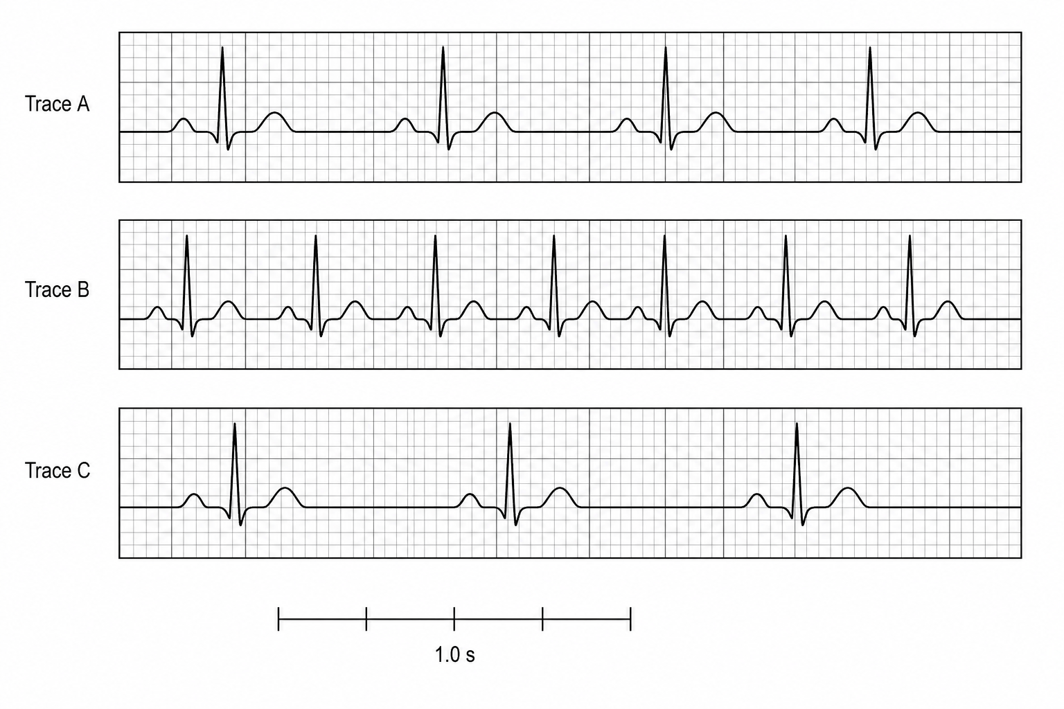
  • Individual 1: A healthy adult with a normal resting heart rate.
  • Individual 2: An adult with tachycardia due to an overactive thyroid gland.
  • Individual 3: An elite marathon runner showing physiological bradycardia.

Three resting ECG traces drawn on a grid where 5 horizontal large squares represent 1.0 s. Trace A shows R-waves (the sharp vertical peaks) spaced exactly 5 large squares apart. Trace B shows R-waves spaced exactly 3 large squares apart. Trace C shows R-waves spaced exactly 7.5 large squares apart. At the bottom, a time scale bar of 5 large squares is labeled 1.0 s.

a.

Calculate the heart rate in beats per minute (bpm) for: (i) Trace A (ii) Trace B

[4]
b.

Identify which individual (1, 2, or 3) corresponds to:

  • Trace A
  • Trace B
  • Trace C
[3]
c.

Explain how the heart of the elite runner (Individual 3) adaptively maintains a normal resting cardiac output despite a much lower resting heart rate.


[3]

Transport in animals Questions

  1. A Level
  2. /Biology
  3. /Transport in animals