What you'll learn
- How a mass spectrometer turns molecules into ions and separates them by mass-to-charge ratio.
- How to read a mass spectrum, including the molecular ion peak and base peak.
- How fragmentation and isotope patterns give clues about structure.
- How high-resolution mass spectrometry can suggest a molecular formula.
The starting point: relative masses and ions
Mass spectrometry is an analytical technique: it helps you find out what a substance is by measuring something about it. In this case, the key measurement is the mass of particles after they have been turned into ions.
Relative molecular mass, Mr
The relative molecular mass, MrM_rMr, is the sum of the relative atomic masses of all the atoms in a molecule. It has no units. Its numerical value is the same as the molar mass in g mol⁻¹.
A mass spectrometer does not usually detect neutral molecules directly. Instead, it makes positive ions and then separates them.
Mass-to-charge ratio
The mass-to-charge ratio, written m/zm/zm/z, is the mass of an ion divided by its charge number. For most A-Level organic mass spectra, ions have charge +1+1+1, so m/zm/zm/z is numerically equal to the ion’s relative mass.
What mass spectrometry really measures
A mass spectrum is a graph of relative abundance against m/zm/zm/z. It shows which positive ions are formed, and how common each ion is compared with the most abundant one.
How a mass spectrometer works
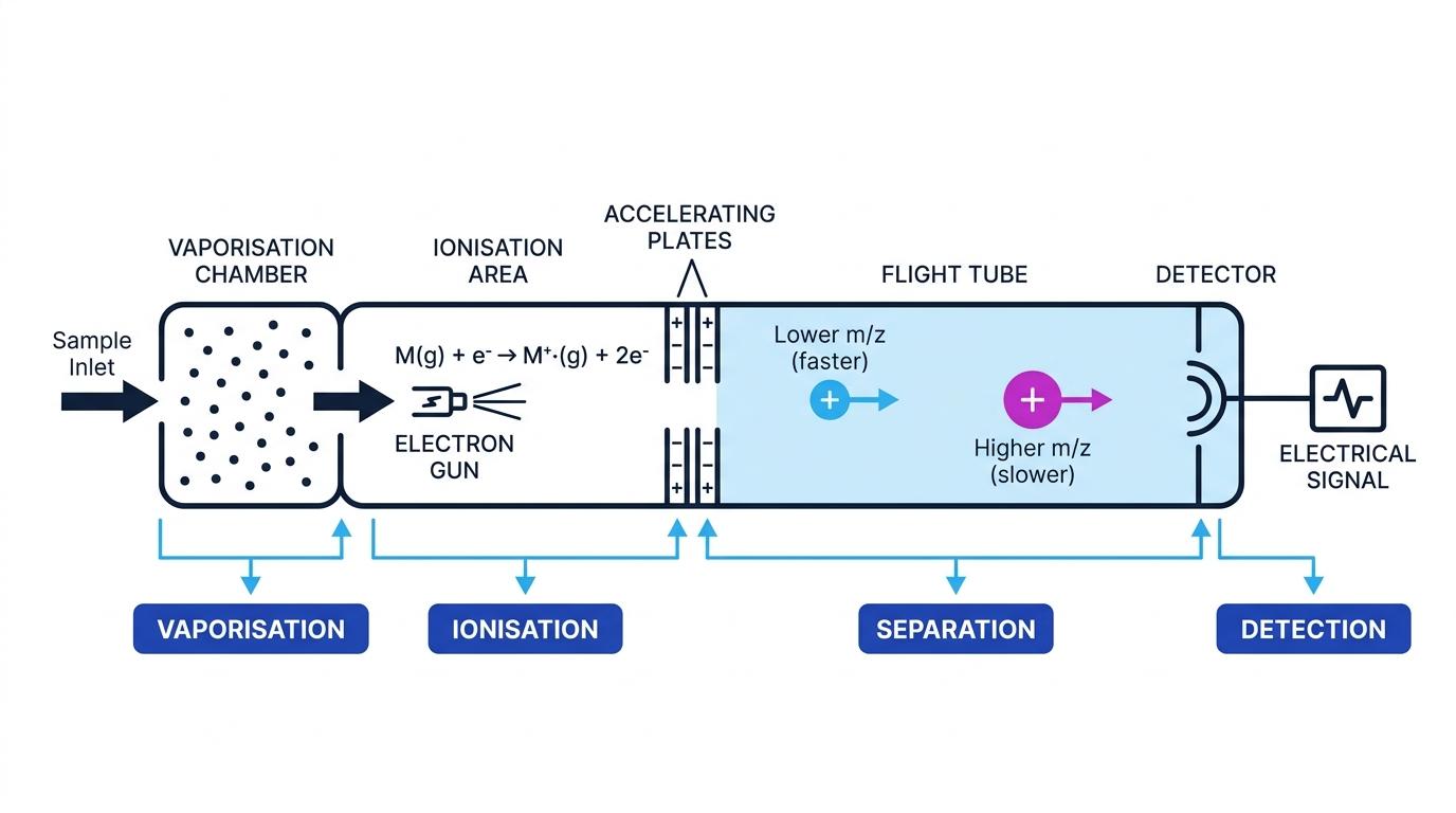
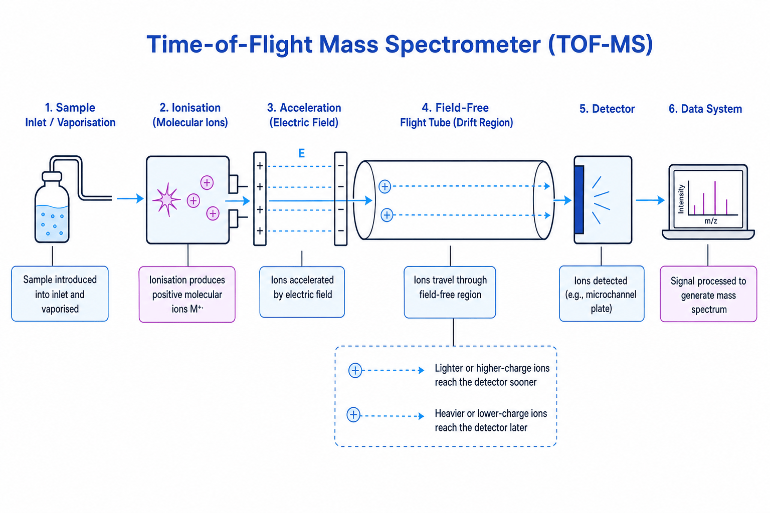
A typical mass spectrometer has four essential stages:
- Vaporisation: the sample is turned into a gas.
- Ionisation: molecules lose electrons to form positive ions.
- Separation: ions are separated according to m/zm/zm/z.
- Detection: ions hit a detector, producing an electrical signal that is converted into a spectrum.
For electron impact ionisation, a molecule is hit by high-energy electrons:
M(g)+e−→M+⋅(g)+2e−\text{M}(g) + e^- \to \text{M}^{+\cdot}(g) + 2e^-M(g)+e−→M+⋅(g)+2e−The ion M+⋅\text{M}^{+\cdot}M+⋅ is called the molecular ion. The dot shows that it has an unpaired electron, so it is a radical cation.
Forgetting the sample must be ionised
The detector only detects charged particles. Neutral molecules or neutral fragments are not recorded directly in the mass spectrum.
Reading a low-resolution mass spectrum
A low-resolution mass spectrum usually gives peaks at whole-number m/zm/zm/z values. This is enough to find the relative molecular mass and recognise common fragments.
Base peak and molecular ion peak
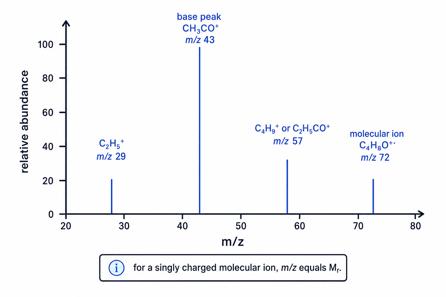
The base peak is the tallest peak in the spectrum and is assigned 100% relative abundance. The molecular ion peak is caused by the whole molecule after losing one electron; for a singly charged molecular ion, its m/zm/zm/z gives MrM_rMr.
The molecular ion peak is often at the highest significant m/zm/zm/z value, but small isotope peaks can appear just to its right.
Finding Mr from a molecular ion peak
A low-resolution mass spectrum shows a molecular ion peak at m/z=74m/z=74m/z=74 and a small isotope peak at m/z=75m/z=75m/z=75. Find MrM_rMr.
- Identify the molecular ion peak, not the small isotope peak. The peak at m/z=74m/z=74m/z=74 is the molecule itself; the peak at m/z=75m/z=75m/z=75 is mainly due to one heavier isotope, such as carbon-13.
- Use the fact that the molecular ion is singly charged, so its m/zm/zm/z value is numerically equal to its relative mass.
- Therefore, Mr=74M_r=74Mr=74. If you needed the molar mass, it would be 74 g mol⁻¹.
Base peak does not mean molecular ion peak
The tallest peak is the most abundant ion, not necessarily the whole molecule. Always look for the molecular ion peak when finding MrM_rMr.
Fragmentation: why there are lots of peaks
Ionisation can give the molecule enough energy to break covalent bonds. This produces fragment ions and neutral fragments.
Fragment ion
A fragment ion is a smaller positive ion formed when the molecular ion breaks apart. Only the charged fragment is detected.
Common fragment ions include:
- m/z=15m/z=15m/z=15: CH3+\text{CH}_3^+CH3+
- m/z=29m/z=29m/z=29: C2H5+\text{C}_2\text{H}_5^+C2H5+
- m/z=43m/z=43m/z=43: C3H7+\text{C}_3\text{H}_7^+C3H7+ or CH3CO+\text{CH}_3\text{CO}^+CH3CO+
- m/z=57m/z=57m/z=57: C4H9+\text{C}_4\text{H}_9^+C4H9+ or C2H5CO+\text{C}_2\text{H}_5\text{CO}^+C2H5CO+
Notice that different ions can have the same nominal mass in low-resolution spectra. You must use the rest of the evidence, especially functional group information from techniques such as infrared spectroscopy.
Using a fragment peak
Butanone, CH3COCH2CH3\text{CH}_3\text{COCH}_2\text{CH}_3CH3COCH2CH3, has a molecular ion peak at m/z=72m/z=72m/z=72 and a strong peak at m/z=43m/z=43m/z=43. Explain the m/z=43m/z=43m/z=43 peak.
- Test a plausible carbonyl-containing fragment: CH3CO+\text{CH}_3\text{CO}^+CH3CO+ has mass 12+3(1)+12+16=4312+3(1)+12+16=4312+3(1)+12+16=43, so it matches the peak.
- Check the missing neutral piece: 72−43=2972-43=2972−43=29, which corresponds to a neutral ethyl fragment, C2H5⋅\text{C}_2\text{H}_5\cdotC2H5⋅.
- The peak at m/z=43m/z=43m/z=43 therefore supports fragmentation next to the carbonyl group, forming the stable acylium ion CH3CO+\text{CH}_3\text{CO}^+CH3CO+.
Fragmentation checks
When suggesting a fragment, make sure the ion has a positive charge and that the masses add up to the original molecular ion.
Isotope patterns
Atoms of the same element can have different numbers of neutrons. These are isotopes. Some isotopes create recognisable patterns in mass spectra.
A molecule containing one carbon-13 atom gives a small M+1 peak, one unit above the molecular ion peak. The size of the M+1 peak increases as the number of carbon atoms increases.
Halogens are especially useful:
- Chlorine gives molecular ion peaks two units apart in an approximate 3:1 ratio.
- Bromine gives molecular ion peaks two units apart in an approximate 1:1 ratio.
Recognising a bromine pattern
A mass spectrum has two molecular ion peaks at m/z=108m/z=108m/z=108 and m/z=110m/z=110m/z=110 with approximately equal heights. What does this suggest?
- The peaks are two m/zm/zm/z units apart, so they are likely caused by isotopes that differ by two mass units.
- Equal-height M and M+2 peaks match bromine, because bromine-79 and bromine-81 have similar abundances.
- The compound is likely to contain one bromine atom. The lower-mass peak contains bromine-79, and the higher-mass peak contains bromine-81.
High-resolution mass spectrometry
A high-resolution mass spectrum measures m/zm/zm/z much more accurately, often to four decimal places. This can distinguish compounds with the same whole-number mass but different molecular formulae.
For example, C3H8O\text{C}_3\text{H}_8\text{O}C3H8O and C2H4O2\text{C}_2\text{H}_4\text{O}_2C2H4O2 both have nominal Mr=60M_r=60Mr=60, but they do not have exactly the same mass because atoms do not all have exact integer masses.
Choosing a molecular formula from accurate mass
A high-resolution molecular ion peak is found at m/z=60.0575m/z=60.0575m/z=60.0575. Choose the best formula from C3H8O\text{C}_3\text{H}_8\text{O}C3H8O, C2H4O2\text{C}_2\text{H}_4\text{O}_2C2H4O2 and C2H8N2\text{C}_2\text{H}_8\text{N}_2C2H8N2.
-
Calculate the accurate mass of each candidate using exact isotope masses.
C3H8O:3(12.000000)+8(1.007825)+15.994915=60.057515C2H4O2:2(12.000000)+4(1.007825)+2(15.994915)=60.021130C2H8N2:2(12.000000)+8(1.007825)+2(14.003074)=60.068748\begin{aligned} \text{C}_3\text{H}_8\text{O}:&\quad 3(12.000000)+8(1.007825)+15.994915=60.057515 \\ \text{C}_2\text{H}_4\text{O}_2:&\quad 2(12.000000)+4(1.007825)+2(15.994915)=60.021130 \\ \text{C}_2\text{H}_8\text{N}_2:&\quad 2(12.000000)+8(1.007825)+2(14.003074)=60.068748 \end{aligned}C3H8O:C2H4O2:C2H8N2:3(12.000000)+8(1.007825)+15.994915=60.0575152(12.000000)+4(1.007825)+2(15.994915)=60.0211302(12.000000)+8(1.007825)+2(14.003074)=60.068748 -
Compare each value with the measured mass, 60.0575. The closest value is 60.057515 for C3H8O\text{C}_3\text{H}_8\text{O}C3H8O.
-
The best molecular formula is C3H8O\text{C}_3\text{H}_8\text{O}C3H8O. This gives the formula, but not the exact structure, because propan-1-ol, propan-2-ol and methoxyethane all have this formula.
High-resolution mass does not prove the structure
High-resolution mass spectrometry can identify a molecular formula, but structural isomers can share the same formula. You usually combine mass spectrometry with IR and NMR evidence.
Pulling the evidence together
In exam questions, mass spectrometry usually gives you one or more of these clues:
- The molecular ion peak gives MrM_rMr.
- Fragment peaks suggest parts of the molecule.
- M+1 and M+2 peaks suggest isotope information.
- High-resolution data can identify the molecular formula.
Mass spectrometry is powerful, but it works best as part of a toolkit. Think of it as giving the “mass and pieces” of the molecule, while other techniques help arrange those pieces into a structure.
In the exam
- Find the molecular ion peak first, and be careful not to mistake small M+1 or M+2 isotope peaks for the molecular ion.
- Unless told otherwise, assume the ions are singly charged, so the molecular ion m/zm/zm/z gives MrM_rMr.
- For fragments, propose a positive ion and check that its mass matches the peak.
- For high-resolution data, calculate accurate masses for the possible formulae and choose the closest match.
Check yourself
- Why does the molecular ion peak give MrM_rMr for a singly charged ion?
- A spectrum has M and M+2 peaks in a 3:1 ratio. What element is likely to be present?
- Why can high-resolution mass spectrometry distinguish formulae but not always distinguish structural isomers?