What you'll learn
- How to read an organic mass spectrum using the molecular ion peak and base peak.
- How fragmentation produces useful structural clues.
- How high-resolution mass spectrometry can identify a molecular formula.
- How isotope patterns help spot carbon count, chlorine and bromine.
Starting point: what a mass spectrum shows
A mass spectrum is a graph showing the abundance of positive ions produced from a sample, separated according to their mass-to-charge ratio, written as m/zm/zm/z.
For most A-Level organic mass spectra, the ions have a single positive charge, so m/zm/zm/z is usually numerically equal to the relative mass of the ion.
Mass-to-charge ratio
The mass-to-charge ratio, m/zm/zm/z, compares the mass of an ion with its charge. If the ion has charge +1, then its m/zm/zm/z value is the same as its relative mass.
The vertical axis shows relative abundance. The tallest peak is always assigned 100% abundance, even if it is not the molecular ion.
Base peak
The base peak is the tallest peak in a mass spectrum. It is the most abundant ion detected and is assigned a relative abundance of 100%.
Ionisation: making positive ions
In many organic mass spectra, molecules are ionised by electron impact ionisation. A high-energy electron knocks an electron out of a gaseous molecule, forming a positive ion.
M(g)+e−→M+⋅(g)+2e−\mathrm{M(g) + e^- \to M^{+\cdot}(g) + 2e^-}M(g)+e−→M+⋅(g)+2e−The ion M+⋅\mathrm{M^{+\cdot}}M+⋅ is called the molecular ion. The dot shows that it is also a radical, but at A-Level you are usually focused on its mass.
Molecular ion
The molecular ion is the positive ion formed when a molecule loses one electron without breaking apart. Its peak gives the relative molecular mass, MrM_rMr, of the compound.
Base peak is not always the molecular ion
The tallest peak is the base peak, not necessarily the molecular ion peak. The molecular ion is usually the peak with the highest relevant m/zm/zm/z value, ignoring small isotope peaks such as M+1.
Reading the molecular ion peak
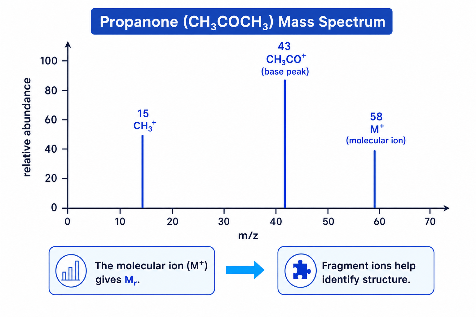
The molecular ion peak, often labelled M+\mathrm{M^+}M+, gives the relative molecular mass of the molecule.
For example, if the molecular ion peak is at m/z=58m/z = 58m/z=58, then the molecule has Mr=58M_r = 58Mr=58.
Using a molecular ion peak with an empirical formula
A compound has empirical formula C2H5O\mathrm{C_2H_5O}C2H5O and a molecular ion peak at m/z=90m/z = 90m/z=90. Find its molecular formula.
-
Calculate the empirical formula mass:
(2×12.0)+(5×1.0)+16.0=45.0(2 \times 12.0) + (5 \times 1.0) + 16.0 = 45.0(2×12.0)+(5×1.0)+16.0=45.0 -
Compare the molecular mass from the molecular ion peak with the empirical formula mass:
9045.0=2\frac{90}{45.0} = 245.090=2 -
Multiply every atom in the empirical formula by 2:
C2H5O×2=C4H10O2\mathrm{C_2H_5O} \times 2 = \mathrm{C_4H_{10}O_2}C2H5O×2=C4H10O2
Fragmentation: why there are many peaks
The molecular ion is often unstable. It can break into smaller pieces. This process is called fragmentation.
A typical fragmentation forms:
- one positive fragment ion, which is detected
- one neutral radical or molecule, which is not detected
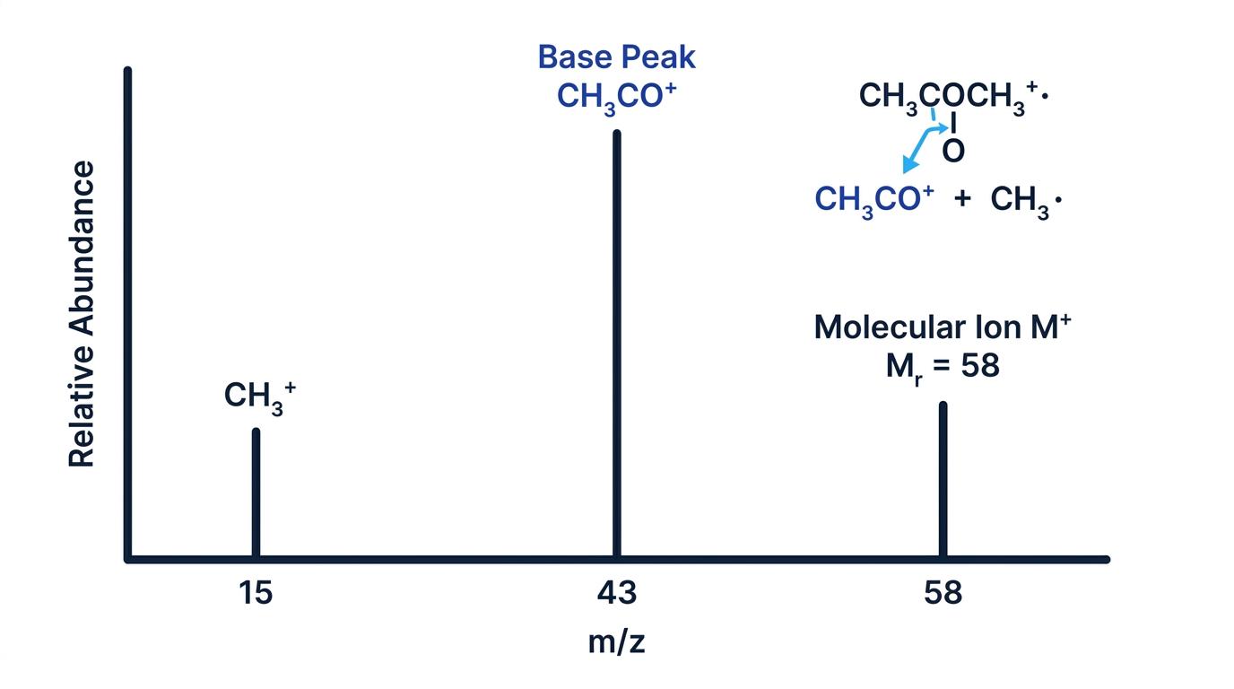
For example, propanone can fragment to form an acylium ion:
CH3COCH3+⋅→CH3CO++CH3⋅\mathrm{CH_3COCH_3^{+\cdot} \to CH_3CO^+ + CH_3^\cdot}CH3COCH3+⋅→CH3CO++CH3⋅The peak at m/z=43m/z = 43m/z=43 is due to CH3CO+\mathrm{CH_3CO^+}CH3CO+.
Only charged particles are detected
Mass spectrometers detect ions, not neutral fragments. When a molecule breaks apart, the peak you see comes from the fragment that keeps the positive charge.
Common fragment ions
You are not expected to memorise every possible fragment, but some common ones are very useful:
- m/z=15m/z = 15m/z=15: CH3+\mathrm{CH_3^+}CH3+
- m/z=29m/z = 29m/z=29: often C2H5+\mathrm{C_2H_5^+}C2H5+ or CHO+\mathrm{CHO^+}CHO+
- m/z=31m/z = 31m/z=31: often CH2OH+\mathrm{CH_2OH^+}CH2OH+, useful for alcohols
- m/z=43m/z = 43m/z=43: often CH3CO+\mathrm{CH_3CO^+}CH3CO+ or C3H7+\mathrm{C_3H_7^+}C3H7+
- m/z=57m/z = 57m/z=57: often C4H9+\mathrm{C_4H_9^+}C4H9+
Because different ions can have the same nominal mass, fragmentation gives clues rather than a complete structure by itself.
Using fragment peaks to suggest a structure
A compound has a molecular ion peak at m/z=58m/z = 58m/z=58 and strong fragment peaks at m/z=15m/z = 15m/z=15 and m/z=43m/z = 43m/z=43. Explain why propanone is a reasonable suggestion.
-
The molecular ion peak at m/z=58m/z = 58m/z=58 means the compound has Mr=58M_r = 58Mr=58.
-
Propanone, CH3COCH3\mathrm{CH_3COCH_3}CH3COCH3, has relative molecular mass:
(3×12.0)+(6×1.0)+16.0=58.0(3 \times 12.0) + (6 \times 1.0) + 16.0 = 58.0(3×12.0)+(6×1.0)+16.0=58.0 -
The peak at m/z=43m/z = 43m/z=43 can be explained by the fragment ion CH3CO+\mathrm{CH_3CO^+}CH3CO+, formed by breaking a C-C bond next to the carbonyl group.
-
The peak at m/z=15m/z = 15m/z=15 can be explained by CH3+\mathrm{CH_3^+}CH3+, showing that methyl fragments are present.
High-resolution mass spectrometry
A low-resolution mass spectrum gives whole-number m/zm/zm/z values. This can identify MrM_rMr, but different molecular formulae can have the same nominal mass.
High-resolution mass spectrometry measures masses much more precisely. This allows you to distinguish between possible molecular formulae.
High-resolution mass spectrometry
High-resolution mass spectrometry measures the accurate mass of an ion to several decimal places, allowing molecular formulae with the same nominal mass to be distinguished.
For accurate mass calculations, use precise isotopic masses, usually based on the most abundant isotopes:
- 12C=12.0000\mathrm{^{12}C} = 12.000012C=12.0000
- 1H=1.0078\mathrm{^1H} = 1.00781H=1.0078
- 16O=15.9949\mathrm{^{16}O} = 15.994916O=15.9949
- 14N=14.0031\mathrm{^{14}N} = 14.003114N=14.0031
Choosing a molecular formula from accurate mass
A molecular ion has accurate mass 88.0524. Three possible formulae are C4H8O2\mathrm{C_4H_8O_2}C4H8O2, C3H4O3\mathrm{C_3H_4O_3}C3H4O3 and C5H12O\mathrm{C_5H_{12}O}C5H12O. Identify the formula.
-
Calculate the accurate mass of C4H8O2\mathrm{C_4H_8O_2}C4H8O2:
Mr=(4×12.0000)+(8×1.0078)+(2×15.9949)=48.0000+8.0624+31.9898=88.0522\begin{aligned} M_r &= (4 \times 12.0000) + (8 \times 1.0078) + (2 \times 15.9949) \\ &= 48.0000 + 8.0624 + 31.9898 \\ &= 88.0522 \end{aligned}Mr=(4×12.0000)+(8×1.0078)+(2×15.9949)=48.0000+8.0624+31.9898=88.0522 -
Calculate the accurate mass of C3H4O3\mathrm{C_3H_4O_3}C3H4O3:
Mr=(3×12.0000)+(4×1.0078)+(3×15.9949)=36.0000+4.0312+47.9847=88.0159\begin{aligned} M_r &= (3 \times 12.0000) + (4 \times 1.0078) + (3 \times 15.9949) \\ &= 36.0000 + 4.0312 + 47.9847 \\ &= 88.0159 \end{aligned}Mr=(3×12.0000)+(4×1.0078)+(3×15.9949)=36.0000+4.0312+47.9847=88.0159 -
Calculate the accurate mass of C5H12O\mathrm{C_5H_{12}O}C5H12O:
Mr=(5×12.0000)+(12×1.0078)+15.9949=60.0000+12.0936+15.9949=88.0885\begin{aligned} M_r &= (5 \times 12.0000) + (12 \times 1.0078) + 15.9949 \\ &= 60.0000 + 12.0936 + 15.9949 \\ &= 88.0885 \end{aligned}Mr=(5×12.0000)+(12×1.0078)+15.9949=60.0000+12.0936+15.9949=88.0885 -
Compare each value with 88.0524. The closest match is C4H8O2\mathrm{C_4H_8O_2}C4H8O2, so this is the molecular formula.
Accurate mass strategy
If several formulae have the same whole-number MrM_rMr, calculate each accurate mass using the precise isotope masses given in the question, then choose the closest match to the measured molecular ion.
Isotope peaks: M+1 and M+2
Real samples contain naturally occurring isotopes, so a molecule can give small extra peaks near the molecular ion peak.
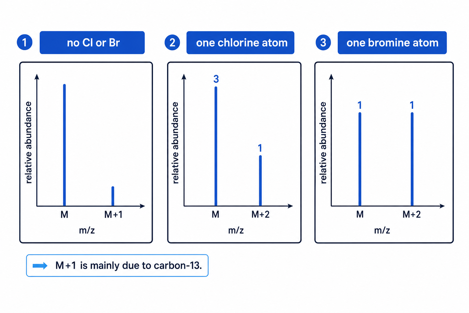
The M peak is the molecular ion made from the most common isotopes. The M+1 peak is one mass unit higher, often mainly due to one 13C\mathrm{^{13}C}13C atom instead of one 12C\mathrm{^{12}C}12C atom.
Since carbon-13 has a natural abundance of about 1.1%, the M+1 peak can estimate the number of carbon atoms:
number of carbon atoms≈M+1 abundance relative to M1.1\text{number of carbon atoms} \approx \frac{\text{M+1 abundance relative to M}}{1.1}number of carbon atoms≈1.1M+1 abundance relative to MEstimating the number of carbon atoms from M+1
A molecular ion peak has relative abundance 100. The M+1 peak has relative abundance 6.6. Estimate the number of carbon atoms in the molecule.
-
Use the carbon-13 approximation:
number of C atoms≈6.61.1\text{number of C atoms} \approx \frac{6.6}{1.1}number of C atoms≈1.16.6 -
Calculate the value:
6.61.1=6\frac{6.6}{1.1} = 61.16.6=6 -
The molecule is likely to contain 6 carbon atoms.
Chlorine and bromine patterns
Chlorine and bromine give very distinctive M and M+2 patterns because their common isotopes differ by two mass units.
- One chlorine atom gives M : M+2 of about 3 : 1.
- One bromine atom gives M : M+2 of about 1 : 1.
More than one halogen
The simple 3 : 1 chlorine and 1 : 1 bromine patterns apply to molecules with one chlorine or one bromine atom. With two halogen atoms, you get a cluster of M, M+2 and M+4 peaks.
Pulling the evidence together
Mass spectrometry is most powerful when combined with other techniques.
A good structure-deduction sequence is often:
- Use the molecular ion peak to find MrM_rMr.
- Use high-resolution mass spectrometry to find the molecular formula.
- Use M+1 and M+2 peaks to check for carbon count and halogens.
- Use fragmentation peaks to suggest parts of the structure.
- Combine with IR spectroscopy and NMR spectroscopy to confirm functional groups and the carbon-hydrogen framework.
What mass spectrometry can and cannot do
Mass spectrometry is excellent for molecular mass, molecular formula and fragment clues, but it rarely proves a full organic structure on its own. Use it alongside IR and NMR evidence.
In the exam
- Start with the molecular ion peak: it gives MrM_rMr, but check whether nearby M+1 or M+2 isotope peaks are present.
- For accurate mass questions, use the isotope masses given in the question and compare calculated values to the measured mass.
- Treat fragment ions as evidence for possible groups, not absolute proof of a whole structure.
- Watch for chlorine and bromine: M : M+2 ratios of about 3 : 1 and 1 : 1 are very high-value clues.
Check yourself
- Why does the molecular ion peak give the relative molecular mass of the compound?
- A compound has M and M+2 peaks in a 1 : 1 ratio. What element is strongly suggested?
- How could high-resolution mass spectrometry distinguish between two formulae with the same nominal mass?