What you'll learn
- The mechanics and strict regulation of the cell cycle, mitosis, and meiosis.
- How to prepare, stain, and calculate the mitotic index of plant root tissues.
- The adaptations of specialized animal and plant cells and how they form tissues.
- How stem cells differentiate into specialized cells and their promising roles in medicine.
The Eukaryotic Cell Cycle
In multicellular organisms, cells do not simply divide continuously. Instead, they progress through a highly regulated, continuous sequence of growth and division known as the cell cycle.
Cell Cycle
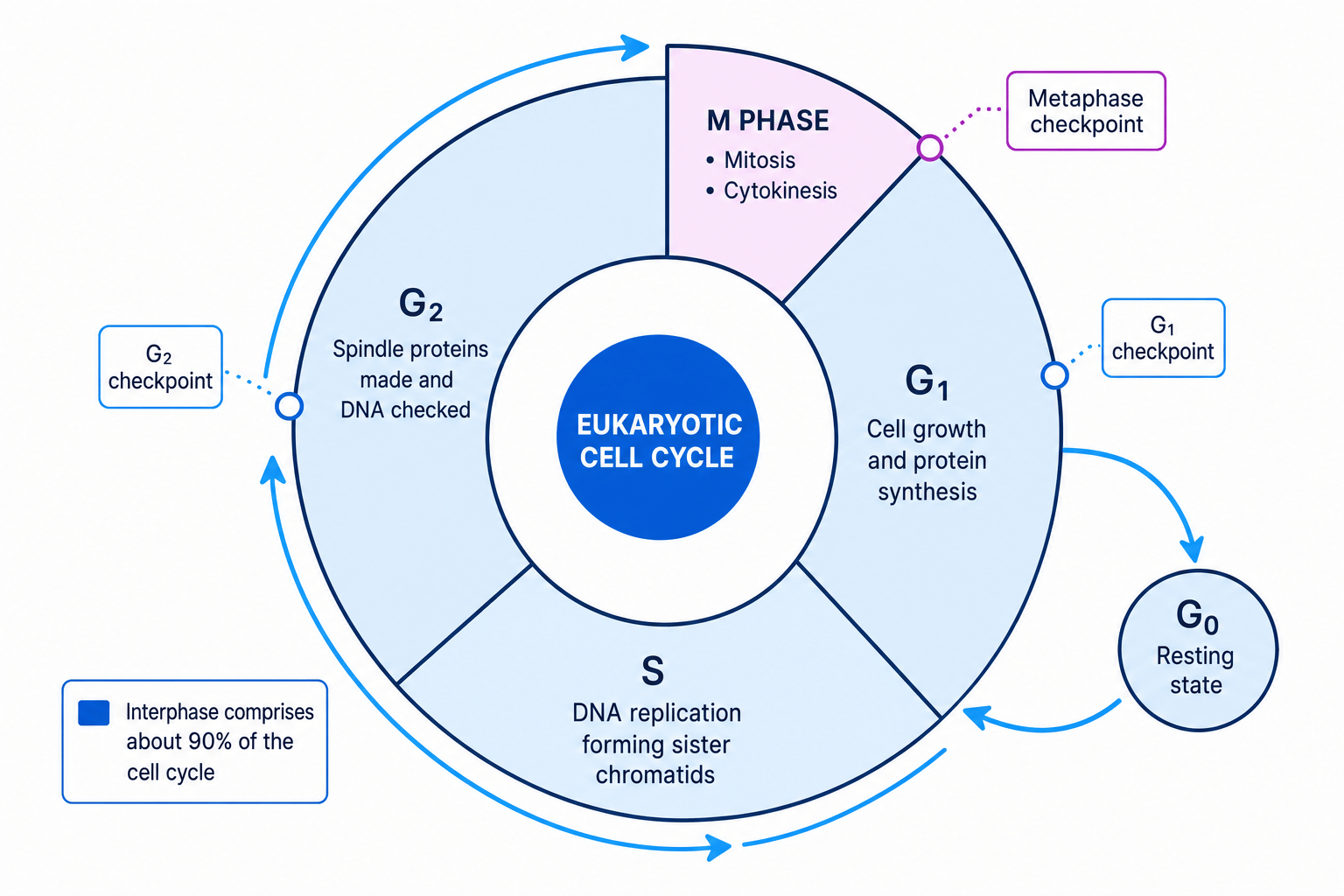
The cell cycle is the highly ordered sequence of events that takes place in a cell from its formation to its division into daughter cells. It is split into three main stages: interphase, mitosis (nuclear division), and cytokinesis (cytoplasmic division).
While mitosis is often the most visual part of the cycle, cells spend approximately 90% of their life in interphase. Interphase is an active period of growth, protein synthesis, and DNA replication. It is divided into three distinct sub-phases:
- G1G_1G1 (Gap 1) phase: The cell grows in size. Organelles replicate, and transcription and translation occur to produce proteins necessary for cell survival and division.
- SSS (Synthesis) phase: DNA replication occurs. Every chromosome is copied, resulting in two identical sister chromatids joined at a central region called the centromere. This is critical because it ensures that each daughter cell receives an exact, complete copy of the genetic material.
- G2G_2G2 (Gap 2) phase: The cell continues to grow and synthesizes proteins specifically needed for division (such as spindle tubulin). Energy stores (ATP) are replenished, and the replicated DNA is checked for errors.
Regulating the Cycle: Checkpoints
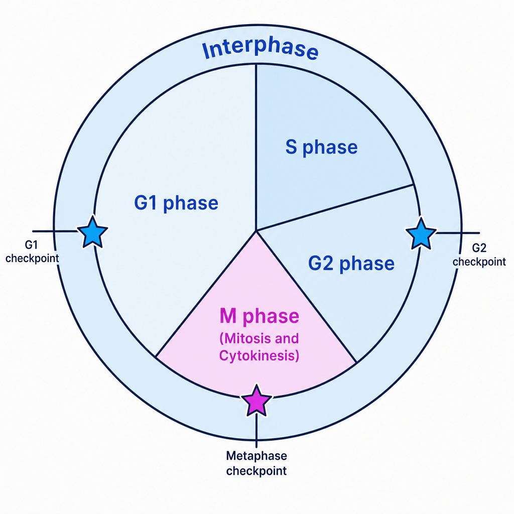
To prevent uncontrolled cell division (which can lead to tumors and cancer) and to ensure DNA is only replicated when undamaged, the cell cycle is controlled by checkpoints. These are chemical control points where the cell monitors its own progress before moving to the next phase.
- The G1G_1G1 checkpoint (Restriction point): Positioned near the end of G1G_1G1. It checks for cell size, nutrients, growth factors, and DNA damage. If the cell passes this checkpoint, it is committed to replication (SSS phase). If it does not, it enters a non-dividing state called G0G_0G0.
- The G2G_2G2 checkpoint: Located at the transition between G2G_2G2 and mitosis. It verifies that DNA replication in SSS phase was complete and checks for any DNA replication errors. If errors are found, division is halted until repairs are made.
- The Metaphase checkpoint (Spindle assembly checkpoint): Occurs during metaphase in mitosis. It checks that all chromosomes are correctly attached to the spindle fibers and aligned at the equator. This prevents unequal distribution of chromosomes (aneuploidy) in daughter cells.
Mitosis: Nuclear Division
Once a cell passes the G2G_2G2 checkpoint, it enters mitosis (MMM phase). The purpose of mitosis is to separate the replicated chromosomes, ensuring that each of the two new nuclei is genetically identical to the parent cell.
The Four Stages of Mitosis (PMAT)
Mitosis is a continuous process, but it is split into four distinct stages for ease of study: Prophase, Metaphase, Anaphase, and Telophase.
1. Prophase
- Chromatin condenses; chromosomes shorten and thicken, becoming visible under a light microscope.
- The nucleolus disappears and the nuclear envelope begins to break down.
- In animal cells, tiny organelles called centrioles migrate to opposite poles of the cell.
- Centrioles begin laying down a network of protein microtubules called spindle fibers.
2. Metaphase
- The nuclear envelope is completely broken down.
- Spindle fibers attach to the centromere of each chromosome.
- Spindle fibers contract, pulling the chromosomes until they align along the center of the cell, known as the metaphase plate (or equator).
3. Anaphase
- The centromere of each chromosome splits.
- Spindle fibers shorten, pulling the sister chromatids (now referred to as individual chromosomes) apart.
- The chromatids are pulled centromere-first to opposite poles of the cell, forming a characteristic 'V' shape.
4. Telophase
- The chromosomes reach the opposite poles of the cell and begin to decondense (uncoil), returning to diffuse chromatin.
- A new nuclear envelope reforms around each of the two sets of chromosomes.
- The nucleoli reappear and the spindle apparatus breaks down.
Chromatids vs. Chromosomes
During SSS phase, a chromosome replicates to form two sister chromatids. They are called chromatids as long as they are physically joined at the centromere. The moment they separate during anaphase, they are referred to as individual chromosomes. Do not call them chromatids once they have split!
Cytokinesis: Dividing the Cytoplasm
While mitosis is the division of the nucleus, cell division is not complete until cytokinesis divides the entire cytoplasm and organelles to form two distinct cells.
- In animal cells: A ring of actin filaments forms under the cell membrane. This contracts to create a cleavage furrow, pinching the membrane inwards until the cytoplasm splits into two.
- In plant cells: Because plants have a rigid cell wall, they cannot pinch inwards. Instead, vesicles from the Golgi apparatus align along the old metaphase plate and fuse to form a cell plate. This plate matures into a new cell membrane and middle lamella, upon which new cellulose cell walls are laid down.
Practical Skills (PAG 1): Preparing a Plant Root Tip Squash
To observe mitosis in action under a microscope, we use actively dividing plant tissues. In plants, cell division is restricted to specialized growth zones called meristems. The root tip is an excellent source of apical meristematic tissue.
Why do we carry out these specific steps?
- Using the very tip of the root (the apical meristem): This is where cells are actively undergoing mitosis. Cells further up the root are elongating and will be in interphase.
- Treating with hydrochloric acid (HCl): The acid hydrolyzes the middle lamella (the pectin "glue" holding plant cell walls together). This allows the tissue to separate easily into a single layer of cells.
- Staining (e.g., acetic orcein or toluidine blue): DNA is colorless. Stains bind specifically to chromatin/chromosomes (which are acidic), making them stand out in dark purple or blue against the cytoplasm.
- Squashing the sample: Pressing firmly down on the coverslip spreads the cells out into a single, thin layer. This ensures light can pass through the specimen under a light microscope.
Do not twist!
When squashing the root tip under the coverslip, press straight down firmly with your thumb. Never twist the coverslip, as this will shear the fragile chromosomes and break the cells, ruining your slide.
Calculating the Mitotic Index
The mitotic index is a quantitative measure of the proportion of cells in a tissue sample that are actively dividing.
Mitotic Index=Number of cells containing visible chromosomesTotal number of cells in the field of view \text{Mitotic Index} = \frac{\text{Number of cells containing visible chromosomes}}{\text{Total number of cells in the field of view}} Mitotic Index=Total number of cells in the field of viewNumber of cells containing visible chromosomesMultiply the result by 100 if you want to express it as a percentage.
Calculating the Mitotic Index
A student prepared a garlic root tip squash and counted the cells in a single field of view under a light microscope. They recorded the following raw data:
- Interphase: 154 cells
- Prophase: 16 cells
- Metaphase: 8 cells
- Anaphase: 4 cells
- Telophase: 6 cells
Calculate the mitotic index for this tissue sample, expressing your answer as a percentage to 3 significant figures.
- Sum the cells that are in any stage of active mitosis:
- Calculate the total number of cells observed in the field of view:
- Substitute the values into the Mitotic Index formula and calculate the percentage:
Rounding to 3 significant figures gives 18.1%18.1\%18.1%.
The Significance of Mitosis in Life Cycles
Mitosis is vital for life cycles across plants, animals, and fungi. It serves three principal biological roles:
- Growth: Multicellular organisms start as a single diploid cell (zygote). Mitosis allows this cell to divide repeatedly to produce millions of genetically identical cells, allowing the organism to increase in size.
- Tissue Repair: When tissues are damaged or worn out, new cells must replace them. Mitosis produces cells that are genetically identical to the original tissue cells to restore function. (Note: Mitosis replaces damaged cells; it does not repair the damaged cells themselves).
- Asexual Reproduction: Many organisms reproduce without gametes. Plants (e.g., runners in strawberries, bulbs in daffodils), fungi (e.g., budding in yeast), and some animals (e.g., budding in Hydra) use mitosis to produce genetically identical clones of the parent organism.
Meiosis and Genetic Variation
While mitosis produces genetically identical diploid cells, meiosis is a specialized form of cell division that produces genetically distinct haploid gametes.
Haploid and Diploid
- Diploid (2n2n2n): Cells containing two complete sets of chromosomes, one inherited from each parent. In humans, 2n=462n = 462n=46.
- Haploid (nnn): Cells containing a single set of unpaired chromosomes. Gametes (sperm and egg cells) are haploid. In humans, n=23n = 23n=23.
If gametes were diploid, the chromosome number would double with every generation. Meiosis halves the chromosome number, ensuring that when fertilisation occurs, the diploid number (2n2n2n) is restored in the zygote.
Introducing Genetic Variation
To ensure survival of a species in a changing environment, meiosis introduces genetic variation. This happens through two primary mechanisms:
1. Crossing Over (Prophase I)
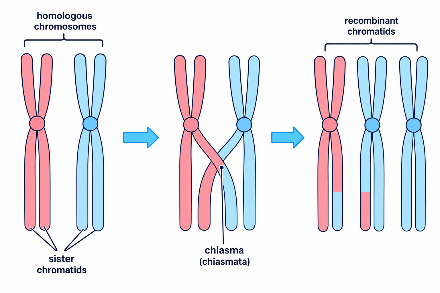
During Prophase I, maternal and paternal chromosomes pair up to form homologous pairs (also called bivalents). Non-sister chromatids wrap around each other and can break at points called chiasmata (singular: chiasma). The broken segments swap over and rejoin, resulting in a new combination of maternal and paternal alleles on the recombinant chromatids.
2. Independent Assortment
- In Metaphase I: Homologous pairs align randomly along the equator. Whether the maternal or paternal chromosome of a pair faces a particular pole is completely random. When they separate in Anaphase I, a random mix of maternal and paternal chromosomes goes to each pole.
- In Metaphase II: The sister chromatids align along the equator. Because of crossing over, the sister chromatids are no longer genetically identical. Their alignment and subsequent separation in Anaphase II is also completely random, generating further variation.
The Stages of Meiosis
Meiosis involves one round of DNA replication followed by two successive nuclear divisions: Meiosis I and Meiosis II.
Meiosis I: Reduction Division
In Meiosis I, the homologous pairs of chromosomes are separated, halving the chromosome number from diploid (2n2n2n) to haploid (nnn).
- Prophase I: Chromosomes condense. Homologous chromosomes pair up to form bivalents. Crossing over occurs at chiasmata. The nucleolus and nuclear envelope disappear, and the spindle forms.
- Metaphase I: Homologous pairs align side-by-side along the metaphase plate. Spindle fibers attach to the centromeres. Independent assortment of chromosomes occurs.
- Anaphase I: Spindle fibers contract, pulling the homologous chromosomes apart to opposite poles. Crucially, the centromeres do not split, so each chromosome still consists of two sister chromatids.
- Telophase I: Nuclear envelopes reform around the two new nuclei, and chromosomes decondense. Cytokinesis follows, producing two haploid (nnn) cells.
Meiosis II: Mitotic-like Division
In Meiosis II, which occurs in both of the newly formed cells, the sister chromatids are separated.
- Prophase II: Chromosomes re-condense, nuclear envelopes break down, and new spindle fibers set up at right angles to the original spindle.
- Metaphase II: Individual chromosomes align along the equator. Independent assortment of sister chromatids occurs.
- Anaphase II: Centromeres split, and sister chromatids are pulled apart to opposite poles of the cells.
- Telophase II: Nuclear envelopes reform, chromosomes decondense, and cytokinesis occurs. This results in four genetically distinct haploid (nnn) daughter cells.
Mitosis vs. Meiosis Summary
Keep this clean distinction clear in your mind for comparative questions:
- Mitosis: 1 division, produces 2 diploid cells, genetically identical clones, no pairing of homologous chromosomes, no crossing over.
- Meiosis: 2 divisions, produces 4 haploid cells, genetically unique cells, homologous chromosomes pair up and cross over.
Cellular Specialisation and Organisation
Multicellular organisms are large and complex. To survive, they rely on cellular specialisation (differentiation) and structural organisation. Cells do not work in isolation; they are organized into hierarchically structured units:
Specialised Cells⟶Tissues⟶Organs⟶Organ Systems⟶Whole Organism \text{Specialised Cells} \longrightarrow \text{Tissues} \longrightarrow \text{Organs} \longrightarrow \text{Organ Systems} \longrightarrow \text{Whole Organism} Specialised Cells⟶Tissues⟶Organs⟶Organ Systems⟶Whole OrganismSpecialised Animal Cells
- Erythrocytes (Red Blood Cells): Adapted for oxygen transport. They have a biconcave disc shape to maximise surface area-to-volume ratio, contain no nucleus or internal organelles to leave maximum space for haemoglobin, and are highly flexible to squeeze through narrow capillaries.
- Neutrophils (White Blood Cells): Key to the immune response. They have a multi-lobed nucleus which allows them to deform easily to squeeze through junctions in capillary walls (diapedesis). Their cytoplasm contains many lysosomes packed with hydrolytic enzymes to digest engulfed pathogens.
- Squamous Epithelial Cells: Very flat, thin, plate-like cells that form a single layer lining surfaces like the alveoli. This structure provides a very short diffusion pathway for rapid gas exchange.
- Ciliated Epithelial Cells: Columnar cells found in the trachea and bronchi. They have tiny hair-like projections called cilia on their apical surface, which beat in a coordinated rhythm to sweep mucus (containing trapped dust and pathogens) up and away from the lungs.
- Sperm Cells: Male gametes. They feature a flagellum (tail) for motility, a middle section packed with mitochondria to generate ATP for swimming, an acrosome (head vesicle) containing hydrolytic enzymes to penetrate the egg's outer layer, and a haploid nucleus.
Specialised Plant Cells
- Palisade Mesophyll Cells: Located in leaves for photosynthesis. They are long and cylindrical, packing together tightly. They contain a high density of chloroplasts and a large central vacuole that pushes chloroplasts to the edge of the cell, minimising the diffusion distance for carbon dioxide.
- Root Hair Cells: Located near growing root tips. They have a long hair-like extension that dramatically increases the surface area for absorbing water (by osmosis) and mineral ions (by active transport). They feature thin cell walls and a high concentration of mitochondria to fuel active transport.
- Guard Cells: Found in pairs surrounding stomatal pores. Their inner cell walls are thick and rigid, while their outer walls are thin and flexible. When they take up water, they swell unevenly and bend outward, opening the stoma to allow gas exchange.
Organisation into Tissues, Organs, and Organ Systems
A tissue is a collection of differentiated cells that work together to perform a specific function.
Key Animal Tissues
- Squamous Epithelium: A single layer of flat cells forming a smooth, ultra-thin lining (e.g., in alveoli and blood vessels) to facilitate rapid diffusion.
- Ciliated Epithelium: A layer of ciliated cells interspaced with mucus-secreting goblet cells (e.g., in the trachea) to trap and remove inhaled particulates.
- Cartilage: A firm, flexible connective tissue made of chondrocyte cells embedded in an extracellular matrix of collagen and elastin fibers. It provides structural support (e.g., keeping airways open) and prevents friction at joints.
- Muscle: Highly cellular tissue containing contractile fibers (actin and myosin filaments) that slide over each other to shorten cells, allowing movement.
Key Plant Tissues
- Xylem Tissue: Composed of dead, hollow vessels aligned end-to-end with no end walls to form continuous tubes. The walls are reinforced with waterproof lignin, which provides mechanical support and allows the transport of water and dissolved minerals from roots to leaves.
- Phloem Tissue: Responsible for transporting organic assimilates (translocation). It consists of sieve tube elements (living cells with heavily reduced cytoplasm and no nucleus, joined by perforated sieve plates) and metabolically active companion cells loaded with mitochondria to actively load sucrose into the tubes.
Stem Cells and Differentiation
Multicellular organisms start as a single undifferentiated cell. To build specialized tissues, they require stem cells.
Stem Cell
A stem cell is an undifferentiated cell that is capable of dividing by mitosis an unlimited number of times (self-renewal) and has the potential to differentiate into a variety of specialised cell types.
Potency of Stem Cells
The range of cell types a stem cell can differentiate into is called its potency:
- Totipotent: Can differentiate into any cell type, including extra-embryonic tissues like the placenta (e.g., a zygote).
- Pluripotent: Can differentiate into any cell type that forms the body of the embryo, but not extra-embryonic tissues.
- Multipotent: Adult stem cells that can only differentiate into a limited range of cell types within a specific tissue (e.g., bone marrow stem cells).
Case Study 1: Animal Differentiation in the Bone Marrow
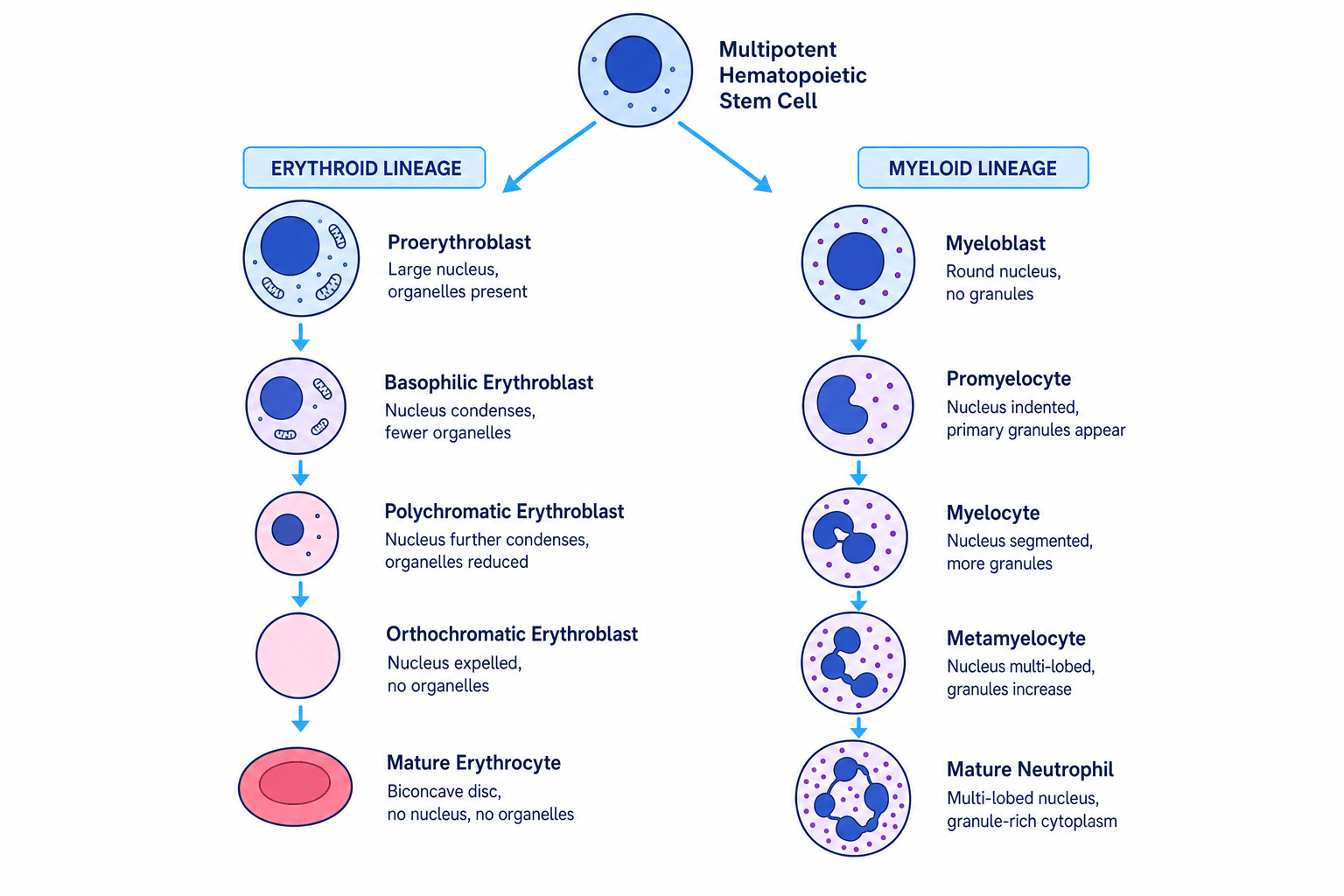
Adult stem cells in the red bone marrow are multipotent hematopoietic stem cells. They divide continuously to produce millions of cells every second, which differentiate into distinct blood cells depending on chemical signals:
- To become erythrocytes, the cell synthesizes massive quantities of haemoglobin, gradually shrinks, ejects its nucleus and organelles, and collapses into a biconcave shape.
- To become neutrophils, the cell develops a multi-lobed nucleus and synthesizes thousands of lysosomal granules in its cytoplasm.
Case Study 2: Plant Differentiation in the Meristem
In plants, stem cells are located in meristems. A layer of unspecialized cells called the vascular cambium sits between the xylem and phloem in vascular bundles:
- Cells that differentiate inwards toward the center of the stem become xylem vessels. Lignin is deposited in their walls, making them waterproof. This blocks the entry of nutrients, causing the cell contents to die and clear out, leaving a hollow tube.
- Cells that differentiate outwards become phloem sieve tubes or companion cells. Sieve tube elements lose most of their organelles but remain alive, supported by their adjacent companion cells.
Stem Cells in Research and Medicine
Because stem cells can replace damaged or dead cells, they hold enormous promise for modern medicine and research:
- Repair of Damaged Tissues: Stem cells can be used to grow skin grafts for severe burn victims, or to regenerate damaged heart muscle tissue following a myocardial infarction (heart attack).
- Treatment of Neurological Conditions: Neurological diseases involve the irreversible loss of specialized neurons. Stem cells can be directed to differentiate into functional neurons to treat conditions like Parkinson's disease (by replacing dopamine-producing cells) or Alzheimer's disease.
- Research into Developmental Biology: By studying how stem cells differentiate into specialized tissues in the lab, scientists can understand how organisms grow, how birth defects occur, and how cancer cells bypass cell cycle controls.
In the exam
- Be specific with terminology: Never say mitosis is "cell division". It is the division of the nucleus. The division of the cell itself is cytokinesis.
- State adaptations clearly: When describing specialized cells, always state the structural feature and connect it explicitly to its functional benefit (e.g., "Many mitochondria to synthesise ATP for active transport of ions").
- Show your workings: For mitotic index calculations, show every step of your calculation clearly. Marks are awarded for the correct intermediate steps even if you make a simple typing error on your calculator.
- Meiosis chromosome counts: Always check whether a question is asking for the diploid (2n2n2n) or haploid (nnn) number of chromosomes.
Check yourself
- Why does the S phase of interphase have to occur before mitosis can begin?
- How does crossing over in Prophase I differ from independent assortment in Metaphase I?
- Describe two key structural differences between a mature erythrocyte and a mature neutrophil.