

What you'll learn
- Why membranes are essential barriers, reaction surfaces and signalling structures.
- How the fluid mosaic model explains membrane structure and function.
- How substances cross membranes by diffusion, facilitated diffusion, active transport, endocytosis and exocytosis.
- How temperature, solvents and water potential affect membranes in practical investigations.
1. Why cells need membranes
A biological membrane is a thin structure made mainly from lipids and proteins. The membrane at the edge of a cell is the plasma membrane or cell surface membrane. Eukaryotic cells also contain membranes around and inside organelles, such as mitochondria, chloroplasts, the endoplasmic reticulum and the Golgi apparatus.
Partially permeable membrane
A partially permeable membrane allows some substances to pass through more easily than others. This lets cells control their internal conditions rather than simply becoming the same as their surroundings.
Membranes have three big roles.
Barriers and compartments
At the cell surface, the plasma membrane separates the cytoplasm from the external environment. It controls what enters and leaves the cell, helping maintain a stable internal environment.
Inside cells, membranes separate organelles from the cytoplasm and can also form compartments within organelles. For example, mitochondria have folded inner membranes, and chloroplasts contain thylakoid membranes. These compartments allow different reactions to happen in different places.
Sites of chemical reactions
Some membrane proteins are enzymes. This means membranes can act as surfaces where chemical reactions happen efficiently. For example, many reactions in respiration occur on the inner mitochondrial membrane.
Cell communication
Membranes also help cells communicate. Some membrane proteins act as receptors, which are molecules with a specific shape that bind to signalling molecules such as hormones. Many medicinal drugs also work by binding to membrane-bound receptors.
Why membranes matter
Membranes are not just “bags” around cells. They control exchange, organise reactions into compartments and allow cells to respond to signals.
2. The fluid mosaic model
The accepted model of membrane structure is the fluid mosaic model.
Fluid mosaic model
The fluid mosaic model describes membranes as a fluid phospholipid bilayer with proteins and other molecules scattered through it. “Fluid” means many components can move sideways. “Mosaic” means the membrane is made of many different parts.
A phospholipid is a lipid molecule with a hydrophilic phosphate head and two hydrophobic fatty acid tails. Hydrophilic means attracted to water. Hydrophobic means repelled by water.
In water, phospholipids form a bilayer. The phosphate heads face the watery cytoplasm and extracellular fluid, while the fatty acid tails point inwards away from water.
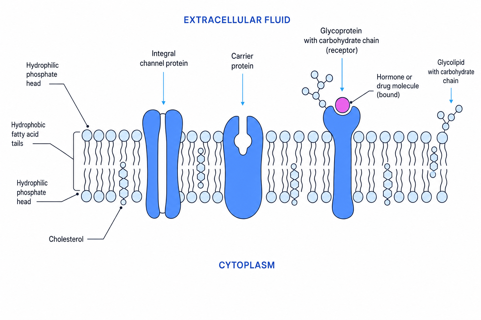
Main membrane components
Phospholipids form the basic bilayer. The hydrophobic centre of the bilayer makes it difficult for ions and large polar molecules to pass through directly.
Cholesterol fits between phospholipids in animal cell membranes. It helps regulate membrane fluidity and stability. It also reduces permeability to small water-soluble molecules.
Membrane proteins include channel proteins, carrier proteins, enzymes and receptors. Some proteins sit on one side of the membrane; others span the whole bilayer.
Glycoproteins are proteins with carbohydrate chains attached. Glycolipids are lipids with carbohydrate chains attached. These carbohydrate chains can help with cell recognition, adhesion and signalling.
Membranes are not static
Do not describe the membrane as a fixed, solid wall. In the fluid mosaic model, phospholipids and some proteins can move sideways within the bilayer.
Predicting the effect of changing cholesterol content
- Cholesterol normally helps stabilise the bilayer and reduces excessive movement of phospholipids.
- If cholesterol content falls, the membrane may become less stable and more permeable, especially to small molecules that would usually be restricted.
- Therefore, a cell with too little cholesterol may have poorer control over what enters and leaves through its membrane.
3. Factors affecting membrane structure and permeability
Permeability means how easily substances can pass through a membrane. Anything that disrupts the phospholipid bilayer or membrane proteins can change permeability.
Temperature
At low temperatures, phospholipids have less kinetic energy, so the membrane is less fluid.
As temperature rises, phospholipids move more. This can increase permeability because the bilayer becomes more fluid.
At high temperatures, membrane proteins can denature, meaning their shape changes. The bilayer can also become disrupted. This often causes a sharp increase in permeability.
Solvents
Some solvents, such as ethanol, dissolve or disrupt lipids. Because membranes contain many phospholipids, solvents can damage the bilayer and increase leakage from cells.
Investigating membrane permeability
A common practical uses beetroot tissue. Beetroot cells contain a red pigment called betalain inside the vacuole. If membranes are damaged, pigment leaks out into the surrounding solution.
You can investigate the effect of temperature or ethanol concentration by measuring the colour intensity of the solution using a colorimeter. Higher absorbance usually means more pigment has leaked out, so membrane permeability is greater.
Good control variables include:
- same size and surface area of beetroot pieces
- same volume of solution
- same exposure time
- same pH
- washing beetroot pieces first to remove pigment released by cutting
- repeating each treatment and calculating a mean
Absorbance versus transmission
In a beetroot colorimeter practical, higher absorbance means more pigment in solution. Higher percentage transmission means less light was absorbed, so it may indicate less pigment. Check which measurement the question gives you.
Interpreting beetroot permeability results
- Suppose beetroot in 20 °C water gives a low absorbance, but beetroot in 70 °C water gives a much higher absorbance.
- The higher absorbance shows that more red pigment has leaked from the beetroot cells into the water.
- This suggests the higher temperature disrupted membranes, increasing permeability, partly because membrane proteins may have denatured and the bilayer became more fluid.
4. Movement of molecules across membranes
Substances move across membranes in several ways. The key question is whether movement is down a gradient or against a gradient.
A concentration gradient is a difference in concentration between two regions. Movement down a concentration gradient means movement from higher concentration to lower concentration.
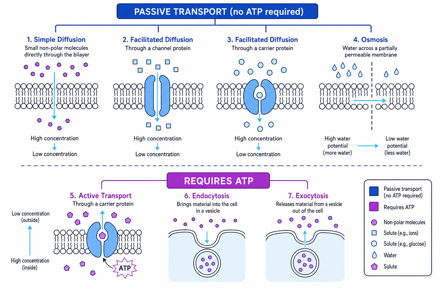
Diffusion
Diffusion
Diffusion is the net movement of particles from a region of higher concentration to a region of lower concentration, down a concentration gradient, due to random movement.
Diffusion is passive, meaning it does not require ATP. Small non-polar molecules such as oxygen and carbon dioxide can diffuse directly through the phospholipid bilayer.
The rate of diffusion is affected by factors including:
- steepness of the concentration gradient
- surface area
- diffusion distance
- temperature
- size of molecule
Facilitated diffusion
Facilitated diffusion is also passive. It is the movement of substances down a concentration gradient through membrane proteins.
Channel proteins form hydrophilic pores that allow specific ions or molecules through. Carrier proteins bind to a specific molecule, change shape, and release it on the other side of the membrane.
Because facilitated diffusion depends on proteins, it can become limited when all available proteins are being used.
Active transport
Active transport
Active transport is the movement of substances across a membrane against a concentration gradient, using ATP as the immediate energy source.
ATP stands for adenosine triphosphate. In active transport, ATP provides energy for carrier proteins to change shape and move substances from lower concentration to higher concentration.
Endocytosis and exocytosis
Some materials are too large to move through proteins in the membrane.
Endocytosis brings material into a cell. The membrane folds inwards around the material and forms a vesicle, which is a small membrane-bound sac.
Exocytosis releases material from a cell. A vesicle fuses with the plasma membrane and releases its contents outside the cell.
Both processes require ATP because membranes and vesicles must be moved and reshaped.
Choosing the correct transport mechanism
- If a molecule moves from higher concentration to lower concentration without a membrane protein, it is likely to be simple diffusion.
- If it moves down its concentration gradient but needs a channel or carrier protein, it is facilitated diffusion.
- If it moves from lower concentration to higher concentration, it is active transport and must use ATP.
5. Investigating diffusion in model cells
A model cell is a simplified system used to represent a real cell. In diffusion investigations, agar cubes are often used as model cells because their size and surface area can be controlled.
One method uses agar containing an indicator. When the agar is placed in acid or alkali, the colour change shows how far the substance has diffused.
Smaller cubes have a larger surface area to volume ratio, so diffusion reaches the centre more quickly. This helps explain why small cells can exchange substances more efficiently than large cells.
Calculating diffusion rate in agar
- Convert the diffusion distance into metres: 3.0 mm is 3.0×10⁻³ m3.0 \times 10^{-3}\text{ m}3.0×10⁻³ m.
- Use the rate equation:
- Substitute the values:
6. Osmosis and water potential
Osmosis is a special case of diffusion involving water.
Osmosis
Osmosis is the net movement of water molecules from a region of higher water potential to a region of lower water potential, across a partially permeable membrane.
Water potential, given the symbol ψ\psiψ, is a measure of the tendency of water molecules to move. Pure water has the highest water potential, defined as 0 kPa. Adding solute lowers water potential, making it more negative.
So water moves from less negative water potential to more negative water potential.
Effects on animal cells
Animal cells do not have cell walls.
If an animal cell is placed in a solution with higher water potential than its cytoplasm, water enters by osmosis. The cell may swell and burst. Bursting is called lysis.
If an animal cell is placed in a solution with lower water potential than its cytoplasm, water leaves by osmosis. The cell shrinks. In red blood cells, this is called crenation.
Effects on plant cells
Plant cells have cellulose cell walls.
If a plant cell is placed in a solution with higher water potential than its cell sap, water enters the vacuole by osmosis. The vacuole swells and pushes the cytoplasm against the cell wall. The cell becomes turgid.
If a plant cell is placed in a solution with lower water potential than its cell sap, water leaves by osmosis. The vacuole shrinks and the plasma membrane pulls away from the cell wall. This is plasmolysis.
Use water potential language
For OCR, explain osmosis using a water potential gradient across a partially permeable membrane. Avoid only saying “water moves from dilute to concentrated” unless you also link it to water potential.
7. Investigating water potential in cells
A common plant practical uses potato cylinders placed in different sucrose concentrations. You measure initial and final mass, then calculate percentage change in mass.
If mass increases, water has entered the potato cells by osmosis. If mass decreases, water has left. The sucrose concentration where percentage change is zero is the isotonic point, where there is no net movement of water.
Animal cells can also be investigated using red blood cells in solutions of different water potential. In high water potential solutions, haemolysis may occur. In low water potential solutions, cells may crenate.
Good practical design includes:
- using equal-sized tissue pieces
- blotting tissue dry before weighing
- using the same solution volume and exposure time
- controlling temperature
- using repeats and calculating a mean
- plotting percentage change against solution concentration
Calculating percentage change in mass
- Use the percentage change formula:
- Substitute the values for a potato cylinder with initial mass 2.40×10⁻³ kg2.40 \times 10^{-3}\text{ kg}2.40×10⁻³ kg and final mass 2.28×10⁻³ kg2.28 \times 10^{-3}\text{ kg}2.28×10⁻³ kg:
- Calculate and interpret:
The negative value means the potato lost mass, so water moved out of the cells into a solution with lower water potential.
In the exam
- For transport questions, state the direction of movement and whether ATP is required.
- For osmosis questions, use the terms water potential gradient and partially permeable membrane.
- For practical questions, link the independent variable to membrane damage or diffusion rate, then mention controls, repeats and a suitable graph.
Check yourself
- Why can oxygen diffuse directly through the phospholipid bilayer, but ions usually cannot?
- How does ethanol increase membrane permeability?
- What happens to a plant cell placed in a solution with lower water potential than its cell sap?
