What you'll learn
- Why surface area to volume ratio affects whether diffusion alone is enough.
- The shared features of efficient exchange surfaces.
- How mammalian lungs ventilate and exchange gases, including spirometer data.
- How bony fish and insects ventilate and exchange gases, plus practical microscopy and dissection skills.
Why organisms need exchange surfaces
All cells need substances to enter and leave. Oxygen enters many cells for aerobic respiration, while carbon dioxide leaves as a waste product. Small molecules often move by diffusion, which is the net movement of particles from a region of higher concentration to a region of lower concentration.
A concentration gradient is the difference in concentration between two areas. A steeper gradient gives faster diffusion.
Exchange surface
An exchange surface is a specialised boundary across which substances move between an organism and its environment, or between two parts of the body, usually by diffusion.
Single-celled organisms
A single-celled organism is small, so it has a relatively large surface area compared with its volume. Its cell membrane can usually exchange enough oxygen, carbon dioxide, nutrients, and waste by diffusion alone.
The diffusion distance is also short: substances do not need to travel far to reach the middle of the cell.
Multicellular organisms
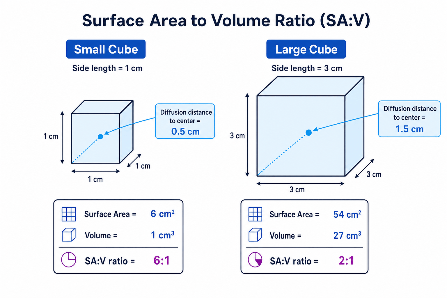
As organisms become larger, their surface area to volume ratio decreases. This means there is less surface available for exchange per unit volume of living tissue.
Large animals also often have high metabolic activity, meaning their cells carry out many chemical reactions per second. Active tissues need oxygen quickly and produce carbon dioxide quickly, so diffusion across the outer body surface is not enough.
That is why large, active organisms need:
- specialised exchange surfaces,
- ventilation systems to refresh the exchange medium,
- transport systems, such as blood, to move substances around the body.
Surface area to volume ratio
The surface area to volume ratio compares how much outer surface an object has with how much internal volume it contains.
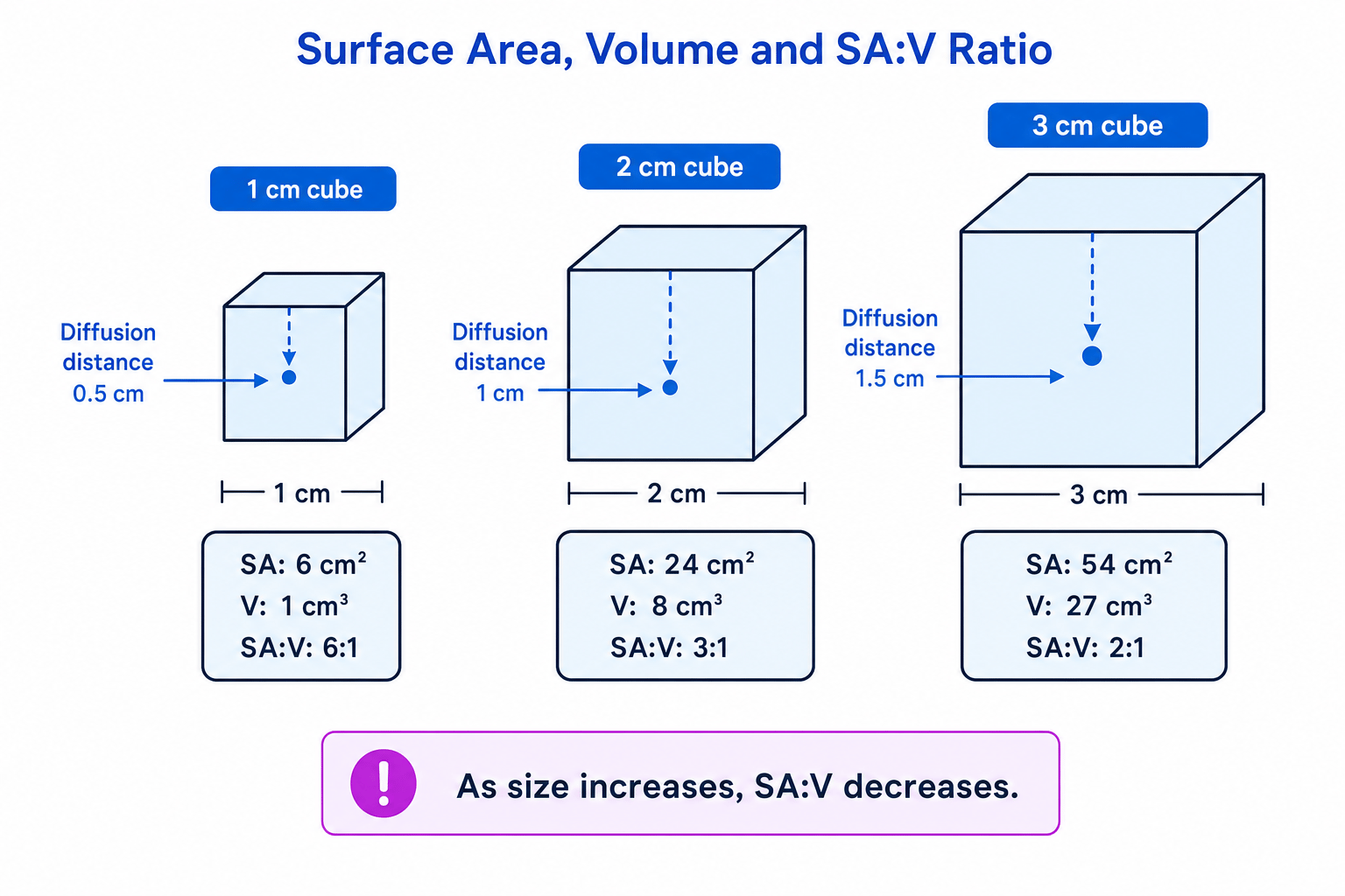
surface area to volume ratio=surface areavolume\text{surface area to volume ratio} = \frac{\text{surface area}}{\text{volume}}surface area to volume ratio=volumesurface areaAs size increases, volume increases faster than surface area. This makes diffusion less effective for supplying all the cells inside a large organism.
The diagram shows how increasing cube size decreases surface area to volume ratio and increases diffusion distance.
Calculating surface area to volume ratio
- For a cube with side length ℓ=2.0 cm\ell = 2.0\ \text{cm}ℓ=2.0 cm, calculate surface area using 6ℓ26\ell^26ℓ2:
- Calculate volume using ℓ3\ell^3ℓ3:
- Divide surface area by volume:
- This is commonly written as an SA:V ratio of 3:1. A smaller cube has a higher SA:V ratio, so it has more surface available for exchange per unit volume.
Why size matters
Small organisms may rely on diffusion across their body surface, but large or very active organisms need specialised exchange surfaces because their SA:V ratio is too low and their metabolic demand is too high.
Features of an efficient exchange surface
Efficient exchange surfaces usually have the same core adaptations.
Large surface area
A larger surface area allows more particles to diffuse at the same time.
Examples include:
- root hair cells, which are plant epidermal cells with long projections that increase surface area for absorbing water and mineral ions,
- many tiny alveoli in mammalian lungs,
- many gill filaments and gill lamellae in fish,
- highly branched tracheoles in insects.
Thin exchange barrier
A thin layer gives a short diffusion distance, so molecules cross quickly. In the lungs, the alveolar epithelium and capillary endothelium are each one cell thick.
Steep concentration gradient
A steep gradient is maintained when substances are constantly supplied on one side and removed on the other.
In mammals, ventilation brings fresh air into the alveoli, while blood flow removes oxygen and brings carbon dioxide. In fish, ventilation moves water over the gills, while blood flow carries oxygen away.
Moist and permeable surface
Gases must dissolve before diffusing across cell membranes, so gas exchange surfaces are moist. The membranes must also be permeable to the exchanged substances.
Only saying ‘large surface area’
For full explanations, link the feature to the effect: for example, “many alveoli give a large surface area, so more oxygen molecules can diffuse into the blood per second.”
Mammalian gaseous exchange system
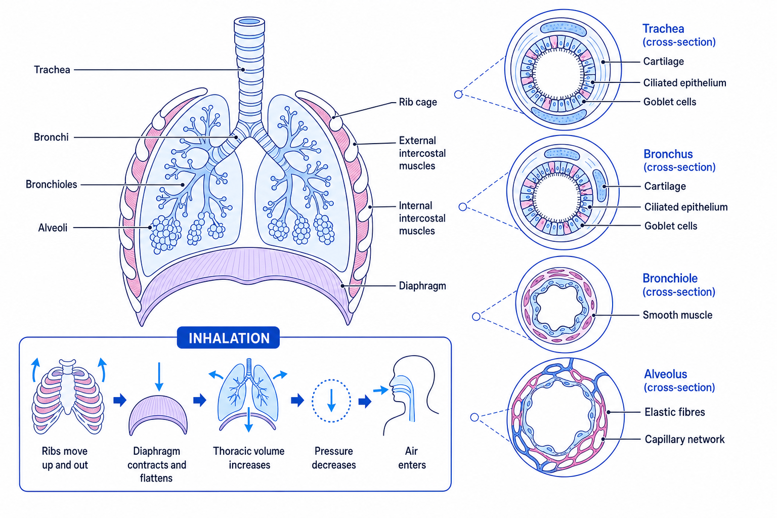
In mammals, air moves through the trachea, bronchi, bronchioles, and into the alveoli. The alveoli are the main gas exchange surfaces, surrounded by dense capillary networks.
This diagram shows the main structures of the mammalian gaseous exchange system, the tissues in their walls, and the sequence of inhalation.
Key tissues and their functions
A ciliated epithelium is a layer of cells with tiny hair-like cilia. The cilia beat to move mucus towards the throat, where it can be swallowed. Goblet cells secrete mucus, which traps dust, pollen, pathogens, and other particles.
| Structure | Main tissues present | Function |
|---|---|---|
| Trachea | Cartilage, ciliated epithelium, goblet cells, smooth muscle, elastic fibres | Cartilage keeps the airway open; mucus traps particles; cilia move mucus away from lungs |
| Bronchi | Cartilage, ciliated epithelium, goblet cells, smooth muscle, elastic fibres | Carry air into each lung and help clean, warm, and moisten it |
| Bronchioles | Smooth muscle, elastic fibres, ciliated epithelium; little or no cartilage | Control airflow by changing diameter |
| Alveoli | Thin squamous epithelium, elastic fibres, capillary network | Provide a thin, large surface for diffusion and recoil during exhalation |
Cartilage is found in the trachea and bronchi, but not in bronchioles. This matters because bronchioles can narrow or widen more easily due to smooth muscle contraction or relaxation.
Putting cartilage in the alveoli
Alveoli do not contain cartilage. They need thin walls for rapid diffusion, plus elastic fibres to stretch during inhalation and recoil during exhalation.
Ventilation in mammals
Ventilation
Ventilation is the mass movement of air into and out of the lungs. It maintains steep concentration gradients for oxygen and carbon dioxide at the alveoli.
Inhalation
During inhalation:
- the external intercostal muscles contract,
- the internal intercostal muscles relax,
- the ribs move upwards and outwards,
- the diaphragm contracts and flattens,
- the volume of the thorax increases,
- pressure in the lungs falls below atmospheric pressure,
- air moves into the lungs.
Exhalation
During quiet exhalation:
- the external intercostal muscles relax,
- the diaphragm relaxes and becomes dome-shaped,
- elastic recoil of lung tissue helps reduce lung volume,
- thoracic volume decreases,
- pressure in the lungs rises above atmospheric pressure,
- air moves out.
During forced exhalation, the internal intercostal muscles contract, pulling the ribs down and in more strongly.
Predicting airflow during breathing
-
If thoracic volume decreases, the same amount of air is compressed into a smaller space, so pressure inside the lungs increases.
-
Air moves from a region of higher pressure to a region of lower pressure, so air moves out of the lungs.
-
This is exhalation. At rest, it mainly involves relaxation of the diaphragm and external intercostal muscles; during forced exhalation, the internal intercostal muscles contract.
Vital capacity, tidal volume, breathing rate, and oxygen uptake
A spirometer or data logger can record changes in lung volume over time.
Key terms:
- Tidal volume: the volume of air breathed in or out in one normal breath.
- Breathing rate: the number of breaths per minute.
- Vital capacity: the maximum volume of air that can be breathed out after the deepest possible breath in.
- Oxygen uptake: the volume of oxygen absorbed by the body per unit time.
Minute ventilation can be estimated using:
ventilation rate=tidal volume×breathing rateoxygen uptake=decrease in gas volumetime\begin{aligned} \text{ventilation rate} &= \text{tidal volume} \times \text{breathing rate} \\ \text{oxygen uptake} &= \frac{\text{decrease in gas volume}}{\text{time}} \end{aligned}ventilation rateoxygen uptake=tidal volume×breathing rate=timedecrease in gas volumeIn a closed spirometer, carbon dioxide is absorbed by soda lime. Therefore, the gradual fall in the trace is mainly due to oxygen being removed from the air and taken up by the body.
Calculating breathing rate and oxygen uptake
- A spirometer trace shows 6 breaths in 30 s. Convert to breaths per minute:
- If tidal volume is 0.50 dm3 breath−10.50\ \text{dm}^3\ \text{breath}^{-1}0.50 dm3 breath−1, calculate ventilation rate:
- If the trace baseline falls from 3.40 dm33.40\ \text{dm}^33.40 dm3 to 3.10 dm33.10\ \text{dm}^33.10 dm3 in 2.0 min, calculate oxygen uptake:
Reading spirometer traces
Use peak-to-trough height for tidal volume, count complete breaths for breathing rate, and use the downward slope of the baseline for oxygen uptake.
Gas exchange in bony fish
Bony fish use gills because water contains less oxygen than air and is harder to move. The buccal cavity is the mouth cavity. The operculum is the bony flap covering the gills.
Water is ventilated over the gills by pressure changes:
- The mouth opens, the operculum closes, and the buccal cavity volume increases, lowering pressure so water enters.
- The mouth closes, the buccal cavity volume decreases, and pressure rises.
- The operculum opens, so water is forced over the gills and out.
Gills have many gill filaments, which carry many thin gill lamellae or gill plates. Lamellae give a large surface area, short diffusion distance, and rich blood supply.
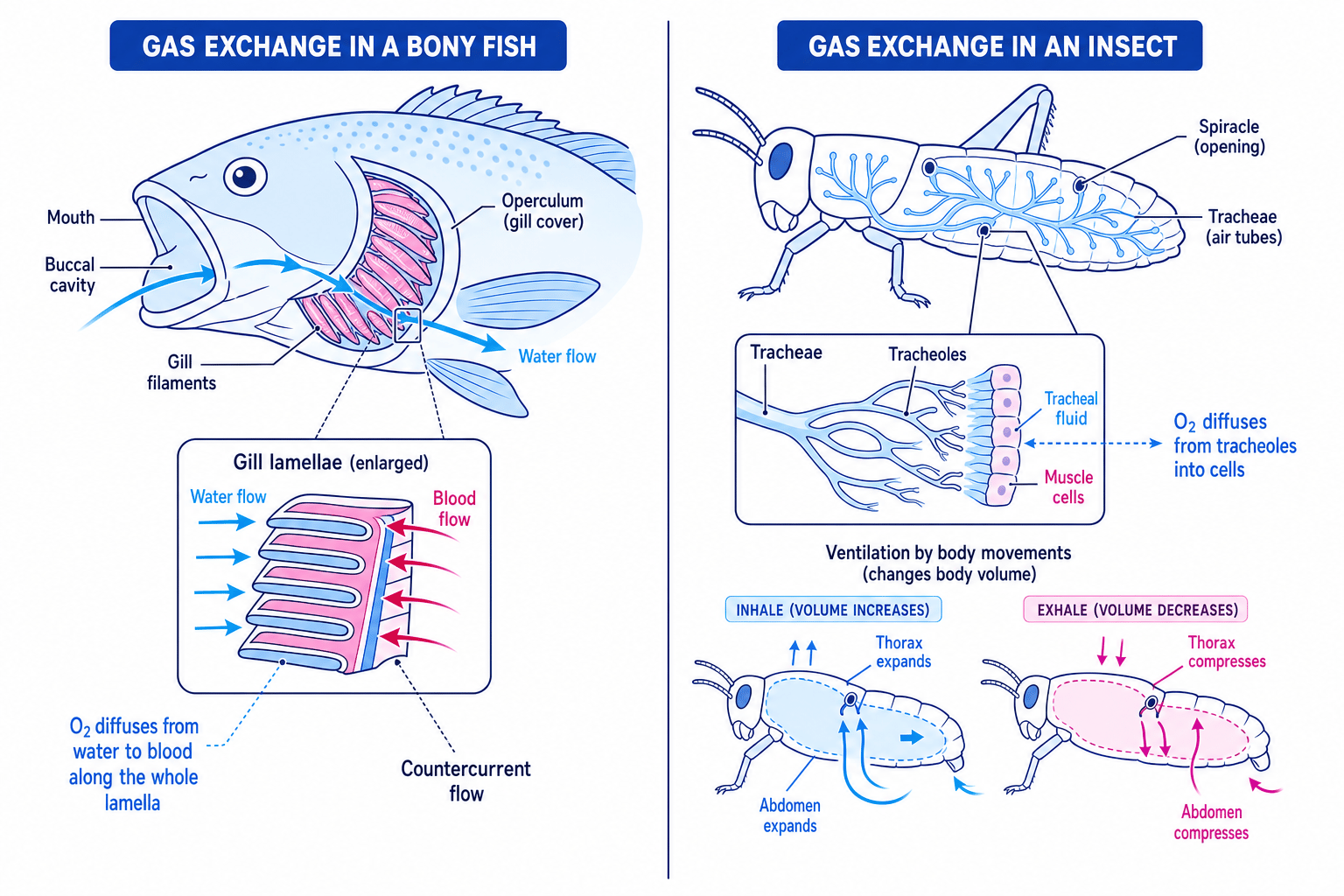
Fish and insects use very different exchange systems. Fish use gills with countercurrent blood and water flow; insects use air-filled tubes that take gases directly to tissues.
Countercurrent flow
In countercurrent flow, blood and water flow in opposite directions across the gill lamellae. This maintains a concentration gradient along the whole length of the lamella, so oxygen continues to diffuse from water into blood.
Interpreting countercurrent flow
-
At one end of a lamella, water has a higher oxygen concentration than the blood beside it, so oxygen diffuses into the blood.
-
Because blood flows in the opposite direction to water, blood that has already gained oxygen still meets water with an even higher oxygen concentration.
-
The gradient is maintained along the whole lamella, so more oxygen is absorbed than if water and blood flowed in the same direction.
Gas exchange in insects
Insects do not rely on blood to carry oxygen. Instead, they have a tracheal system.
Spiracles are openings on the body surface. They lead into larger air tubes called tracheae, which branch into smaller tracheoles. Tracheoles extend close to individual cells, so diffusion distance is short.
Gas exchange occurs between air in the tracheoles, tracheal fluid at the ends of the tracheoles, and the cells. Oxygen dissolves in the tracheal fluid and diffuses into cells; carbon dioxide diffuses in the opposite direction.
Insects can ventilate the system by thoracic and abdominal movements. Changing body volume helps move air in and out through the spiracles. Spiracles can also close to reduce water loss.
Practical work: dissection, drawing, and microscopy
For a bony fish dissection, you may examine the gills by lifting or removing the operculum and observing the gill arches, filaments, and lamellae using a hand lens or microscope. For insect tracheae, small sections can be mounted on a slide and observed under a light microscope.
Use safe practical technique: handle scalpels carefully, wear eye protection if instructed, wash hands after handling biological material, and keep tissues moist so delicate structures do not dry out.
Histology means the microscopic study of tissues. When examining slides of exchange surfaces, link what you see to function: thin layers reduce diffusion distance, many folds increase surface area, and capillaries indicate a good blood supply.
Biological drawings
- Use a sharp pencil and clear single lines.
- Do not shade or sketch roughly.
- Make the drawing large enough to show detail.
- Use straight label lines that do not cross.
- Include a title and magnification or scale bar if available.
In the exam
-
Link every structural feature to its effect on diffusion, such as surface area, diffusion distance, or concentration gradient.
-
For calculations, write the formula, substitute values with units, then give the final answer with units.
-
For ventilation questions, use the sequence: muscle action → volume change → pressure change → movement of air or water.
-
Keep organisms separate: mammals use alveoli and blood, fish use gills and countercurrent flow, insects use spiracles, tracheae, and tracheoles.
Check yourself
- Why does surface area to volume ratio decrease as an organism gets larger?
- Describe inhalation in mammals from muscle contraction to air entering the lungs.
- How does countercurrent flow help a fish absorb more oxygen from water?