What you'll learn
- How prokaryotic cells — cells without a nucleus — differ from eukaryotic cells — cells with a nucleus.
- The key structures found in prokaryotic cells, including their DNA, ribosomes, cell wall and optional extra features.
- Why viruses are acellular, meaning not made of cells, and why they are described as non-living.
- How to compare cell and virus sizes using micrometres (µm) and nanometres (nm).
Start point: what makes something a cell?
A cell is the basic unit of structure and function in living organisms. In earlier cell-structure work, you met eukaryotic cells: cells whose DNA — deoxyribonucleic acid, the genetic material carrying instructions — is enclosed in a nucleus. A nucleus is a membrane-bound compartment.
A membrane-bound organelle is a specialised structure inside a cell that is surrounded by its own membrane, such as a mitochondrion or chloroplast. Prokaryotic cells are different because they do not have these internal membrane-bound compartments.
Prokaryotic cell
A prokaryotic cell is a cell with cytoplasm — the jelly-like contents where many reactions occur — but no nucleus and no membrane-bound organelles. Its DNA is free in the cytoplasm.
Most A-Level questions use bacteria as the main examples of prokaryotic cells. Prokaryotic cells are much smaller than eukaryotic cells: many bacteria are about 1 micrometre long, while many animal and plant cells are around 10–100 micrometres across.
The basic prokaryotic cell plan
A prokaryotic cell still has a boundary, cytoplasm, genetic material and ribosomes. The important point is that these are arranged much more simply than in a eukaryotic cell.
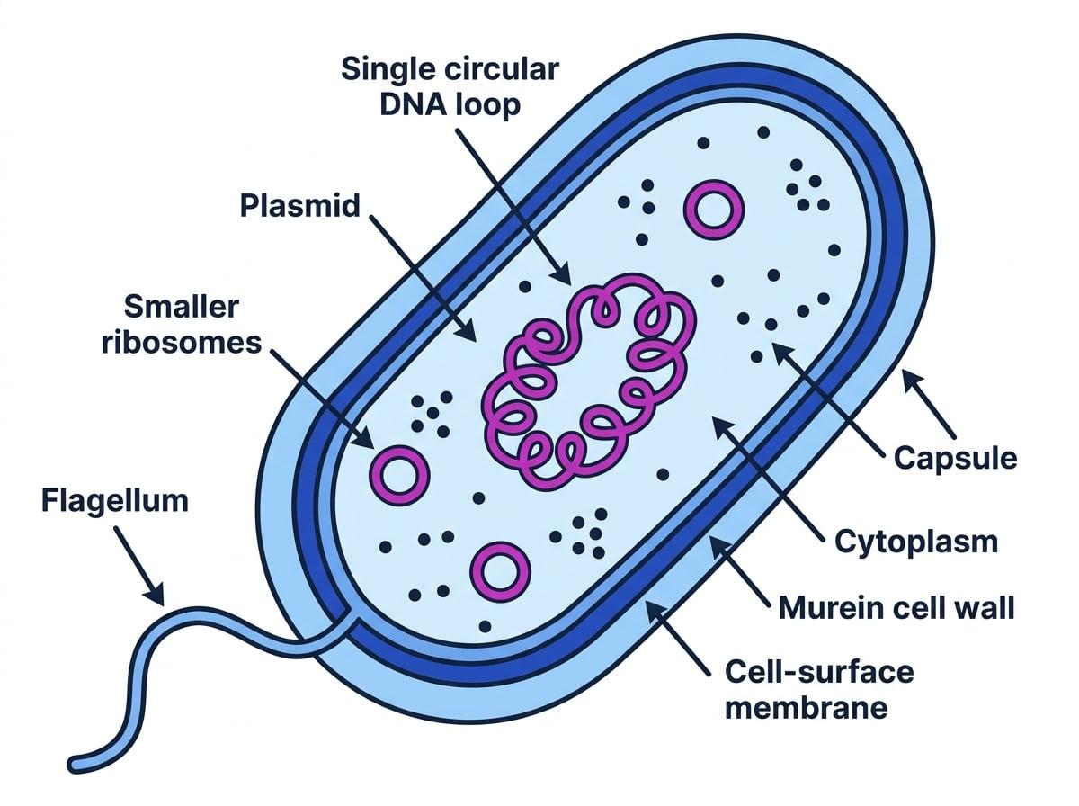
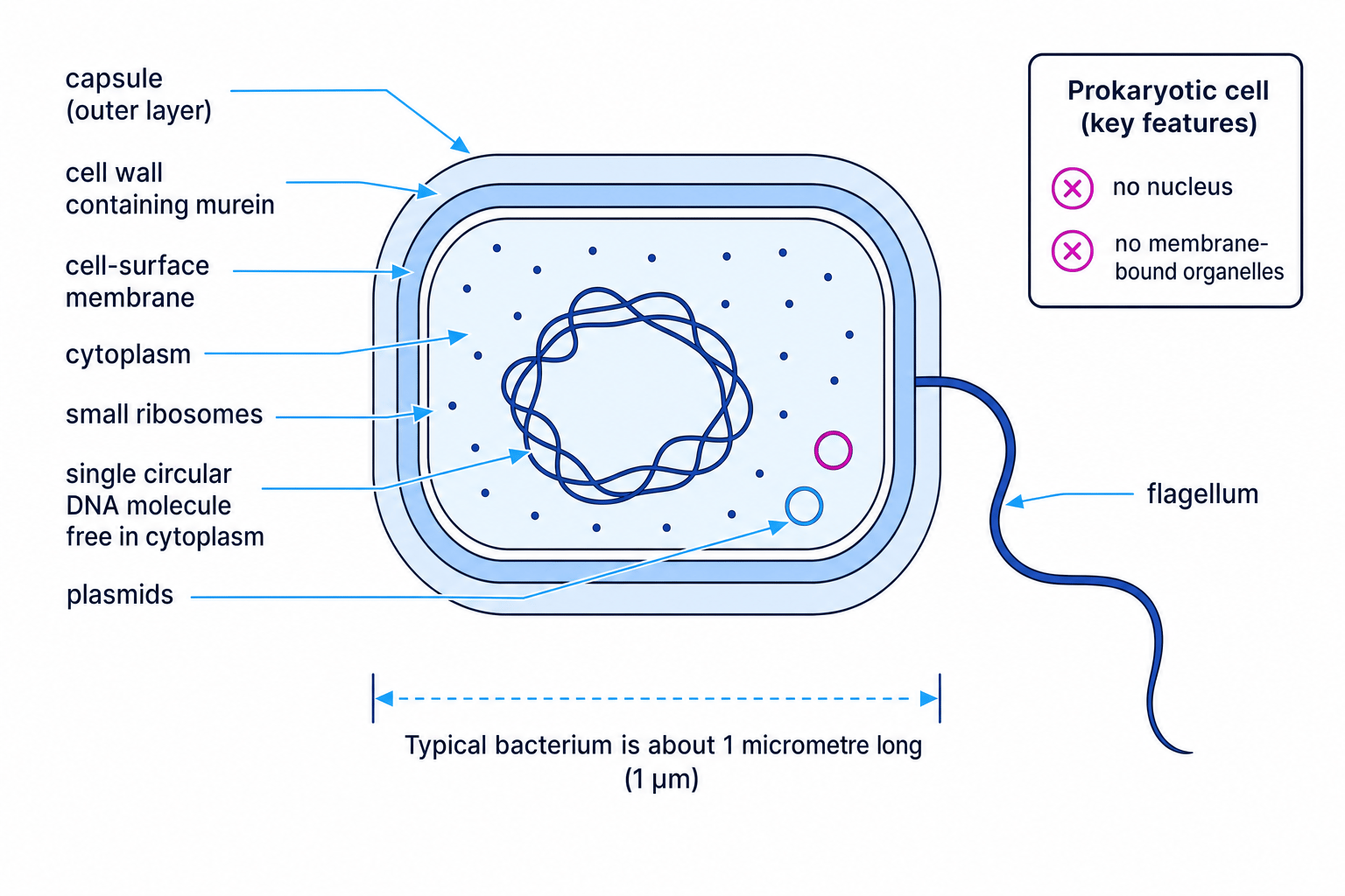
The labelled diagram below shows the structures AQA expects you to recognise.
Cytoplasm with no membrane-bound organelles
The cytoplasm of a prokaryotic cell lacks membrane-bound organelles. This means there is no nucleus, mitochondria, chloroplasts, Golgi apparatus or rough endoplasmic reticulum.
However, prokaryotic cells can still carry out metabolic reactions. Many reactions happen in the cytoplasm, and some processes use enzymes in the cell-surface membrane, the membrane that controls movement of substances into and out of the cell.
No membrane-bound organelles does not mean no ribosomes
Ribosomes are not membrane-bound, so prokaryotic cells can still have ribosomes even though they lack membrane-bound organelles.
Smaller ribosomes
A ribosome is a small structure where proteins are made. Prokaryotic cells have smaller ribosomes than eukaryotic cells.
For this part of the specification, you do not need detailed ribosome types or sizes. The key exam phrase is simply smaller ribosomes.
No nucleus: circular DNA free in the cytoplasm
A prokaryotic cell has no nucleus. Instead, it has a single circular DNA molecule free in the cytoplasm. This DNA is not associated with proteins, using the wording required by the specification.
You may see the DNA region called a nucleoid, which means the area of cytoplasm containing the main DNA, but it is not surrounded by a membrane.
Cell wall containing murein
A cell wall is a rigid layer outside the cell-surface membrane that helps support and protect the cell. Prokaryotic cell walls contain murein, a glycoprotein. A glycoprotein is a molecule containing both carbohydrate and protein components.
This is different from plant cell walls, which contain cellulose. It is also different from fungal cell walls, which contain chitin.
Wall material shortcut
For AQA, match the wall material to the cell type: plant cell walls contain cellulose, fungal cell walls contain chitin, and prokaryotic cell walls contain murein.
Extra structures in many prokaryotic cells
Not every prokaryotic cell has every extra structure. The specification says many prokaryotic cells have these features, so avoid writing as if they are always present.
Plasmids
A plasmid is a small circular loop of DNA separate from the main circular DNA molecule. A prokaryotic cell may have one or more plasmids.
Plasmids often carry useful genes, such as genes for antibiotic resistance, but the key structural point is that they are extra small rings of DNA.
Capsule
A capsule is a layer surrounding the outside of some prokaryotic cells. It can help protect the cell and help it stick to surfaces, but the detailed structure is not required here.
Flagella
A flagellum is a long, tail-like structure used for movement. The plural is flagella. Some prokaryotic cells have one or more flagella.
Core prokaryote facts
A prokaryotic cell is much smaller than a eukaryotic cell and has cytoplasm with no membrane-bound organelles, smaller ribosomes, no nucleus, a single circular DNA molecule free in the cytoplasm, and a cell wall containing murein.
Viruses are not cells
A virus is not a prokaryotic cell. It is not any kind of cell. Viruses are described as acellular, meaning they are not made of cells, and non-living, meaning they do not carry out all the life processes independently.
Viruses do not have cytoplasm, ribosomes or a cell wall. They cannot replicate by themselves; they must infect a host cell, which is a living cell used by the virus to make more virus particles.
Virus particle
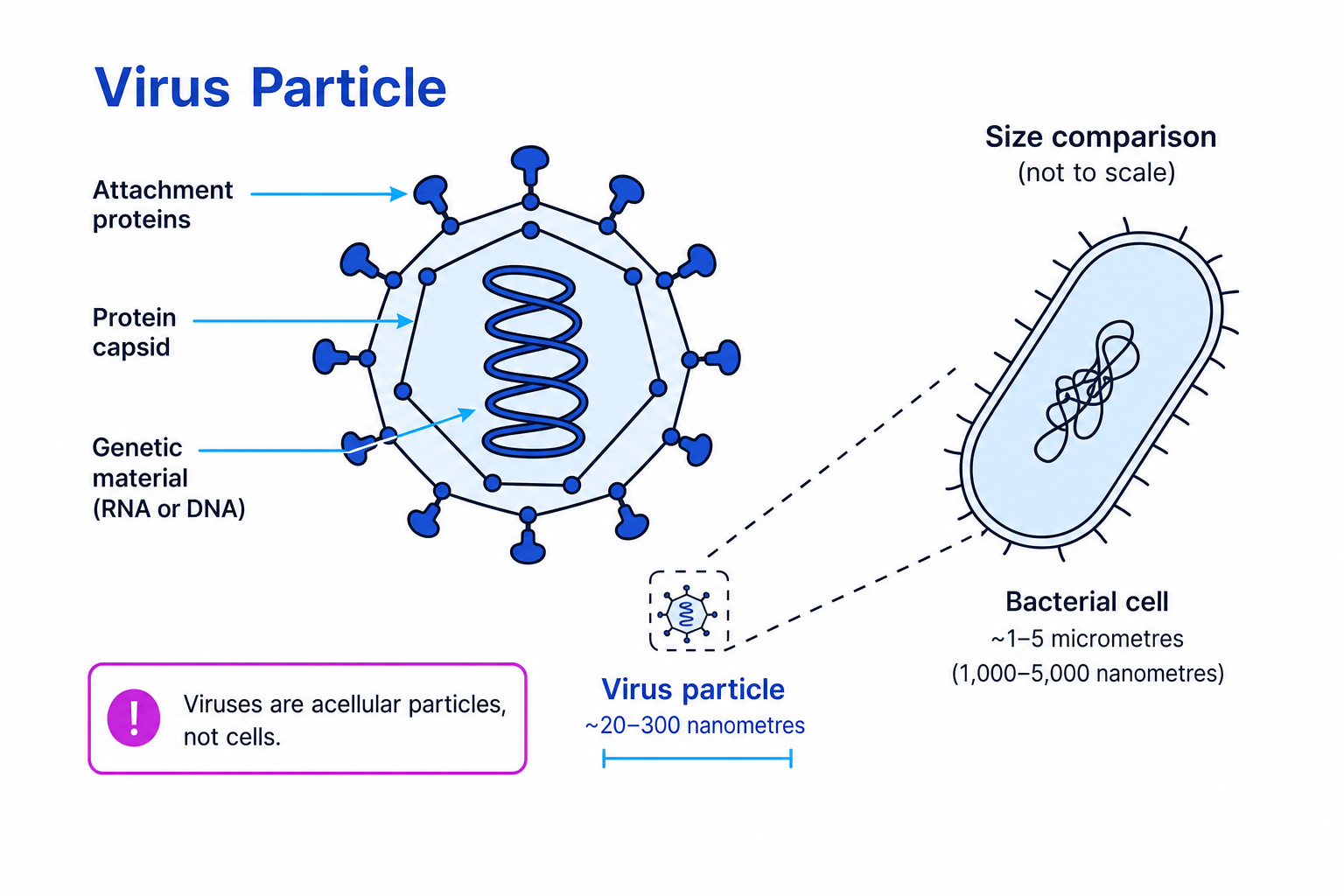
A virus particle is an acellular, non-living structure made from genetic material surrounded by a protein capsid, with attachment proteins on the outside.
The diagram below shows the three virus structures you need: genetic material, capsid and attachment proteins.
Genetic material
Viral genetic material carries the instructions needed to make new virus particles. In different viruses, this genetic material may be DNA or RNA. RNA, ribonucleic acid, is another type of nucleic acid involved in storing or transferring genetic information.
Capsid
A capsid is the protein coat surrounding the viral genetic material. It helps protect the genetic material and gives the virus particle its shape.
Attachment proteins
Attachment proteins are proteins on the outside of a virus particle that bind to specific molecules on a host cell. These host molecules are called receptors, meaning cell-surface molecules with a shape that can bind to a particular attachment protein.
This helps explain why viruses often infect only certain cell types: the attachment protein must fit the receptor.
Prokaryotic cells versus viruses
| Feature | Prokaryotic cell | Virus particle |
|---|---|---|
| Cellular structure | A cell | Not a cell; acellular |
| Living status | Living organism | Non-living particle |
| Cytoplasm | Present | Absent |
| Ribosomes | Smaller ribosomes present | No ribosomes |
| Genetic material | Single circular DNA molecule free in cytoplasm; may also have plasmids | Genetic material inside a capsid |
| Outer structure | Cell wall containing murein; may have a capsule | Protein capsid with attachment proteins |
| Replication | Cell divides to reproduce | Replicates only inside a host cell |
Comparing sizes: micrometres and nanometres
A micrometre is one millionth of a metre: 1 μm=1×10⁻⁶ m1\,\mu\text{m} = 1 \times 10^{-6}\,\text{m}1μm=1×10⁻⁶m.
A nanometre is one billionth of a metre: 1 nm=1×10⁻⁹ m1\,\text{nm} = 1 \times 10^{-9}\,\text{m}1nm=1×10⁻⁹m.
The conversion you will use most here is:
1 μm=1000 nm1\,\mu\text{m} = 1000\,\text{nm}1μm=1000nmProkaryotic cells are usually measured in micrometres. Viruses are much smaller and are usually measured in nanometres.
Comparing a virus with a bacterium
A bacterium is 2.0 µm long. A virus particle is 100 nm across. How many virus diameters fit across the length of the bacterium?
- Convert the bacterium length into nanometres so both measurements use the same unit.
- Divide the bacterium length by the virus diameter.
- Interpret the result: about 20 virus particles, each 100 nm across, could fit end-to-end across the length of this 2.0 µm bacterium.
In the exam
- For prokaryotic cells, focus on the required differences: no nucleus, no membrane-bound organelles, smaller ribosomes, circular DNA free in cytoplasm, and a murein cell wall.
- Do not call viruses prokaryotes. Describe them as acellular and non-living, with genetic material, a capsid and attachment proteins.
- For size comparisons, convert all measurements into the same unit before dividing; remember that 1 µm equals 1000 nm.
Check yourself
- Which features show that a cell is prokaryotic rather than eukaryotic?
- What is the difference between the main circular DNA molecule and a plasmid?
- Why are viruses described as acellular and non-living?