

What you'll learn
- How optical microscopes, TEMs and SEMs produce images — and what each can and cannot show.
- The difference between magnification and resolution, including how to calculate real sizes.
- How to measure cells using an optical microscope and calibrated graticule.
- How cell fractionation and ultracentrifugation separate organelles from cells.
Why we need methods to study cells
Cells and organelles are usually too small to see clearly with the naked eye. Many animal and plant cells are around 10–100 micrometres (µm) across, while organelles such as ribosomes are much smaller, often measured in nanometres (nm).
A microscope helps in two different ways: it makes objects appear larger, and — if it has good resolving power — it lets you distinguish tiny structures that are close together.
Microscope specimen
A specimen is the material being viewed under a microscope, such as a thin slice of tissue, a smear of cells, or a prepared sample of organelles.
Magnification and resolution
Magnification
Magnification is how many times larger the image is than the real object.
The A-Level formula is:
magnification=size of imagesize of real object\text{magnification} = \frac{\text{size of image}}{\text{size of real object}}magnification=size of real objectsize of imageYou can rearrange it depending on what you need:
M=IOI=M×OO=IM\begin{aligned} M &= \frac{I}{O} \\ I &= M \times O \\ O &= \frac{I}{M} \end{aligned}MIO=OI=M×O=MIwhere MMM is magnification, III is image size, and OOO is real object size.
Resolution
Resolution is the ability to distinguish two separate points as separate. If resolution is poor, two close structures blur into one.
The big distinction
Magnification makes an image bigger. Resolution determines whether extra detail can actually be seen. Increasing magnification without improving resolution only gives a bigger blurry image.
Calculating real cell size
A cell appears 35 mm long in a photograph. The photograph has a magnification of ×700\times 700×700. Find the real length of the cell in micrometres.
-
Use the rearranged magnification equation:
O=IMO = \frac{I}{M}O=MI -
Convert the image size into micrometres so the final answer is in cell-sized units:
35 mm=35 000 μm35\text{ mm} = 35\,000\ \mu\text{m}35 mm=35000 μm -
Substitute into the equation:
O=35 000 μm700=50 μmO = \frac{35\,000\ \mu\text{m}}{700} = 50\ \mu\text{m}O=70035000 μm=50 μm -
State the real size: the cell is 50 µm long.
Using mixed units
The magnification equation only works if the image size and real object size are in the same units before you divide. Convert first, then calculate.
Measuring objects with an optical microscope
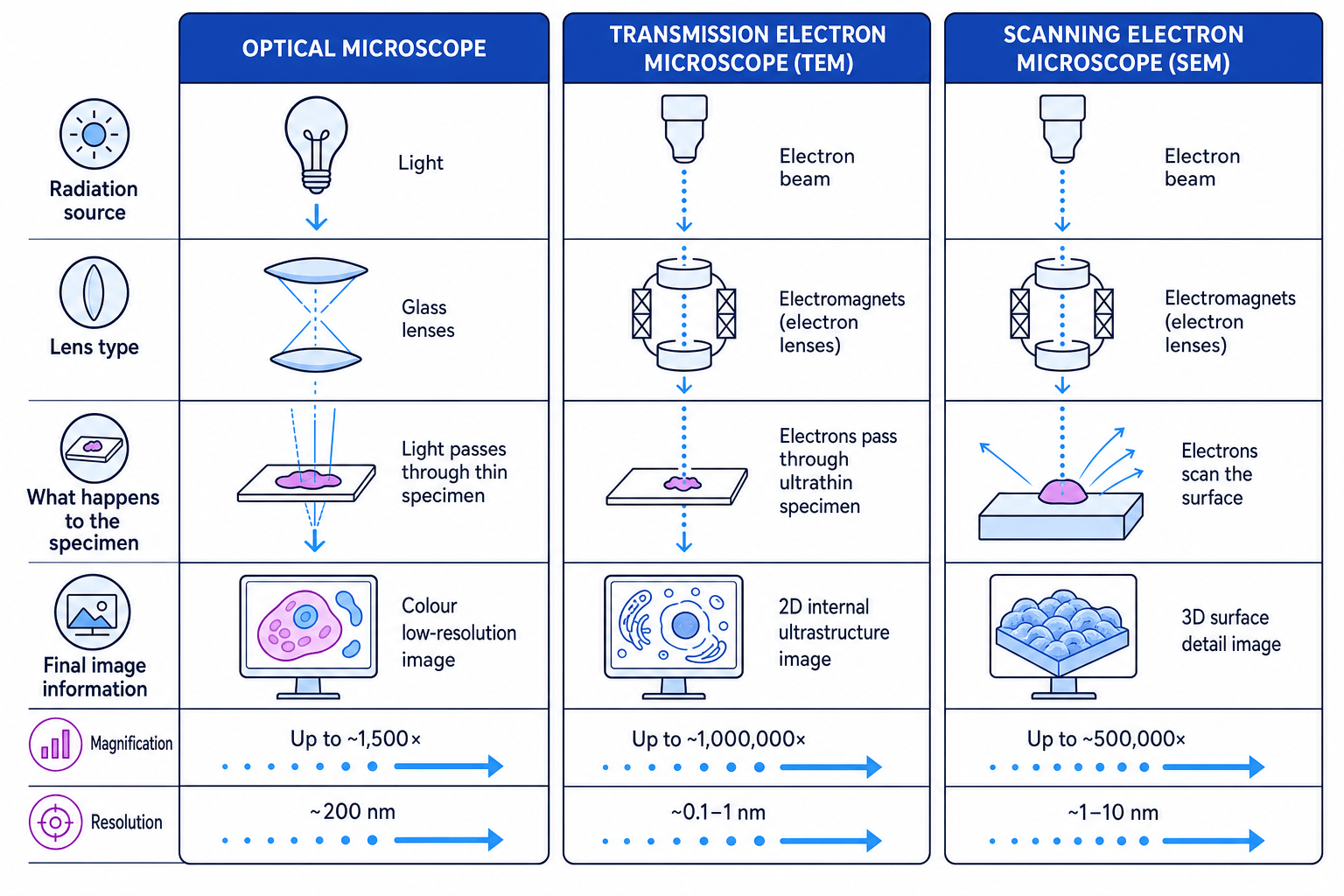
An optical microscope uses visible light and glass lenses to produce a magnified image. In school practicals, you may view cells on a slide and measure them using an eyepiece graticule.
Eyepiece graticule
An eyepiece graticule is a small scale fitted into the eyepiece lens. Its divisions are arbitrary until you calibrate them.
Stage micrometer
A stage micrometer is a microscope slide with a tiny, accurately marked scale. It is used to work out what one eyepiece graticule division represents at a particular magnification.
The method is:
- Place the stage micrometer on the microscope stage.
- Line up the eyepiece graticule scale with the stage micrometer scale.
- Work out the real length of one eyepiece unit.
- Replace the micrometer with the specimen.
- Measure the specimen in eyepiece units, then convert to micrometres.
Calibrating an eyepiece graticule
At a particular objective lens setting, 40 eyepiece units line up with 100 µm on a stage micrometer. A plant cell then measures 18 eyepiece units. Find the cell length.
-
Find the value of one eyepiece unit:
100 μm40=2.5 μm\frac{100\ \mu\text{m}}{40} = 2.5\ \mu\text{m}40100 μm=2.5 μm -
Convert the cell measurement into micrometres:
18×2.5 μm=45 μm18 \times 2.5\ \mu\text{m} = 45\ \mu\text{m}18×2.5 μm=45 μm -
So the plant cell is 45 µm long.
Calibration changes
If you change the objective lens, you must recalibrate the eyepiece graticule. The graticule divisions do not have one fixed real-life value.
Optical microscopes
Optical microscopes use light passing through, or reflecting from, the specimen. The lenses bend the light to form a magnified image.
They are useful because they can show whole cells, stained tissues and sometimes living specimens. For example, iodine in potassium iodide solution can be used to identify starch grains in plant cells; starch turns blue-black.
Their main limitation is resolution. A typical optical microscope resolves structures about 200 nm apart. That means it can show nuclei, chloroplasts and cell walls, but not fine internal details such as ribosomes or the detailed structure of membranes.
Electron microscopes: TEM and SEM
Electron microscopes use beams of electrons rather than light. Electrons have a much shorter wavelength than visible light, so electron microscopes can achieve much higher resolution. They use electromagnets instead of glass lenses.
Transmission electron microscope
A transmission electron microscope, or TEM, passes electrons through an extremely thin specimen. Dense parts of the specimen absorb or scatter more electrons, producing contrast in the image.
TEMs are best for viewing internal cell ultrastructure, such as mitochondria, rough endoplasmic reticulum and membranes. They can have very high resolution, around 0.1–1 nm, and very high magnification.
Scanning electron microscope
A scanning electron microscope, or SEM, scans a beam of electrons across the surface of the specimen. Electrons scattered from the surface are detected to build an image.
SEMs are best for showing surface detail and giving a 3D-looking image, such as the surface of a cell or tissue. Their resolution is usually lower than a TEM, often around 1–10 nm, but still much better than an optical microscope.
TEM versus SEM
TEM shows internal detail in a thin specimen. SEM shows surface detail. If the question says “3D surface image”, think SEM.
Limitations of electron microscopes
Electron microscopes have important limitations:
- Specimens must be placed in a vacuum, so living cells cannot usually be viewed.
- Samples often need complex preparation, including dehydration, staining and slicing.
- The image is produced in black and white, although false colour may be added later.
- Preparation can damage or distort structures.
Artefact
An artefact is a visible feature produced by the preparation or imaging method, rather than a real structure in the living cell.
Early cell biologists had to be cautious. For a considerable period, scientists debated whether some structures seen in micrographs were genuine organelles or artefacts caused by staining, dehydration or sectioning. Confidence increased when the same structures were repeatedly seen using different methods and linked to cell functions.
Cell fractionation and ultracentrifugation
Microscopes let you look at structures. Cell fractionation lets you separate those structures so they can be studied chemically.
Cell fractionation
Cell fractionation is the process of breaking open cells and separating the different organelles and cell components.
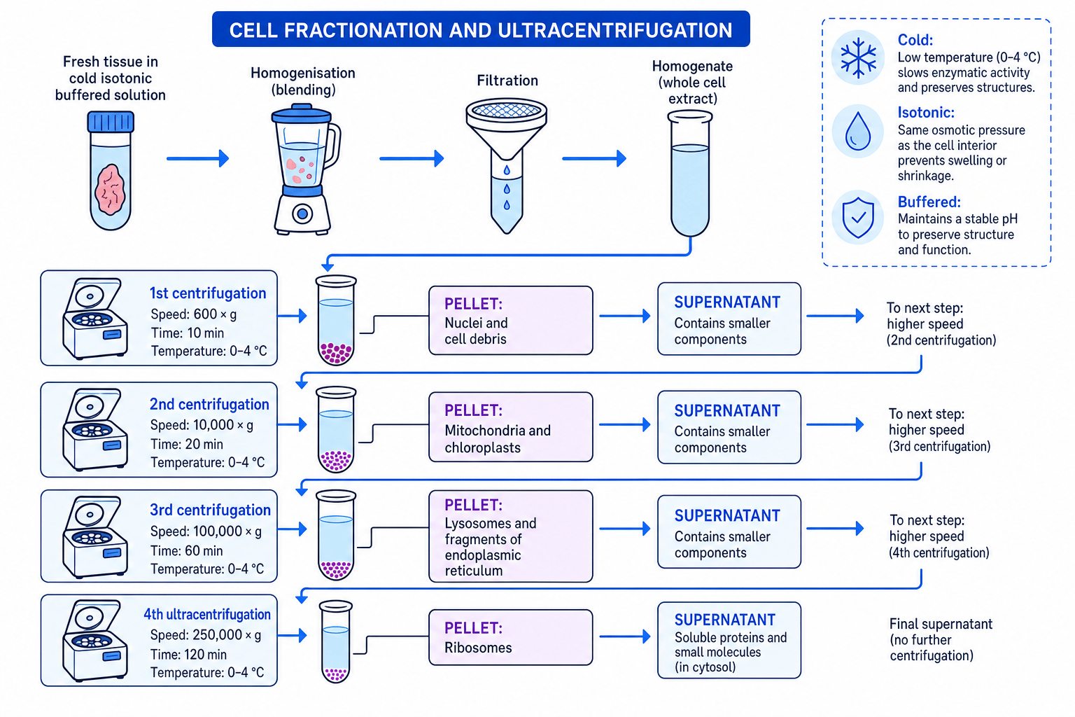
The first stage is homogenisation, where tissue is broken up to release organelles into a liquid. The resulting mixture is called a homogenate.
The tissue is kept in a cold, isotonic, buffered solution:
- Cold slows enzyme activity, reducing organelle damage.
- Isotonic means the solution has the same water potential as the cells, preventing organelles from bursting or shrinking by osmosis.
- Buffered means the pH is kept stable, helping proteins and organelles maintain their structure.
The homogenate is filtered to remove large debris. It is then spun in a centrifuge.
Ultracentrifugation
Ultracentrifugation uses very high-speed spinning to separate cell components according to size and density.
During centrifugation, heavier or denser components form a pellet at the bottom of the tube. The liquid above it is the supernatant. The supernatant is poured off and spun again at a higher speed. Smaller components pellet only at higher speeds.
Order of separation
In differential centrifugation, large dense structures pellet first. A typical order is nuclei, then mitochondria and chloroplasts, then lysosomes and endoplasmic reticulum fragments, then ribosomes.
Choosing the correct fraction
A scientist wants to study enzymes found in mitochondria. A homogenate is centrifuged at low speed, producing a first pellet and a first supernatant. The supernatant is then centrifuged at a higher speed, producing a second pellet.
-
The first low-speed pellet is likely to contain the largest structures, such as nuclei and cell debris, so this is not the best fraction for mitochondrial enzymes.
-
Mitochondria are smaller than nuclei but larger than ribosomes, so they usually pellet after the first supernatant is spun at a higher speed.
-
The scientist should collect the second pellet, because this is most likely to be enriched with mitochondria.
Fractions are not perfectly pure
Centrifugation enriches fractions; it does not always produce completely pure organelle samples. Some contamination between fractions can occur.
In the exam
- For microscope comparisons, link the principle to the image: optical uses light, TEM transmits electrons through a thin specimen, SEM scans the surface.
- For size calculations, convert units before using M=IOM = \frac{I}{O}M=OI, then check whether the answer is sensible for a cell or organelle.
- For fractionation questions, explain the purpose of cold, isotonic and buffered conditions, then describe increasing centrifugation speed and the pellet/supernatant sequence.
Check yourself
- What is the difference between magnification and resolution?
- Why can a TEM show smaller structures than an optical microscope?
- In cell fractionation, why are mitochondria not usually found in the first pellet?
