What you'll learn
- The main structures inside plant and animal cells.
- The function of each structure, including the nucleus, mitochondria, ribosomes, chloroplasts and vacuole.
- How to compare plant and animal cells in diagrams.
- Why cell differentiation and stem cells are important in medicine.
Cells: the basic starting point
All living organisms are made of cells. Some organisms are made of one cell, but animals and plants are multicellular, meaning they are made of many cells working together.
Cells and organelles
- A cell is the smallest unit of life.
- An organelle is a specialised structure inside a cell that has a particular job.
Plant and animal cells are eukaryotic cells: their genetic material is contained inside a nucleus. This is different from bacterial cells, which are covered in a later section.
Structure and function
In Biology, always link what a structure is like to what it does. Cell parts are not random — each one helps the cell carry out life processes.
Animal and plant cell structures
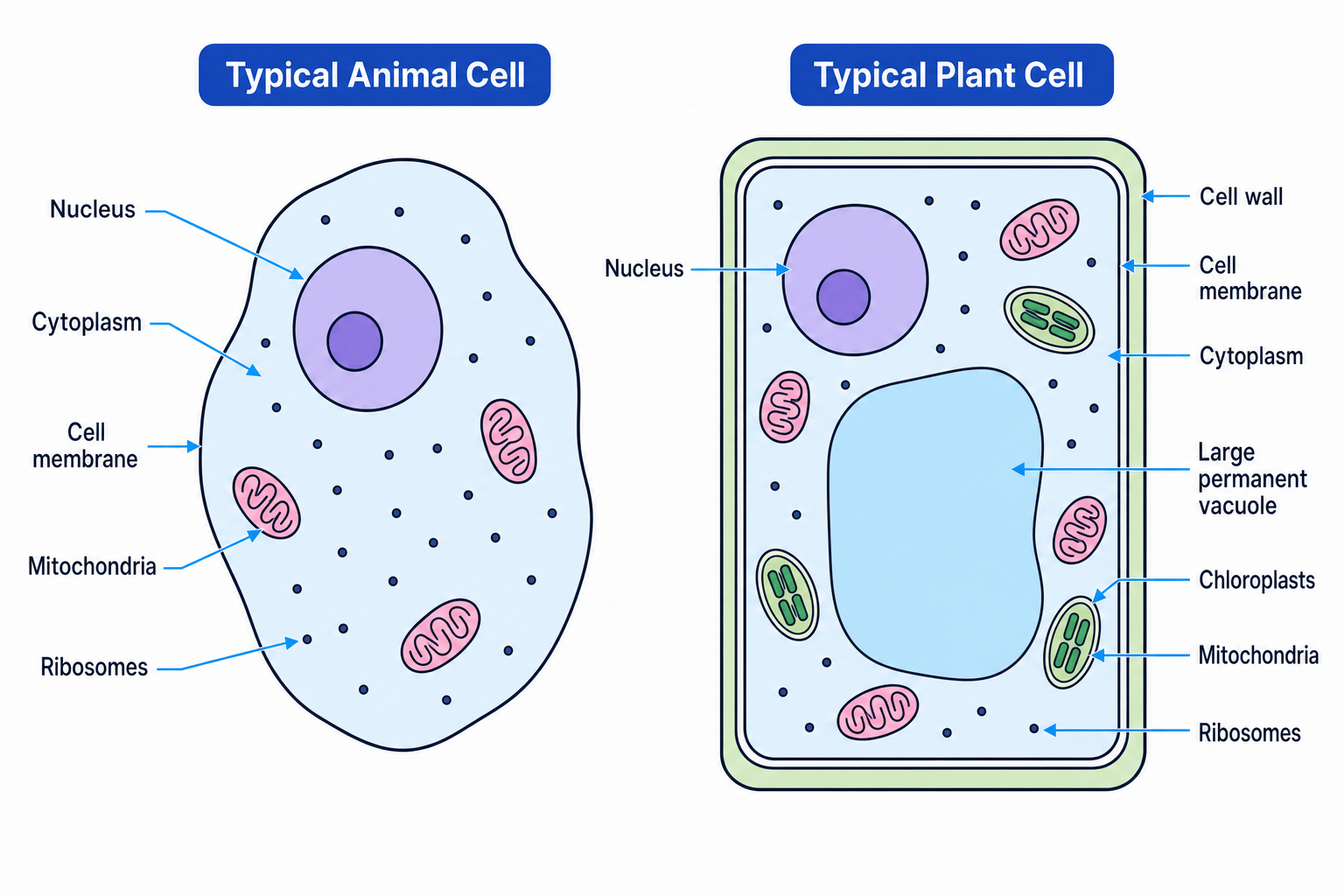
A typical animal cell and a typical plant cell share several structures, but plant cells also have extra features.
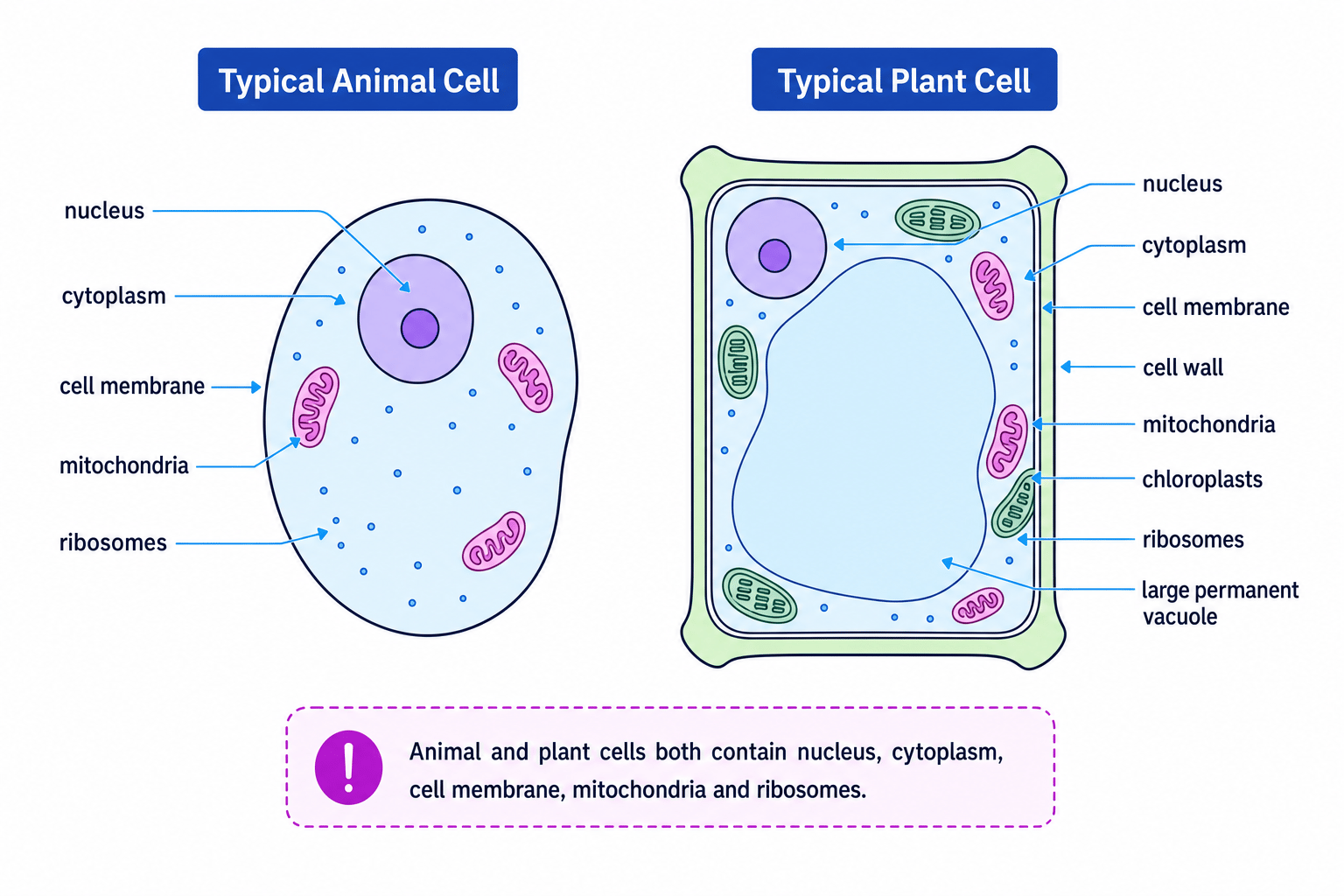
Structures found in both plant and animal cells
Nucleus
The nucleus contains the cell’s genetic material, called DNA. DNA carries instructions for making the cell’s proteins and controlling cell activities.
In diagrams, the nucleus is usually one of the largest organelles.
Cytoplasm
The cytoplasm is a jelly-like substance where many chemical reactions happen. Organelles are suspended in the cytoplasm.
Cell membrane
The cell membrane is a thin boundary around the cell. It controls what enters and leaves the cell, such as glucose, oxygen, water and waste substances.
Cell membrane vs cell wall
Animal cells do have a cell membrane. They do not have a cell wall. The wall is the strong outer support layer found in plant cells.
Mitochondria
Mitochondria are the site of aerobic respiration, which is the release of energy from glucose using oxygen. Cells that need lots of energy often have many mitochondria.
For example, muscle cells need many mitochondria because contraction requires energy.
Ribosomes
Ribosomes are tiny structures where protein synthesis happens. Protein synthesis means making protein molecules.
In cell diagrams, ribosomes are often drawn as tiny dots in the cytoplasm.
Structures typical of plant cells
Cell wall
The cell wall is a strong outer layer made of cellulose. It supports the plant cell, strengthens it and helps stop it bursting when water enters.
Cellulose is a tough carbohydrate found in plant cell walls.
Chloroplasts
Chloroplasts contain chlorophyll, a green pigment that absorbs light energy for photosynthesis. Photosynthesis is the process plants use to make glucose from carbon dioxide and water using light.
Large permanent vacuole
A plant cell usually has a large permanent vacuole filled with cell sap, a watery solution containing dissolved substances. The vacuole helps keep the cell firm.
A plant cell that is full of water is turgid, meaning firm and swollen due to water pressure inside the cell.
Plant cell extras
For IGCSE diagrams, the three big plant-cell-only features to remember are: cell wall, chloroplasts and a large permanent vacuole.
Using magnification with cells
Microscope images and cell drawings are often larger than the real cell. Magnification tells you how many times larger the image is than the actual object.
magnification=image sizeactual size\text{magnification} = \frac{\text{image size}}{\text{actual size}}magnification=actual sizeimage sizeMagnification has no unit because the two sizes cancel out — but they must be in the same unit before you divide.
Calculating magnification
A microscope drawing of a plant cell is 60 mm long. The actual cell is 120 µm long. Calculate the magnification.
- Convert the image size into micrometres: 60 mm×1000=60 000 μm60\ \text{mm} \times 1000 = 60\,000\ \mu\text{m}60 mm×1000=60000 μm.
- Substitute into the formula: M=image sizeactual size=60 000 μm120 μmM = \frac{\text{image size}}{\text{actual size}} = \frac{60\,000\ \mu\text{m}}{120\ \mu\text{m}}M=actual sizeimage size=120 μm60000 μm.
- Calculate the value: M=500M = 500M=500, so the magnification is x500.
Comparing plant and animal cells
| Feature | Animal cell | Plant cell |
|---|---|---|
| Nucleus | Present | Present |
| Cytoplasm | Present | Present |
| Cell membrane | Present | Present |
| Mitochondria | Present | Present |
| Ribosomes | Present | Present |
| Cell wall | Absent | Present |
| Chloroplasts | Absent | Present in photosynthetic cells |
| Large permanent vacuole | Absent | Present |
Plant cells often look more regular or box-shaped because the cell wall gives them support. Animal cells are often more rounded or irregular in shape.
Not every plant cell has chloroplasts
Chloroplasts are found in plant cells that carry out photosynthesis, such as many leaf cells. Root cells usually do not contain chloroplasts because they are underground and do not receive light.
Classifying an unknown cell
A microscope image shows a cell with a nucleus, cytoplasm, cell membrane, mitochondria, ribosomes, a cell wall and a large permanent vacuole. No chloroplasts are visible. Decide whether it is more likely to be a plant or animal cell.
- Compare the features with animal cells: animal cells do not have a cell wall or a large permanent vacuole.
- Compare the features with plant cells: plant cells have a cell wall and usually a large permanent vacuole.
- The missing chloroplasts do not rule out a plant cell, because not all plant cells photosynthesise. It is most likely a plant cell.
Cell differentiation
This part of the specification is Paper 2 only, but it is very important for understanding development.
Cell differentiation
Cell differentiation is the process by which an unspecialised cell becomes a specialised cell with structures adapted for a particular function.
A specialised cell has features that help it do a specific job. For example, a muscle cell is adapted to contract, while a root hair cell is adapted to absorb water and mineral ions.
A stem cell is an unspecialised cell that can divide and differentiate into specialised cells.
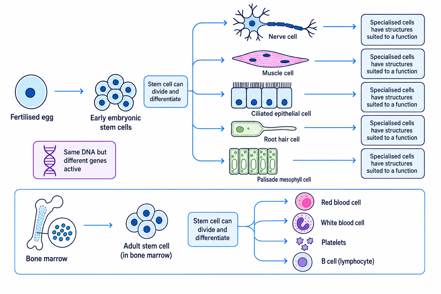
During development, a fertilised egg divides many times. At first, the cells are unspecialised. As the organism develops, cells differentiate so they can form tissues, organs and organ systems.
A gene is a section of DNA with instructions for making a particular protein. Most body cells contain the same DNA, but different genes are active in different specialised cells. This leads to different structures and functions.
Why differentiation matters
Differentiation allows a multicellular organism to have many different types of cells, each adapted for a particular job. Without differentiation, an embryo would just be a mass of similar cells.
Linking structure to function
A palisade mesophyll cell contains many chloroplasts. Explain why this helps the cell carry out its function.
- Identify the function: palisade mesophyll cells carry out photosynthesis in leaves.
- Link the structure to the process: chloroplasts contain chlorophyll, which absorbs light energy.
- Explain the advantage: many chloroplasts allow the cell to absorb more light, helping photosynthesis happen efficiently.
Stem cells in medicine
This section is also Paper 2 only.
Stem cells are useful because they can divide and form specialised cells. In medicine, scientists hope to use stem cells to replace damaged or diseased cells.
Advantages of using stem cells
Stem cells may be used to replace cells lost through injury or disease. For example, stem cells in bone marrow can produce new blood cells.
They could reduce the need for donated organs, because new tissues might be grown from stem cells. If a patient’s own stem cells are used, there may be a lower chance of rejection. Rejection happens when the immune system attacks transplanted cells as foreign.
Stem cells are also useful in research, such as testing new medicines or studying how diseases develop.
Disadvantages of using stem cells
There are ethical concerns about using embryonic stem cells because obtaining them can involve destroying embryos.
There is also a risk that transplanted cells may be rejected by the patient’s immune system if they are not a close match. Stem cells may divide uncontrollably, which could lead to tumours. Treatments can also be expensive, technically difficult and may carry infection risks.
Stem cells are not magic
Do not write that stem cells “cure any disease”. In exams, explain the specific idea: they can divide and differentiate to replace particular damaged cells, but there are risks and limitations.
In the exam
- When naming structures, use precise terms: cell membrane, not just “membrane”; cell wall, not “outer layer”.
- For function questions, connect the structure to its job, such as “mitochondria are the site of aerobic respiration, releasing energy for cell processes”.
- For stem-cell questions, give a balanced answer: include at least one medical benefit and one risk or ethical concern.
Check yourself
- Which five structures are found in both plant and animal cells?
- Why does a plant cell need both a cell wall and a cell membrane?
- How does differentiation help an embryo develop into a multicellular organism?