2.2.2a The heart and blood vessels
The Heart Drives a Double Circulation
Double circulatory system
A circulation in which blood passes through the heart twice for each complete circuit of the body: once around the lungs and once around the rest of the body.
- Your heart is a muscular organ that pumps blood around the body, and it works as two pumps side by side.
- In a double circulatory system, blood passes through the heart twice on each complete lap of the body.
- The right side pumps deoxygenated blood to the lungs, where it gains oxygen and loses carbon dioxide.
- The left side pumps oxygenated blood at high pressure to the rest of the body.
- The loop to the lungs is called the pulmonary circulation, and the loop to the rest of the body is the systemic circulation.
- Blood loses a lot of pressure squeezing through the lungs, so returning it to the heart lets the pressure be raised again before it is sent out to the body.
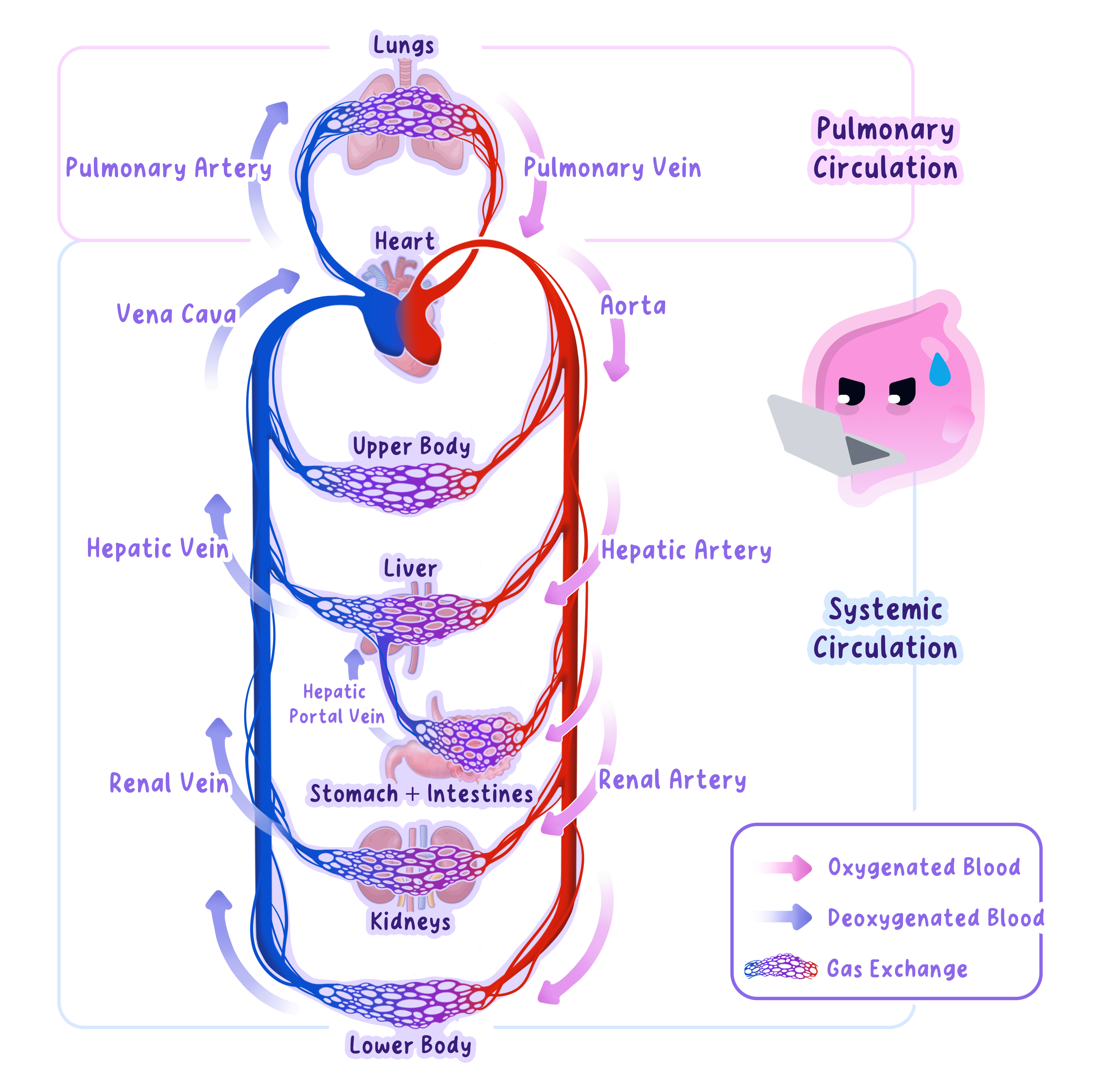
Because blood is re-pressurised after the lungs, oxygenated blood reaches the body cells quickly, supporting a fast rate of respiration and an active life.
Four Chambers: Atria Receive, Ventricles Pump
Atrium
One of the two upper chambers of the heart. It receives blood returning to the heart and pushes it down into the ventricle below. The plural is atria.
- The heart has four chambers, a top and a bottom pair on each side.
- The two upper chambers are the atria, which receive blood returning to the heart.
- The two lower chambers are the ventricles, which pump blood out of the heart.
- Blood flows from each atrium into the ventricle below it, then out through a large vessel.
- Valves between the atria and ventricles, and at the exits of the ventricles, stop blood flowing backwards.
- You do not need to learn the names of the heart valves for AQA, only that valves keep blood flowing one way.
- On a heart diagram the heart is drawn as if facing you, so its left side appears on the right of the picture.
- The lub-dub sound of a heartbeat is the valves snapping shut.
The Septum Keeps the Two Sides Apart
Septum
The wall of muscle that separates the right and left sides of the heart, stopping oxygenated and deoxygenated blood from mixing.
- A muscular wall called the septum runs down the middle of the heart.
- It separates the right side, holding deoxygenated blood, from the left side, holding oxygenated blood.
- This stops the two types of blood from mixing inside the heart.
- If they mixed, less oxygenated blood would reach the body, so the cells would get less oxygen.
Do not say both sides carry the same blood: the septum keeps deoxygenated blood on the right and oxygenated blood on the left.
Name the Vessels by Direction, Not Blood Type
- Vena cava
- A large vein that brings deoxygenated blood from the body into the right atrium.
- Pulmonary artery
- Carries deoxygenated blood from the right ventricle to the lungs.
- Pulmonary vein
- Carries oxygenated blood from the lungs into the left atrium.
- Aorta
- Carries oxygenated blood from the left ventricle to the body.
- Coronary arteries
- Branch over the outside of the heart and supply the heart muscle itself with oxygenated blood.
- Arteries carry blood away from the heart and veins carry it towards the heart, so vessels are named by direction, not by whether the blood carries oxygen.
- The pulmonary artery is the one artery carrying deoxygenated blood, and the pulmonary vein is the one vein carrying oxygenated blood.
Following Blood Through the Heart, Step by Step
- Deoxygenated blood from the body enters the right atrium through the vena cava.
- The right atrium contracts and pushes blood down into the right ventricle.
- The right ventricle contracts and forces blood into the pulmonary artery, which carries it to the lungs for gas exchange.
- Oxygenated blood returns from the lungs through the pulmonary vein into the left atrium.
- The left atrium contracts and pushes blood into the left ventricle.
- The left ventricle contracts hard and forces blood into the aorta, which carries it to the whole body.
Both sides fill and empty at the same time, so the heart beats as one coordinated pump.
Why the Left Ventricle Has the Thickest Wall
Ventricle
One of the two lower chambers of the heart. It has a thick, muscular wall and pumps blood out of the heart; the left ventricle wall is thicker than the right.
- The ventricle walls are thicker and more muscular than the atria, because ventricles pump blood out of the heart, not just into the chamber below.
- The wall of the left ventricle is much thicker than the wall of the right ventricle.
- The left ventricle must generate enough pressure to push blood all the way around the whole body.
- The right ventricle only pumps blood a short way to the nearby lungs, at lower pressure so it does not damage their delicate capillaries.
- The heart is made of cardiac muscle, which contracts continuously without tiring, and the coronary arteries supply it with oxygen and glucose for respiration.
The thick, muscular left ventricle can create enough pressure to drive blood from your chest all the way to your toes and back.
The Pacemaker Sets the Resting Heart Rate
Pacemaker
A group of cells in the right atrium that produces electrical impulses to set the natural resting heart rate and coordinate the heartbeat.
- The natural resting heart rate is controlled by a group of cells in the right atrium that act as a pacemaker.
- These cells send out regular electrical impulses that spread through the heart muscle and make it contract.
- This coordinates the heartbeat and sets the rhythm, around 70 beats per minute at rest.
- During exercise the cells need more oxygen, so the heart rate rises to deliver oxygenated blood faster.
- If the natural pacemaker stops working properly, the heartbeat can become irregular.
- An artificial pacemaker is a small electrical device implanted under the skin, with a wire to the heart, that sends impulses to correct the heart rate.
Arteries Carry Blood Away at High Pressure
Artery
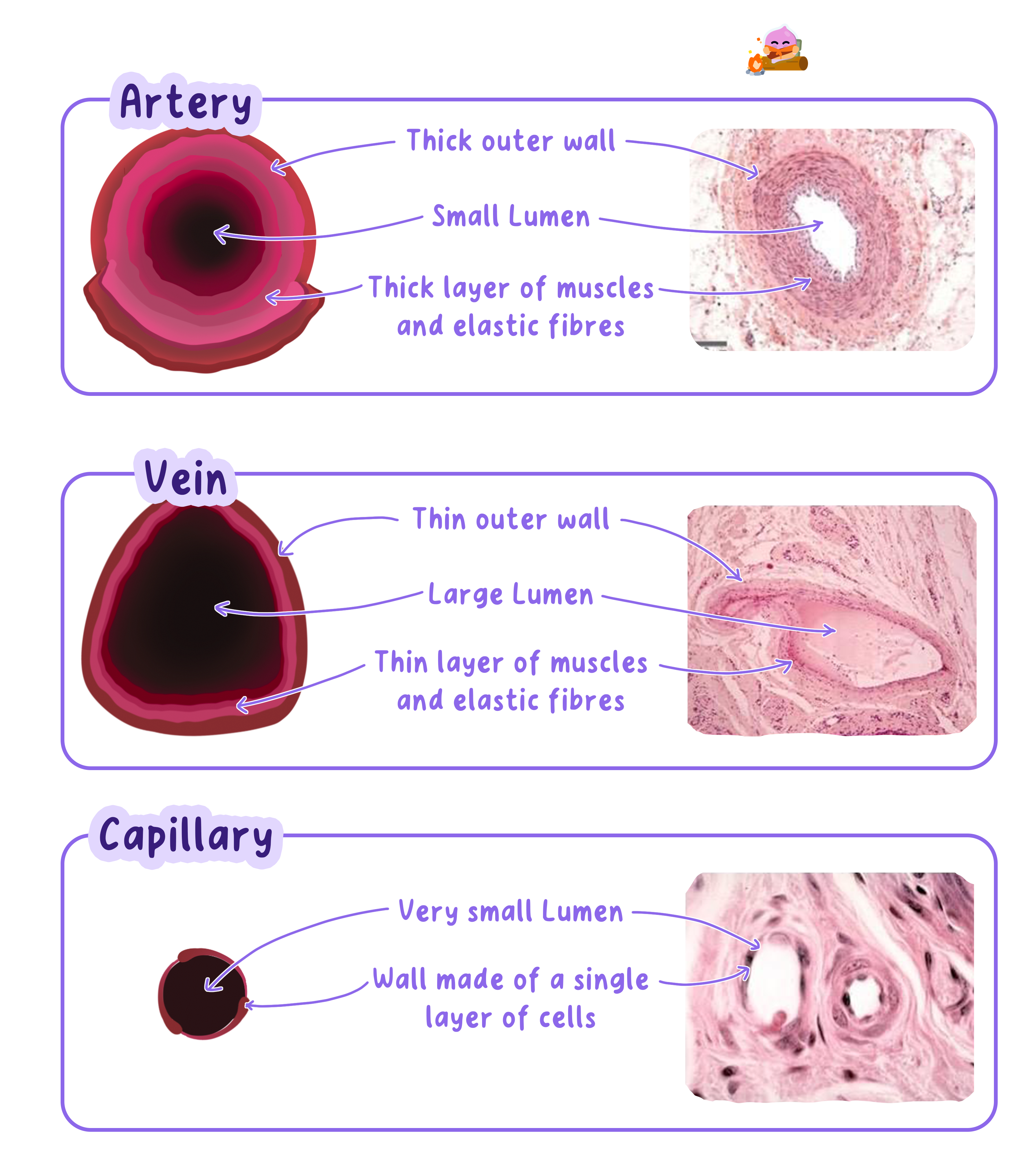
A blood vessel that carries blood away from the heart. It has a thick, muscular, elastic wall and a narrow lumen to withstand and maintain high pressure.
- Blood travels in three types of vessel: arteries, veins and capillaries.
- The lumen is the channel down the middle that blood flows through, and each wall is built for its job.
- Arteries carry blood away from the heart, where it is at high pressure.
- Their thick, muscular, elastic walls stretch and recoil to withstand the surges of pressure from each heartbeat.
- A narrow lumen helps keep the blood pressure high.
- Because this blood is under high pressure, a cut artery bleeds rapidly in spurts, which makes arterial wounds dangerous.
- You can feel your pulse where an artery runs close to the skin, such as your wrist or neck, as the wall stretches with each heartbeat.
- You can see valves at work in the veins of your forearm: push blood along a vein and it will not flow back past a valve.
Veins Return Blood at Low Pressure
Vein
A blood vessel that carries blood towards the heart. It has a thin wall, a wide lumen and valves that stop blood flowing backwards.
- Veins carry blood back towards the heart, where it is at low pressure.
- They have thin walls and a wide lumen, because they do not need to resist high pressure.
- They contain valves that shut to stop blood flowing backwards.
- Nearby skeletal muscles squeeze the veins as you move, helping push blood back to the heart.
Veins do not have a pulse, because the pressure surges from the heartbeat have been smoothed out by the time blood reaches them.
Capillaries Are Where Exchange Happens
Capillary
A tiny blood vessel with a wall one cell thick that links arteries to veins and lets substances be exchanged between the blood and the surrounding cells.
- Capillaries are tiny vessels that link the arteries to the veins inside the tissues.
- Their walls are only one cell thick, giving a very short distance for substances to diffuse.
- Oxygen and glucose diffuse out of the blood to the cells, and carbon dioxide diffuses in.
- They form a huge network, so no cell is far from a supply of blood.
"One cell thick" describes the thin wall; it does not mean the cells have cell walls, because animal cells never have cell walls.
Working Out the Rate of Blood Flow
- The rate of blood flow is the volume of blood passing a point in a given time.
- Work it out with: rate of blood flow=volume of bloodnumber of minutes\text{rate of blood flow} = \dfrac{\text{volume of blood}}{\text{number of minutes}}rate of blood flow=number of minutesvolume of blood.
- For example, if 2460 cm32460\ \text{cm}^32460 cm3 of blood flows through a vessel in 4 minutes, the rate =2460÷4=615 cm3= 2460 \div 4 = 615\ \text{cm}^3=2460÷4=615 cm3 per minute.
- To find the volume in one hour, multiply by 60: 615×60=36 900 cm3615 \times 60 = 36\,900\ \text{cm}^3615×60=36900 cm3.
- Always give your answer in the units the question asks for, converting between minutes and hours where needed.
- What is meant by a double circulatory system?
- Which chambers of the heart pump blood out, and which receive it?
- Why is the pulmonary artery unusual for an artery?
- Explain why the wall of the left ventricle is thicker than the right.
- Give one adaptation of a capillary and how it helps its function.
2.2.2b The respiratory system
The Lungs Are the Body's Gas Exchange Surface
Gas exchange
The swapping of oxygen and carbon dioxide between the air in the alveoli and the blood, which takes place by diffusion.
- The lungs sit in your chest, or thorax, protected by the ribcage.
- They are separated from the organs below by a sheet of muscle called the diaphragm.
- Their job is to bring air and blood very close together so gases can be swapped.
- This swapping of gases is gas exchange, and it happens in tiny air sacs called alveoli.
The lungs bring air and blood close together so oxygen can diffuse into the blood and carbon dioxide can diffuse out.
The Path Air Takes Into the Lungs
Trachea
The windpipe: the tube that carries air from the mouth and nose down towards the lungs.
- Trachea
- The windpipe, which carries air from the throat down towards the lungs.
- Bronchi
- The trachea splits into two bronchi, one carrying air into each lung.
- Bronchioles
- Each bronchus branches into many smaller tubes called bronchioles.
- Alveoli
- Each bronchiole ends in clusters of tiny air sacs called alveoli.
- Capillary network
- A dense network of capillaries wraps around the alveoli, carrying blood right up to the air.
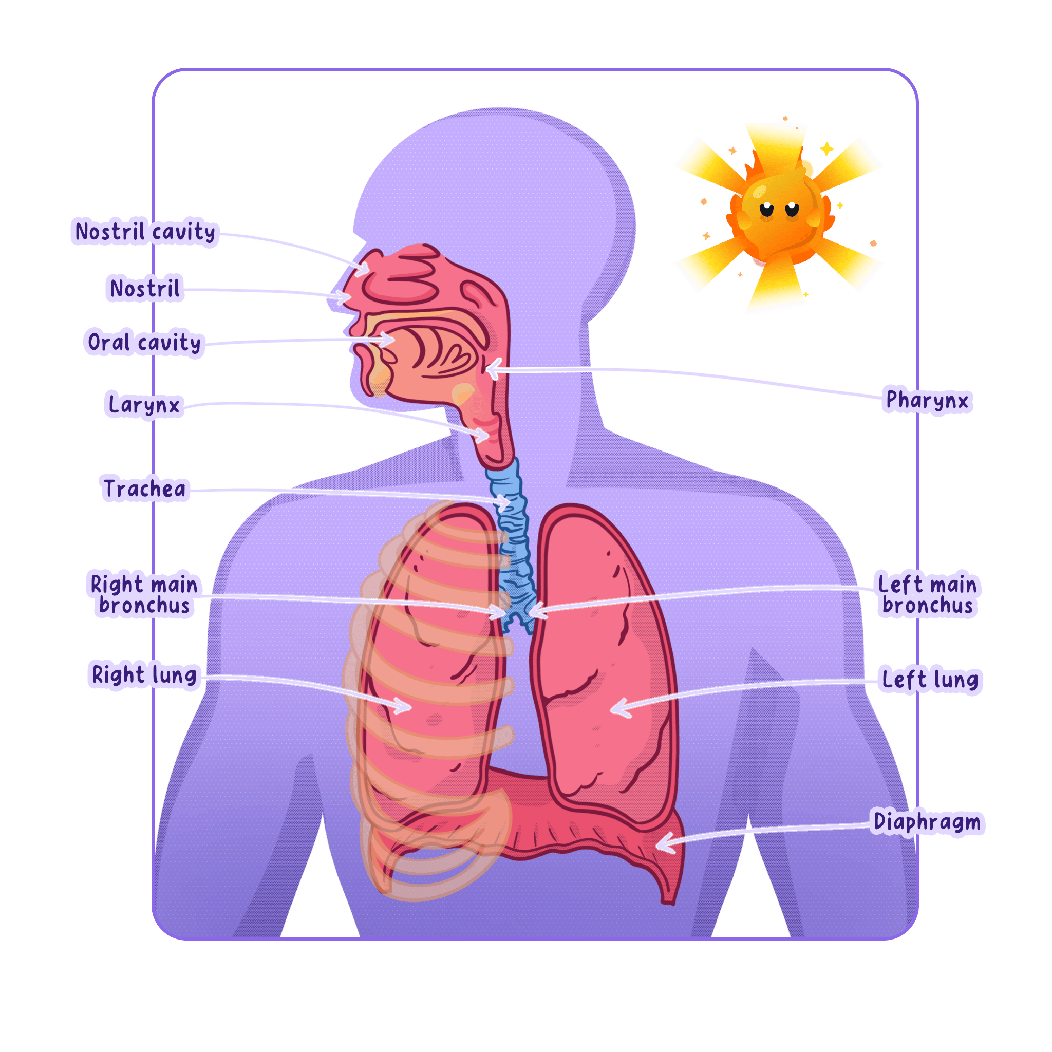
For AQA you need to know only the trachea, bronchi, alveoli and the capillary network around the alveoli.
How You Breathe Air In and Out
Ventilation
The movement of air into and out of the lungs (breathing), which keeps the air in the alveoli fresh and maintains a steep concentration gradient for gas exchange.
Breathing in
- The intercostal muscles between the ribs and the diaphragm contract.
- The ribs move up and out and the diaphragm flattens.
- This increases the volume of the thorax.
- A bigger volume means lower pressure inside the chest than outside.
- Air is drawn into the lungs.
Breathing out
- The intercostal muscles and diaphragm relax.
- The ribs move down and in and the diaphragm domes upwards.
- This decreases the volume of the thorax.
- A smaller volume means higher pressure inside the chest.
- Air is forced out of the lungs.
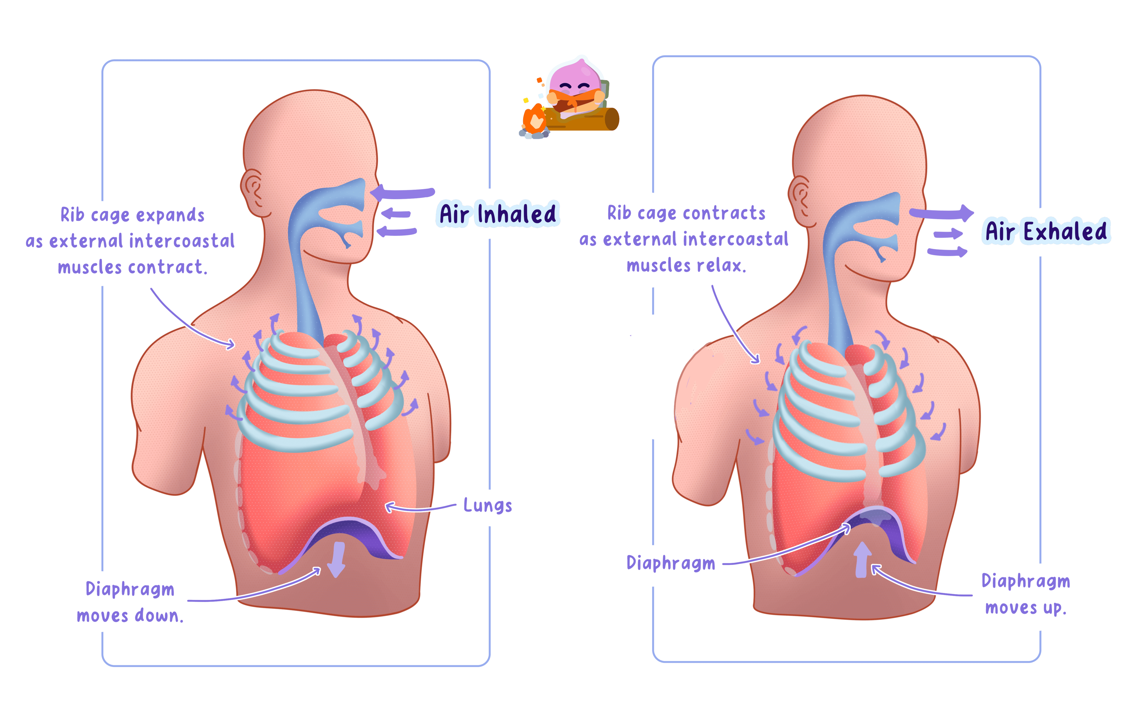
Moving air in and out like this is called ventilation, and it constantly refreshes the air inside the alveoli.
Gas Exchange in the Alveoli Happens by Diffusion
Alveoli
The tiny air sacs at the ends of the airways in the lungs, surrounded by capillaries, where gas exchange takes place. A single one is an alveolus.
- Alveoli are tiny air sacs surrounded by a dense network of capillaries.
- Blood arriving at the lungs is low in oxygen and high in carbon dioxide.
- Oxygen diffuses from the air in the alveoli into the blood.
- Carbon dioxide diffuses from the blood into the air in the alveoli, ready to be breathed out.
- This exchange is diffusion, so it needs a steep concentration gradient to be fast.
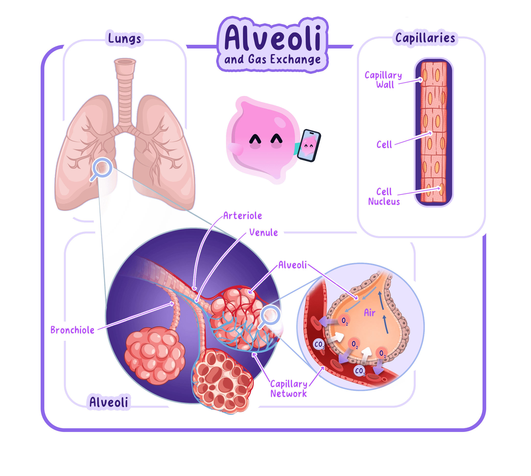
Gas exchange is diffusion, not active pumping, so it only works while there is a difference in concentration across the alveolar wall.
Why the Alveoli Are So Good at Their Job
Capillary
A tiny blood vessel with a wall one cell thick that links arteries to veins and lets substances be exchanged between the blood and the surrounding cells.
- There are millions of alveoli, giving a very large surface area for diffusion.
- Their walls are only one cell thick, giving a short diffusion distance.
- A dense capillary network gives a rich blood supply that keeps removing oxygen and delivering carbon dioxide, maintaining the concentration gradient.
- Ventilation keeps refreshing the air, which also maintains a steep concentration gradient.
- The thin, moist walls let gases dissolve and diffuse across easily.
If the alveoli of your lungs were opened out flat, they would cover an area about the size of 10 to 15 table tennis tables.
Comparing the Air You Breathe In and Out
- Nitrogen
- About 80% breathed in and about 80% breathed out, because the body does not use it.
- Oxygen
- About 20% breathed in, falling to about 16% breathed out, as some is absorbed for respiration.
- Carbon dioxide
- About 0.04% breathed in, rising to about 4% breathed out, as it is a waste product of respiration.
- Exhaled air is also warmer and contains more water vapour than inhaled air.
It is wrong to say you breathe in only oxygen and breathe out only carbon dioxide, because exhaled air is still mostly nitrogen and still contains about 16% oxygen.
How the System Fits Together
- Air travels down the trachea and bronchi to the alveoli.
- Ventilation keeps the air in the alveoli fresh.
- Oxygen diffuses into the blood and carbon dioxide diffuses out, across the thin alveolar walls.
- The large surface area, short distance and good blood supply make this exchange fast and efficient.
- Name the four lung structures you must know for AQA.
- Describe what happens to the ribs and diaphragm when you breathe in.
- Which gas diffuses from the alveoli into the blood?
- Give three ways the alveoli are adapted for efficient gas exchange.
- Roughly what percentage of oxygen is in exhaled air?
