X-rays are high-energy, short-wavelength electromagnetic waves that have revolutionized diagnostic medicine. Because they can penetrate soft tissues but are absorbed by denser materials like bone, they provide a non-invasive way to look inside the human body.
What you'll learn:
- How high-voltage X-ray tubes produce controlled beams of X-ray photons.
- The four quantum mechanisms by which X-ray photons interact with and are attenuated by matter.
- How to quantitatively model X-ray absorption using exponential attenuation equations.
- The clinical roles of iodine and barium contrast media, and how Computerised Axial Tomography (CAT) scans create detailed 3D diagnostic profiles.
1. Production of X-ray Photons
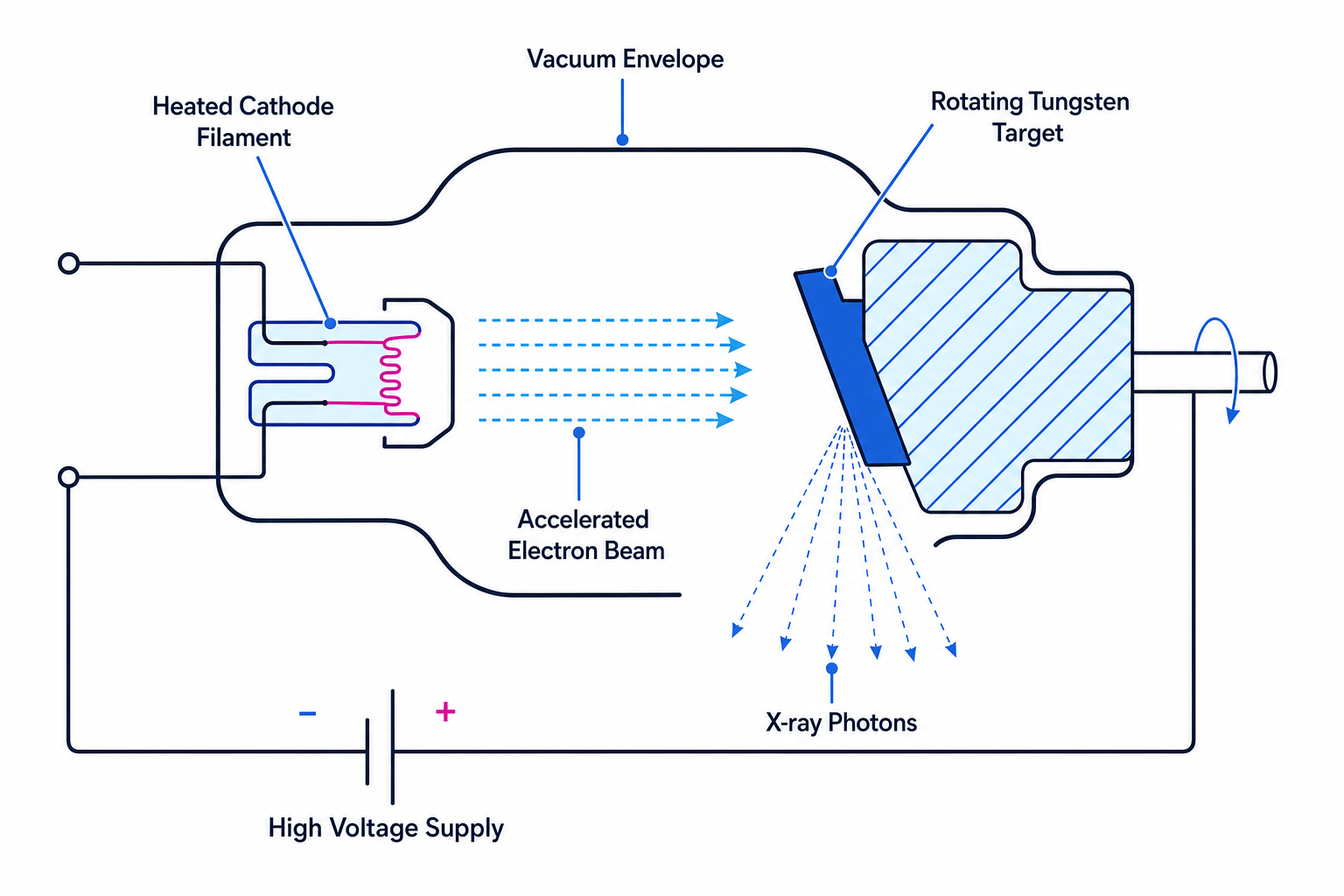
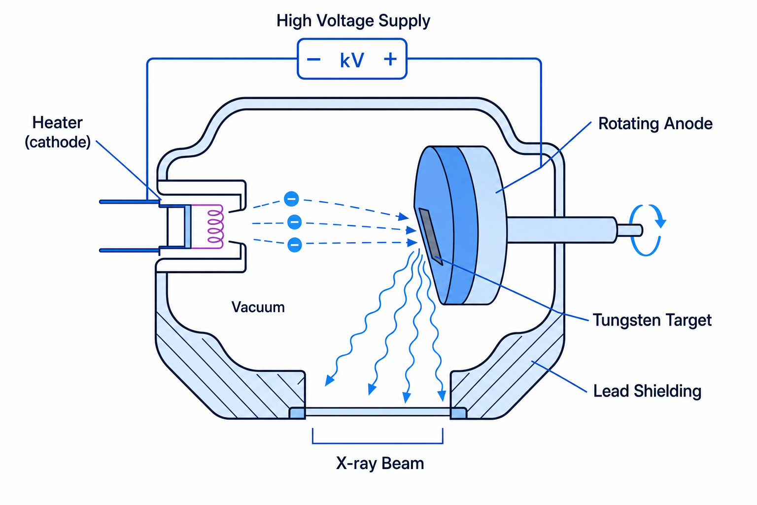
An X-ray tube (often called a Coolidge tube) is an evacuated glass envelope containing a cathode and an anode across which a massive potential difference is applied.
Components of the X-ray Tube
- Heater (Cathode): A low-voltage current passes through a tungsten filament, heating it until it releases electrons through thermionic emission.
- High-Voltage Supply: A very high potential difference (typically 30 kV30\text{ kV}30 kV to 100 kV100\text{ kV}100 kV) is applied between the cathode and the anode. This creates a strong electric field that accelerates the emitted electrons across the vacuum.
- Target Metal (Anode): A target made of a high-melting-point metal, usually tungsten, is embedded in a copper anode. When the high-velocity electrons strike the target, they decelerate rapidly, converting their kinetic energy into electromagnetic radiation.
- Vacuum: The entire process must occur in a vacuum so that the accelerating electrons do not collide with gas molecules and lose kinetic energy before reaching the target.
Energy Conversion and Efficiency
When an electron is accelerated through a potential difference VVV, the maximum kinetic energy EkE_kEk it gains is:
Ek=eV E_k = eV Ek=eVwhere eee is the elementary charge (1.60×10⁻¹⁹ C1.60 \times 10^{-19}\text{ C}1.60×10⁻¹⁹ C).
When these electrons strike the tungsten target, more than 99% of their kinetic energy is converted into thermal energy (heat) in the anode. To prevent the anode from melting, it is made of copper (which has a high thermal conductivity), often rotates to distribute the thermal load, and is cooled using circulating oil or water.
Only about 1% of the kinetic energy is converted into X-ray photons. This conversion happens via two processes:
- Bremsstrahlung (braking radiation): Electrons are decelerated by the electric fields of the tungsten nuclei, releasing a continuous spectrum of X-ray energies.
- Characteristic radiation: Accelerating electrons knock inner-shell electrons out of the tungsten atoms. Outer-shell electrons fall into the vacancies, emitting X-ray photons of discrete, highly specific energies.
Thermionic Emission
The release of electrons from the surface of a heated metal filament due to thermal energy overcoming the work function of the metal.
Maximum Photon Energy
The maximum energy of a single X-ray photon produced cannot exceed the maximum kinetic energy of the accelerated electron that created it:
hfmax=hcλmin=eV hf_{\text{max}} = \frac{hc}{\lambda_{\text{min}}} = eV hfmax=λminhc=eVwhere hhh is Planck's constant and ccc is the speed of light.
2. X-ray Attenuation Mechanisms
When X-rays pass through a material, they are absorbed or scattered. This reduction in the intensity of the X-ray beam as it travels through a medium is known as attenuation.
Depending on the energy of the incident X-ray photons and the atomic number of the material, one of four attenuation mechanisms will dominate.
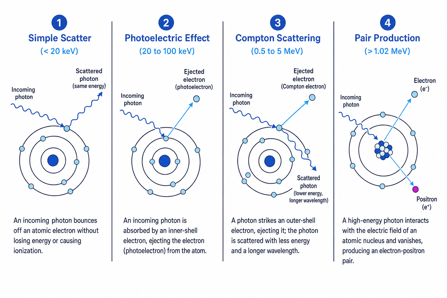
1. Simple Scatter (Low Energy: 1 keV−20 keV1\text{ keV} - 20\text{ keV}1 keV−20 keV)
A low-energy X-ray photon interacts with an electron in an atom but does not have enough energy to remove it. Instead, the photon simply bounces (scatters) off the electron with no change in its energy or wavelength.
2. Photoelectric Effect (Diagnostic Energy: <100 keV< 100\text{ keV}<100 keV)
An X-ray photon is fully absorbed by an inner-shell electron of an atom. The electron escapes the atom as a photoelectron with kinetic energy equal to the photon's energy minus the electron's binding energy. This is the dominant mechanism used in diagnostic medical imaging because the probability of absorption depends strongly on the chemical composition of the tissue.
3. Compton Effect (Therapeutic Energy: 0.5 MeV−5.0 MeV0.5\text{ MeV} - 5.0\text{ MeV}0.5 MeV−5.0 MeV)
A mid-to-high-energy photon interacts with an outer-shell electron. The photon ejects the electron from the atom (ionizing it), but only loses a portion of its energy. The remaining energy is carried away by a lower-energy, scattered X-ray photon with a longer wavelength.
4. Pair Production (High Energy: >1.02 MeV> 1.02\text{ MeV}>1.02 MeV)
An extremely high-energy photon interacts directly with the electric field of an atomic nucleus. The photon completely disappears, and its energy is converted into mass, producing an electron-positron pair. The minimum energy required for this is the sum of the rest-mass energies of the two particles:
Emin=2mec2≈1.02 MeV E_{\text{min}} = 2 m_e c^2 \approx 1.02\text{ MeV} Emin=2mec2≈1.02 MeVThe Diagnostic Sweet Spot
In medical imaging, we primarily utilize the Photoelectric Effect range (20 keV−100 keV20\text{ keV} - 100\text{ keV}20 keV−100 keV). Simple scatter is too weak, while Compton scattering and pair production cause unwanted scattering and tissue damage without providing useful contrast.
3. Mathematical Modelling of Attenuation
The attenuation of a parallel beam of X-rays passing through a uniform material is exponential. The change in intensity depends on the thickness of the material and its specific absorption properties.
Attenuation Equation
The intensity III of an X-ray beam after passing through a thickness xxx of a material is given by:
I=I0e−μx I = I_0 e^{-\mu x} I=I0e−μxWhere:
- I0I_0I0 is the initial intensity of the beam before entering the material (in W m−2\text{W m}^{-2}W m−2).
- III is the transmitted intensity (in W m−2\text{W m}^{-2}W m−2).
- xxx is the thickness of the material (typically in m\text{m}m or mm\text{mm}mm).
- μ\muμ is the attenuation (absorption) coefficient of the material (in m−1\text{m}^{-1}m−1 or mm−1\text{mm}^{-1}mm−1).
Units of x and μ
Always ensure that the units of the thickness xxx and the attenuation coefficient μ\muμ match. If μ\muμ is given in mm−1\text{mm}^{-1}mm−1, then xxx must be in mm\text{mm}mm so that the exponent −μx-\mu x−μx remains dimensionless.
Calculating thickness for shielding
An X-ray beam of initial intensity I0I_0I0 is incident on a lead block with an attenuation coefficient of μ=1.2 mm−1\mu = 1.2\text{ mm}^{-1}μ=1.2 mm−1. Calculate the thickness of lead required to reduce the transmitted intensity to 5.0%5.0\%5.0% of its original value.
- State the relationship between III and I0I_0I0: Since the final intensity is 5.0%5.0\%5.0% of the initial intensity, we can write:
- Substitute this relation into the attenuation equation:
- Divide both sides by I0I_0I0 to isolate the exponential expression:
- Take the natural logarithm (ln\lnln) of both sides:
- Calculate the left-hand side and solve for xxx:
- Round to an appropriate number of significant figures: Since the input values are given to 2 significant figures, the final thickness is 2.5 mm2.5\text{ mm}2.5 mm.
Interchanging Percentage Transmitted and Percentage Absorbed
If a question states that "85% of the X-rays are absorbed", do not substitute 0.850.850.85 for I/I0I/I_0I/I0. If 85% is absorbed, then only 15% is transmitted. You must use I=0.15I0I = 0.15 I_0I=0.15I0.
4. X-ray Imaging with Contrast Media
In standard diagnostic X-rays, image contrast relies on different tissues having different attenuation coefficients. Bone has a high attenuation coefficient because it contains calcium (Z=20Z = 20Z=20), which has a relatively high atomic number. Soft tissues consist mostly of hydrogen, carbon, and oxygen (Z≈6Z \approx 6Z≈6 to 888), which have much lower attenuation coefficients and look virtually identical on an X-ray.
The probability of photoelectric absorption is proportional to Z3Z^3Z3, where ZZZ is the proton (atomic) number. To image soft-tissue structures like blood vessels or the digestive tract, we must artificially increase their attenuation coefficient using contrast media.
Contrast Materials
- Iodine (Z=53Z = 53Z=53): Typically injected into the bloodstream. It is used to view blood vessels (angiography) to detect blockages, heart issues, or organ structure.
- Barium (Z=56Z = 56Z=56): Usually swallowed by the patient as a "barium meal" or "barium swallow". It coats the lining of the stomach and digestive tract, allowing clinicians to image ulcers, blockages, or swallowing issues.
Because both elements have high atomic numbers, they absorb X-rays far more efficiently than surrounding tissue, making the targeted organ systems stand out as stark white areas on the final X-ray image.
5. Computerised Axial Tomography (CAT) Scans
While a standard X-ray produces a quick, low-dose 2D image, it suffers from a major limitation: all 3D structures are superimposed onto a single flat plane. A shadow of a rib can easily obscure a small tumor in the lung behind it.
To overcome this, Computerised Axial Tomography (CAT) scans are used.
Structure and Components of a CAT Scanner
- Rotating X-ray Tube: A high-output X-ray tube rotates rapidly on a circular gantry around a patient lying on a motorized bed.
- Thin Fan-Shaped Beam: The tube produces a very narrow, fan-shaped X-ray beam rather than a broad cone, targeting only a thin slice of the patient's body at any one moment.
- Ring of Detectors: Opposite the X-ray tube is an array of electronic detectors that measure the intensity of the transmitted X-rays after passing through the patient.
- Computer Software and Display: The raw attenuation data from thousands of angles are sent to a computer running complex reconstruction algorithms. This software calculates the attenuation at each point in the slice, building up a 2D cross-sectional image. As the patient is moved slowly through the gantry, multiple 2D slices are combined to build a highly detailed 3D model.
Advantages and Disadvantages of CAT Scans
| Feature | Standard 2D X-ray | CAT Scan |
|---|---|---|
| Dimension | 2D image only (overlapping structures). | Detailed 3D image (no overlap; slices can be viewed from any angle). |
| Soft Tissue Contrast | Poor; difficult to distinguish between tissues of similar densities. | High; can clearly resolve minor differences in soft tissues (e.g., grey vs. white brain matter). |
| Radiation Dose | Very low (typically equivalent to a few days of background radiation). | High (typically equivalent to several years of background radiation); increases cancer risk. |
| Cost & Time | Cheap, rapid, and highly portable. | Expensive, takes longer, requires the patient to remain perfectly still inside a large machine. |
In the exam
- Explain the Z3Z^3Z3 dependency: When discussing why contrast media are used, always state that the probability of photoelectric absorption is proportional to the cube of the atomic number (Z3Z^3Z3). Mention the specific high ZZZ numbers of iodine (Z=53Z=53Z=53) or barium (Z=56Z=56Z=56) compared to soft tissue (Z≈7Z\approx 7Z≈7).
- Describe the CAT mechanism systematically: If asked to describe a CAT scan, list the physical movements clearly: (1) X-ray tube rotates, (2) produces a thin fan-shaped beam, (3) detectors register the transmitted intensity, (4) the table moves the patient slowly through, and (5) software processes the 2D slices into a 3D image.
- Be careful with units in calculations: Attenuation questions love to mix units (e.g., xxx in cm\text{cm}cm and μ\muμ in mm−1\text{mm}^{-1}mm−1). Convert them to a consistent format before substituting them into I=I0e−μxI = I_0 e^{-\mu x}I=I0e−μx.
Check yourself
- A medical physicist uses an X-ray tube operating at 80.0 kV80.0\text{ kV}80.0 kV. What is the maximum frequency of the photons produced?
- Explain why Compton scattering is undesirable during standard medical diagnostic imaging.
- Lead has an attenuation coefficient of μ=1.50 mm−1\mu = 1.50\text{ mm}^{-1}μ=1.50 mm−1 for a specific X-ray beam. By what percentage is the intensity of the beam reduced after passing through 2.00 mm2.00\text{ mm}2.00 mm of lead?
- Why is a contrast medium injected for an angiogram, but swallowed for a digestive tract assessment? State the element used in each case.