

What you'll learn
- How radioactive medical tracers such as technetium-99m and fluorine-18 are chosen and used.
- How a gamma camera forms a medical image from gamma photons.
- How PET scanning uses positron-electron annihilation and coincidence detection.
- How to discuss diagnosis, risk-benefit decisions, and the cost of expensive scanners.
The big idea: imaging function, not just structure
Many medical imaging methods show anatomy: the shape and structure of tissues. Diagnostic methods using radioactive tracers are especially useful because they can show function: how well an organ takes up a substance, how blood flows, or how active certain cells are.
A patient is given a small amount of a radioactive substance. Radiation emitted from inside the body is detected outside the body, and a computer builds an image.
Medical tracer
A medical tracer is a radioactive isotope attached to, or included in, a substance that follows a particular biological pathway in the body. Its emitted radiation is detected to show where the substance has gone.
A good tracer should:
- emit radiation that can escape the body and be detected;
- have a half-life long enough for imaging, but short enough to reduce unnecessary dose;
- be chemically suitable for the organ or process being investigated;
- be given in a small amount and be as non-toxic as possible.
Activity and decay constant
The activity AAA of a radioactive sample is the number of nuclear decays per second, measured in becquerels, Bq. The decay constant λ\lambdaλ is the probability per second that a nucleus decays. They are linked by
A=λNA = \lambda NA=λNwhere NNN is the number of undecayed nuclei.
The number of undecayed nuclei falls exponentially:
N=N0e−λtN = N_0 e^{-\lambda t}N=N0e−λtFor a fixed isotope, activity falls in the same way because A=λNA=\lambda NA=λN.
Calculating remaining tracer activity
A technetium-99m tracer has initial activity 600 MBq. Its half-life is 6.0 h. Calculate the activity after 18 h.
-
Convert the half-life and time into seconds so the decay constant is in per second:
T1/2=6.0 h=2.16×10⁴ sT_{1/2} = 6.0\,\text{h} = 2.16 \times 10^4\,\text{s}T1/2=6.0h=2.16×104s t=18 h=6.48×10⁴ st = 18\,\text{h} = 6.48 \times 10^4\,\text{s}t=18h=6.48×10⁴s -
Find the decay constant using λ=ln2T1/2\lambda = \frac{\ln 2}{T_{1/2}}λ=T1/2ln2:
λ=0.6932.16×104 s=3.21×10⁻⁵ s−1\lambda = \frac{0.693}{2.16 \times 10^4\,\text{s}} = 3.21 \times 10^{-5}\,\text{s}^{-1}λ=2.16×10⁴s0.693=3.21×10⁻⁵s−1 -
Use the exponential decay model for activity:
A=A0e−λtA = A_0 e^{-\lambda t}A=A0e−λt A=600 MBq×e−(3.21×10⁻⁵ s−1)(6.48×10⁴ s)A = 600\,\text{MBq} \times e^{-(3.21 \times 10^{-5}\,\text{s}^{-1})(6.48 \times 10^4\,\text{s})}A=600MBq×e−(3.21×10⁻⁵s−1)(6.48×10⁴s) -
Calculate and quote sensibly:
A=75 MBqA = 75\,\text{MBq}A=75MBqThis also makes sense because 18 h is three half-lives, so the activity is 600÷23=75 MBq600 \div 2^3 = 75\,\text{MBq}600÷23=75MBq.
Half-life is not the scan time
A tracer with a half-life of 6.0 h has not “finished” after 6.0 h. Half of the remaining radioactive nuclei decay in each half-life, so activity decreases exponentially.
Technetium-99m and fluorine-18
Two key medical tracers for OCR H556 are technetium-99m and fluorine-18.
Technetium-99m
Technetium-99m, written 99mTc^{99\text{m}}\mathrm{Tc}99mTc, is widely used with a gamma camera.
Metastable state
The “m” in technetium-99m means metastable: the nucleus is in a long-lived excited state before it emits a gamma photon and moves to a lower energy state.
Technetium-99m is useful because:
- it emits gamma photons, which can escape the body and be detected;
- it has a half-life of about 6 h, giving time for preparation and imaging while limiting dose;
- it can be attached to different compounds to investigate different organs, such as bones, kidneys, or the thyroid.
Fluorine-18
Fluorine-18, written 18F^{18}\mathrm{F}18F, is used in positron emission tomography, usually called PET scanning.
Fluorine-18 is a positron emitter. A positron is the antimatter version of an electron: it has the same mass as an electron but positive charge. Fluorine-18 has a half-life of about 110 min, so it must be produced and transported quickly.
A common PET tracer is fluorodeoxyglucose, often shortened to FDG. It behaves similarly to glucose, so tissues with high glucose uptake show strong tracer uptake.
Why different tracers are chosen
Technetium-99m is suitable for gamma-camera imaging because it directly emits gamma photons. Fluorine-18 is suitable for PET because it emits positrons, which lead to pairs of gamma photons after annihilation.
Gamma camera imaging
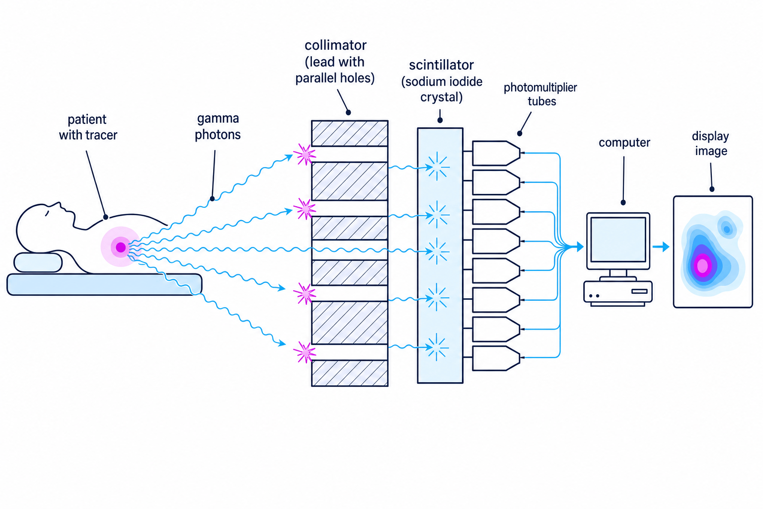
A gamma camera detects gamma photons emitted by a tracer inside the patient and forms a 2D image showing where the tracer is concentrated.
Main components
Collimator
A collimator is a thick lead plate with many narrow holes. It only allows gamma photons travelling in certain directions to reach the detector, while absorbing photons travelling at other angles.
The collimator is essential because gamma photons cannot be focused with ordinary lenses. Without it, photons from different positions would mix together and blur the image.
Scintillator
A scintillator is a material that emits tiny flashes of visible light when gamma photons deposit energy in it. In many gamma cameras, the scintillator is a sodium iodide crystal.
Photomultiplier tube
A photomultiplier tube converts a weak flash of light into an amplified electrical pulse.
The computer uses the pattern and sizes of pulses from the photomultiplier tubes to estimate where each gamma photon struck the scintillator. Many detected photons build up a count-rate image: brighter regions correspond to more detected gamma photons, meaning more tracer activity.
Explaining the purpose of the collimator
A student suggests removing the collimator to make a brighter gamma-camera image. Explain the trade-off.
-
Removing the collimator would allow more gamma photons to reach the scintillator, so the count rate would increase and the image would be formed more quickly.
-
However, photons from many different positions in the patient could hit the same point on the scintillator, so the detector would not know where they came from.
-
The image would therefore be brighter but much less sharp. The collimator reduces count rate, but it improves spatial resolution by preserving directional information.
Brighter does not always mean better
A higher count rate improves signal, but if the photons have lost directional information the image may be blurred and diagnostically poor.
Diagnosis using a gamma camera
Gamma-camera images are used to diagnose problems involving organ function or tracer uptake. For example:
- bone scans can reveal regions of unusually high bone activity;
- kidney scans can show how tracer passes through the kidneys;
- thyroid scans can show abnormal uptake in the thyroid gland;
- heart scans can investigate blood supply to heart muscle.
The doctor compares the tracer distribution with what is expected for a healthy patient. A region of unusually high or low activity may indicate disease, poor blood flow, abnormal metabolism, or tissue damage.
Describe the image physically first
In exam answers, start with the physics: “more tracer uptake gives more gamma photons detected, so a brighter region appears on the image.” Then link that to the medical interpretation.
PET scanning
PET stands for positron emission tomography. Tomography means forming an image of a slice or 3D distribution inside the body.
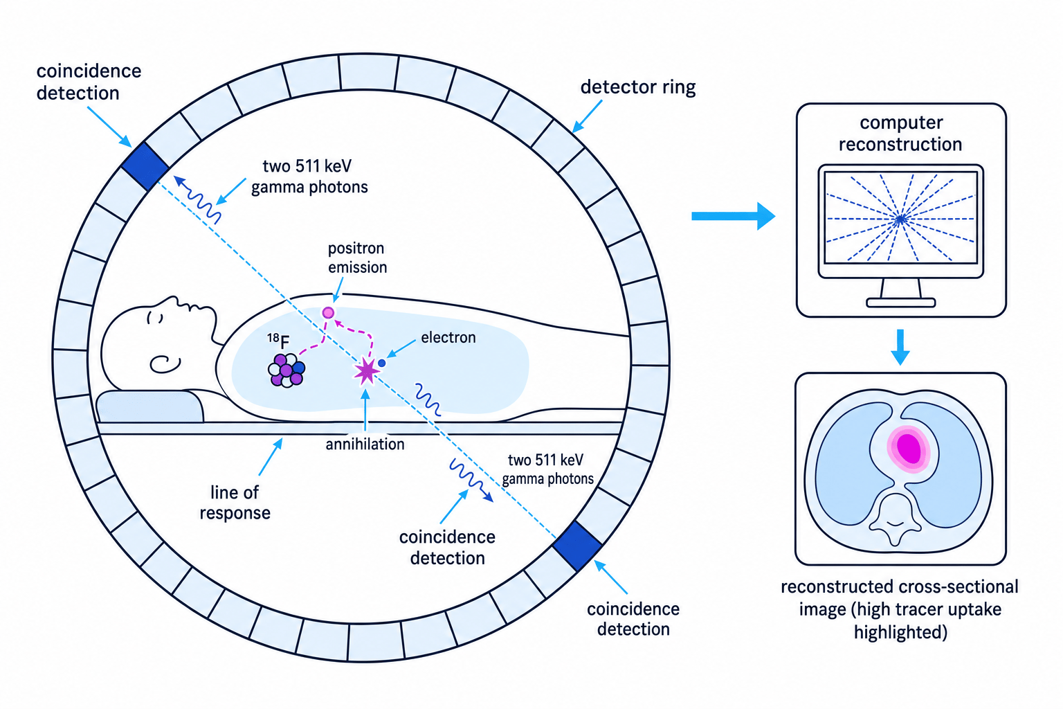
In PET, the patient is given a positron-emitting tracer such as fluorine-18. The positron travels a short distance through tissue, then meets an electron. The positron and electron annihilate.
Annihilation
Annihilation is the process in which a particle and its antiparticle meet and their mass-energy is converted into photons. In PET, a positron and an electron annihilate to produce two gamma photons travelling in opposite directions.
Each annihilation produces two gamma photons of energy 511 keV travelling approximately back-to-back. Detectors in a ring around the patient detect these photons.
Coincidence detection
Coincidence detection
Coincidence detection means recording two gamma photons detected at opposite sides of the scanner within a very short time interval, treating them as coming from the same annihilation event.
The scanner does not know the exact point of annihilation from one pair of photons. It knows the event occurred somewhere along the straight line joining the two detectors. This is called the line of response.
By collecting many coincidence events from many directions, the computer reconstructs the tracer distribution inside the body.
Locating high tracer uptake in PET
A PET scanner records many coincidence events whose lines of response pass through the same small region. What does this imply?
-
Each coincidence event shows that an annihilation occurred somewhere along its line of response.
-
If many different lines of response pass through the same region, that region is a likely source of many annihilation events.
-
Many annihilation events mean many fluorine-18 decays nearby, so the region has high tracer uptake and appears bright in the reconstructed PET image.
PET uses electronic collimation
A gamma camera uses a physical lead collimator to select photon directions. PET uses pairs of opposite detectors and coincidence timing to identify lines of response.
Diagnosis using PET
PET is especially useful for investigating processes involving metabolism. Because FDG behaves like glucose, areas with high glucose uptake can be identified. This is important in applications such as:
- detecting and staging some cancers, because many tumours have high metabolic activity;
- studying brain activity and neurological disorders;
- assessing heart muscle viability and blood flow.
PET images are often combined with anatomical images from another scanner, such as CT or MRI, so doctors can match function to structure. The OCR focus is the PET principle and image formation, not the detailed reconstruction algorithm.
Tracer uptake is not a diagnosis by itself
A bright PET region shows high tracer uptake, but the medical conclusion depends on the tracer, the organ, patient history, and comparison with normal uptake patterns.
Equipping a hospital with an expensive scanner
Installing a PET scanner or advanced gamma-camera system is not only a physics decision. Hospitals must balance benefit, cost, safety, and access.
Important issues include:
- Purchase cost: scanners, shielding, software, and building modifications are expensive.
- Running cost: maintenance, calibration, trained staff, and quality assurance are needed.
- Tracer supply: fluorine-18 has a short half-life, so a hospital may need rapid delivery from a cyclotron facility.
- Patient benefit: the scanner should improve diagnosis, treatment planning, or monitoring enough to justify the cost.
- Throughput: enough patients must be scanned for the equipment to be used effectively.
- Radiation protection: staff and patients need procedures that keep doses as low as reasonably practicable.
- Alternatives: ultrasound, MRI, CT, or gamma-camera imaging may sometimes be cheaper or more appropriate.
- Fair access: expensive scanners can raise questions about which hospitals and patient groups get access.
Risk-benefit thinking
In nuclear medicine, the aim is not “zero radiation”. The aim is to use the smallest suitable activity that gives a clinically useful image, so the diagnostic benefit outweighs the radiation risk.
In the exam
-
For tracer questions, link the isotope to the scanner: technetium-99m emits gamma photons for a gamma camera; fluorine-18 emits positrons for PET.
-
For gamma-camera image formation, name the components in order: collimator, scintillator, photomultiplier tubes, computer and display.
-
For PET, always mention positron-electron annihilation, two opposite 511 keV gamma photons, coincidence detection, and reconstruction from many lines of response.
Check yourself
- Why does a gamma camera need a collimator, even though it reduces the number of photons detected?
- Why is fluorine-18 suitable for PET but technetium-99m is not the usual PET tracer?
- What practical and ethical issues should a hospital consider before buying an expensive PET scanner?
