

What you'll learn
- What ultrasound is, and how a piezoelectric transducer can both emit and receive it.
- How A-scans and B-scans use echo timing and intensity to form medical images.
- How acoustic impedance controls reflection at tissue boundaries, and why gel is needed.
- How Doppler ultrasound can measure the speed of blood in a patient.
Ultrasound: sound above human hearing
Sound is a longitudinal wave: the particles of the medium vibrate parallel to the direction in which energy travels. In body tissue, ultrasound travels as a pattern of compressions and rarefactions.
Ultrasound
Ultrasound is a longitudinal sound wave with frequency greater than 20 kHz, which is above the upper limit of normal human hearing.
Medical ultrasound usually uses frequencies in the megahertz range, much higher than 20 kHz. Higher frequency waves can give better detail, but they are also absorbed more strongly, so they do not travel as far into the body.
Why ultrasound is useful
Ultrasound is non-ionising, so it does not carry enough photon energy to ionise atoms in body tissue. It can be reflected at boundaries between different tissues, allowing internal structures to be imaged.
The piezoelectric effect and ultrasound transducers
A transducer is a device that converts energy from one form to another.
Piezoelectric effect
The piezoelectric effect is the property of some crystals where an applied potential difference causes the crystal to change shape, and a mechanical stress on the crystal produces a potential difference.
An ultrasound transducer uses this in both directions:
- To emit ultrasound, an alternating potential difference is applied to a piezoelectric crystal. The crystal vibrates at the same frequency, producing ultrasound waves.
- To receive ultrasound, returning echoes compress the crystal. This produces an alternating potential difference, which can be processed electronically.
In medical imaging, the transducer sends out short pulses, then waits for echoes. The same device therefore acts as both a transmitter and a receiver.
Think pulse, then listen
Ultrasound imaging is not usually a continuous “shout”. The transducer sends a short pulse, then listens for echoes. The time delay tells you how deep the reflecting boundary is.
Pulse-echo imaging: A-scans and B-scans
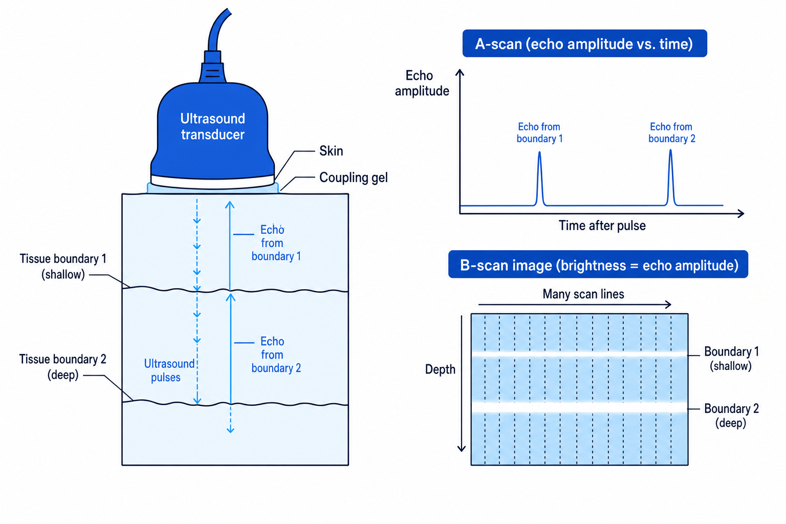
When an ultrasound pulse reaches a boundary between two media, some of the wave is reflected and some is transmitted. The reflected wave is an echo.
If the speed of ultrasound in the tissue is ccc, and an echo returns after time ttt, the boundary depth ddd is found using:
d=ct2d = \frac{ct}{2}d=2ctThe factor of 2 is needed because the ultrasound travels to the boundary and back.
A-scan
A-scan
An A-scan is a one-dimensional ultrasound display showing echo amplitude against time after the emitted pulse.
Each spike represents an echo from a boundary. The position of the spike gives the depth of the boundary, and the height of the spike shows the strength of the reflected signal.
Finding the depth of a reflecting boundary
An ultrasound pulse travels through soft tissue at a speed of 1540 m s⁻¹. An echo is detected 52 µs after the pulse is emitted. Find the depth of the boundary.
-
Convert the time delay into seconds:
t=52 μs=52×10−6 st = 52\ \mu\text{s} = 52 \times 10^{-6}\ \text{s}t=52 μs=52×10−6 s -
Use the pulse-echo depth equation, remembering the return journey:
d=ct2d = \frac{ct}{2}d=2ct -
Substitute the values with units:
d=1540 m s−1×52×10−6 s2d = \frac{1540\ \text{m s}^{-1} \times 52 \times 10^{-6}\ \text{s}}{2}d=21540 m s−1×52×10−6 s -
Calculate the depth:
d=4.0×10⁻² md = 4.0 \times 10^{-2}\ \text{m}d=4.0×10⁻² mSo the boundary is about 4.0 cm below the transducer.
Forgetting the return journey
If you use d=ctd = ctd=ct for pulse-echo ultrasound, you get twice the true depth. The measured time is for the wave travelling there and back.
B-scan
B-scan
A B-scan is a two-dimensional ultrasound image built from many scan lines, where echo brightness represents reflected intensity and position represents depth.
A B-scan is what most people think of as an ultrasound image. The transducer collects many A-scan-like lines across the region. A strong echo is shown as a bright point; a weak echo is shown as a dimmer point.
This is an example of using scientific instrumentation to convert invisible information — echo delay and echo intensity — into a visual image that clinicians can interpret.
Acoustic impedance
Different materials respond differently to ultrasound. This is described using acoustic impedance.
Acoustic impedance
The acoustic impedance ZZZ of a medium is defined by
Z=ρcZ = \rho cZ=ρcwhere ρ\rhoρ is the density of the medium in kg m⁻³ and ccc is the speed of ultrasound in the medium in m s⁻¹.
The unit of acoustic impedance is kg m⁻² s⁻¹.
A large difference in acoustic impedance at a boundary means a large reflection. A small difference means most of the ultrasound is transmitted.
Calculating acoustic impedance
Soft tissue has density 1050 kg m⁻³ and the speed of ultrasound in it is 1540 m s⁻¹. Calculate its acoustic impedance.
-
Start from the definition:
Z=ρcZ = \rho cZ=ρc -
Substitute the density and wave speed:
Z=1050 kg m−3×1540 m s−1Z = 1050\ \text{kg m}^{-3} \times 1540\ \text{m s}^{-1}Z=1050 kg m−3×1540 m s−1 -
Multiply the values and combine the units:
Z=1.62×10⁶ kg m−2s−1Z = 1.62 \times 10^{6}\ \text{kg m}^{-2}\text{s}^{-1}Z=1.62×10⁶ kg m−2s−1
Reflection at a boundary
At a boundary between two media with acoustic impedances Z1Z_1Z1 and Z2Z_2Z2, the fraction of incident ultrasound intensity that is reflected is:
IrI0=(Z2−Z1)2(Z2+Z1)2\frac{I_r}{I_0} = \frac{(Z_2 - Z_1)^2}{(Z_2 + Z_1)^2}I0Ir=(Z2+Z1)2(Z2−Z1)2Here, I0I_0I0 is the incident intensity and IrI_rIr is the reflected intensity.
Impedance difference controls reflection
The greater the difference between Z1Z_1Z1 and Z2Z_2Z2, the greater the fraction of ultrasound reflected at the boundary.
If Z1=Z2Z_1 = Z_2Z1=Z2, then the numerator is zero, so there is no reflection. If the impedances are very different, the reflected fraction can be very close to 1, meaning almost all the ultrasound is reflected.
Calculating the reflected intensity fraction
An ultrasound wave travels from tissue with acoustic impedance 1.63×1061.63 \times 10^61.63×10⁶ kg m⁻² s⁻¹ into another tissue with acoustic impedance 1.38×1061.38 \times 10^61.38×10⁶ kg m⁻² s⁻¹. Calculate the percentage of the incident intensity reflected.
-
Identify the two acoustic impedances:
Z1=1.63×10⁶ kg m−2s−1Z_1 = 1.63 \times 10^6\ \text{kg m}^{-2}\text{s}^{-1}Z1=1.63×10⁶ kg m−2s−1 Z2=1.38×10⁶ kg m−2s−1Z_2 = 1.38 \times 10^6\ \text{kg m}^{-2}\text{s}^{-1}Z2=1.38×10⁶ kg m−2s−1 -
Substitute into the reflection equation:
IrI0=(1.38×10⁶−1.63×10⁶)2(1.38×10⁶+1.63×10⁶)2\frac{I_r}{I_0} = \frac{(1.38 \times 10^6 - 1.63 \times 10^6)^2} {(1.38 \times 10^6 + 1.63 \times 10^6)^2}I0Ir=(1.38×10⁶+1.63×10⁶)2(1.38×10⁶−1.63×10⁶)2 -
Calculate the difference and sum:
IrI0=(−0.25×10⁶)2(3.01×10⁶)2\frac{I_r}{I_0} = \frac{(-0.25 \times 10^6)^2} {(3.01 \times 10^6)^2}I0Ir=(3.01×10⁶)2(−0.25×10⁶)2 -
Square both terms and divide:
IrI0=6.9×10⁻³\frac{I_r}{I_0} = 6.9 \times 10^{-3}I0Ir=6.9×10⁻³ -
Convert the fraction to a percentage:
6.9×10−3×100=0.69%6.9 \times 10^{-3} \times 100 = 0.69\%6.9×10−3×100=0.69%About 0.69% of the incident intensity is reflected.
Using amplitudes instead of intensities
The formula gives a ratio of intensities, IrI0\frac{I_r}{I_0}I0Ir. Do not treat it as an amplitude ratio or forget to square the impedance terms.
Acoustic impedance matching and gel
A thin layer of air between the transducer and the skin would be a big problem. Air has a much smaller acoustic impedance than soft tissue, so almost all the ultrasound would be reflected at the air-skin boundary.
Acoustic impedance matching
Acoustic impedance matching means placing materials with suitable acoustic impedances between two media so that less ultrasound is reflected and more is transmitted.
In ultrasound scanning, a special coupling gel is placed between the transducer and the skin. The gel:
- removes air gaps,
- has an acoustic impedance closer to skin than air does,
- allows much more ultrasound energy to enter the body,
- improves the strength of returning echoes.
Why gel feels practical but matters physically
The gel is not just for comfort or to make the probe slide. Its physics role is to reduce reflection caused by a large acoustic impedance change.
Doppler ultrasound and blood speed
The Doppler effect is the change in observed frequency when there is relative motion between a wave source and an observer.
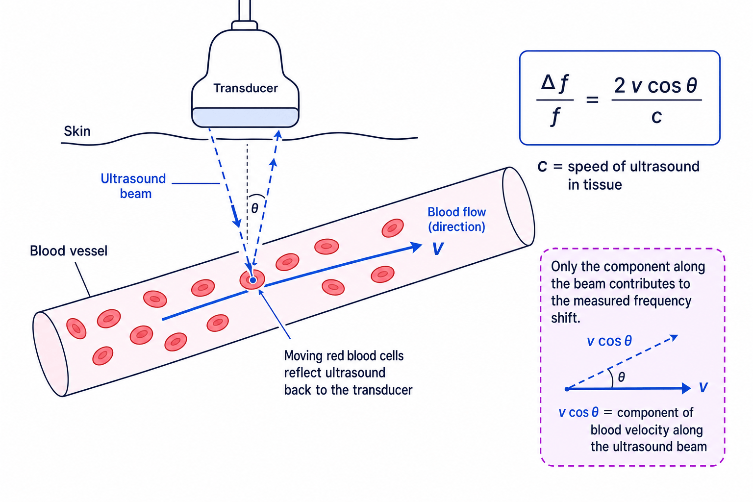
In Doppler ultrasound, ultrasound is reflected from moving blood cells. If the blood cells are moving towards or away from the transducer, the reflected ultrasound has a different frequency from the emitted ultrasound.
For blood speed measurements, OCR uses:
Δff=2vcosθc\frac{\Delta f}{f} = \frac{2v\cos\theta}{c}fΔf=c2vcosθwhere:
- Δf\Delta fΔf is the change in frequency in hertz, Hz,
- fff is the emitted ultrasound frequency in hertz, Hz,
- vvv is the speed of the blood in m s⁻¹,
- θ\thetaθ is the angle between the ultrasound beam and the direction of blood flow,
- ccc is the speed of ultrasound in the tissue in m s⁻¹.
The factor of 2 appears because the ultrasound is Doppler shifted on the way to the moving blood cells and again when reflected back.
Only the along-beam component counts
Doppler ultrasound measures the component of blood velocity along the ultrasound beam. That is why the formula contains vcosθv\cos\thetavcosθ.
Finding blood speed from a Doppler shift
A 5.0 MHz ultrasound wave is reflected from blood moving at angle 60° to the ultrasound beam. The measured frequency shift is 3.2 kHz. The speed of ultrasound in tissue is 1540 m s⁻¹. Calculate the speed of the blood.
-
Convert the frequencies into hertz:
f=5.0×10⁶ Hzf = 5.0 \times 10^6\ \text{Hz}f=5.0×10⁶ Hz Δf=3.2×10³ Hz\Delta f = 3.2 \times 10^3\ \text{Hz}Δf=3.2×10³ Hz -
Rearrange the Doppler equation to make vvv the subject:
v=cΔf2fcosθv = \frac{c\Delta f}{2f\cos\theta}v=2fcosθcΔf -
Substitute the values, using cos60∘=0.500\cos 60^\circ = 0.500cos60∘=0.500:
v=1540 m s−1×3.2×103 Hz2×5.0×106 Hz×0.500v = \frac{1540\ \text{m s}^{-1} \times 3.2 \times 10^3\ \text{Hz}} {2 \times 5.0 \times 10^6\ \text{Hz} \times 0.500}v=2×5.0×106 Hz×0.5001540 m s−1×3.2×103 Hz -
Calculate the speed:
v=0.986 m s−1v = 0.986\ \text{m s}^{-1}v=0.986 m s−1So the blood speed is about 0.99 m s⁻¹.
The angle matters
If θ=90∘\theta = 90^\circθ=90∘, then cosθ=0\cos\theta = 0cosθ=0, so there is no Doppler shift along the beam. In practice, the beam must not be perpendicular to the blood flow if you want to measure speed.
In the exam
- For pulse-echo depth questions, halve the total travel distance using d=ct2d = \frac{ct}{2}d=2ct.
- For impedance questions, calculate Z=ρcZ = \rho cZ=ρc first if needed, then substitute carefully into IrI0=(Z2−Z1)2(Z2+Z1)2\frac{I_r}{I_0} = \frac{(Z_2 - Z_1)^2}{(Z_2 + Z_1)^2}I0Ir=(Z2+Z1)2(Z2−Z1)2.
- For Doppler questions, check that fff and Δf\Delta fΔf are both in hertz, identify θ\thetaθ as the angle to the blood flow, and rearrange for vvv only after writing the full equation.
Check yourself
- Why does an ultrasound echo time need to be divided by 2 when finding depth?
- What would happen to reflection at a boundary if the two acoustic impedances were equal?
- Why does Doppler ultrasound include the factor cosθ\cos\thetacosθ when measuring blood speed?
