

What you'll learn
- How α-amino acids are structured and classified.
- Why amino acids can behave as acids, bases and zwitterions.
- How amino acids join to form dipeptides, polypeptides and proteins.
- How protein structure links to enzyme function in living systems.
1. Starting point: what is an amino acid?
An amino acid is an organic compound containing both an amino group and a carboxyl group.
- The amino group is usually written as –NH₂.
- The carboxyl group is –COOH.
- The rest of the molecule is often represented by R, meaning a variable side chain.
In A-Level Chemistry, the most important type is the α-amino acid.
α-amino acid
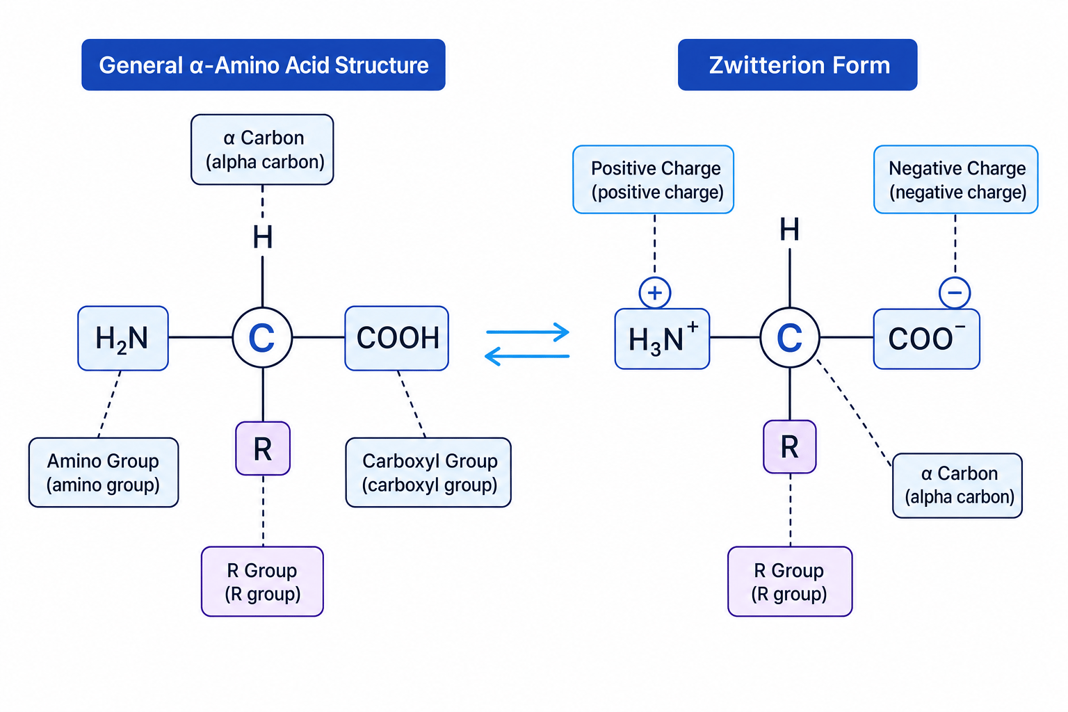
An α-amino acid has both the amino group and the carboxyl group attached to the same carbon atom, called the α-carbon.
Its general formula is:
H₂N–CH(R)–COOH
The R group changes from one amino acid to another. For example, if R is H, the amino acid is glycine. If R is CH₃, it is alanine.
This diagram shows the general α-amino acid structure and the zwitterion form you will meet next.
2. Classification of α-amino acids
You can classify α-amino acids in more than one useful way.
By the position of the amino group
An α-amino acid has the –NH₂ group on the carbon next to the carboxyl carbon. This is the type used to build proteins.
By the nature of the R group
The R group affects properties and protein folding. Amino acids may be described as:
- non-polar, if the R group is mainly hydrocarbon-like
- polar, if the R group can form dipole interactions or hydrogen bonds
- acidic, if the R group contains an extra –COOH group
- basic, if the R group contains an extra –NH₂ or similar basic group
Link to optical isomerism
Most α-amino acids are chiral, meaning they contain a carbon atom bonded to four different groups. The α-carbon is usually bonded to:
- H
- NH₂
- COOH
- R
If all four are different, the molecule can exist as two enantiomers, which are non-superimposable mirror images.
Glycine is the exception
Glycine has R = H, so its α-carbon is bonded to two H atoms. It is not chiral and does not show optical isomerism.
Deciding whether an amino acid is chiral
Compare glycine, H₂N–CH₂–COOH, with alanine, H₂N–CH(CH₃)–COOH.
-
Identify the groups attached to the α-carbon in glycine: NH₂, COOH, H and H. Two groups are the same.
-
Since glycine does not have four different groups around the α-carbon, it is not chiral.
-
Identify the groups attached to the α-carbon in alanine: NH₂, COOH, H and CH₃. These are four different groups.
-
Therefore alanine is chiral and can show optical isomerism.
3. Amphoteric behaviour and zwitterions
A substance is amphoteric if it can behave as both an acid and a base.
Amino acids are amphoteric because they contain:
- an acidic –COOH group, which can donate H⁺
- a basic –NH₂ group, which can accept H⁺
Inside a solid amino acid, and often in aqueous solution, the proton from –COOH transfers to –NH₂. This forms a zwitterion.
Zwitterion
A zwitterion is an ion with both a positive charge and a negative charge in the same molecule, but with no overall charge.
For an α-amino acid, the zwitterion is:
H₃N⁺–CH(R)–COO⁻
Behaviour in acidic and alkaline solution
In acidic solution, there is a high concentration of H⁺ ions. The COO⁻ group can accept H⁺:
H₃N⁺–CH(R)–COO⁻(aq) + H⁺(aq) → H₃N⁺–CH(R)–COOH(aq)
The amino acid now has an overall positive charge.
In alkaline solution, OH⁻ removes H⁺ from the NH₃⁺ group:
H₃N⁺–CH(R)–COO⁻(aq) + OH⁻(aq) → H₂N–CH(R)–COO⁻(aq) + H₂O(l)
The amino acid now has an overall negative charge.
Acid-base behaviour
An amino acid’s charge depends on pH: positive in acidic solution, often neutral as a zwitterion near its isoelectric point, and negative in alkaline solution.
Predicting the charged form at different pH values
For alanine, H₂N–CH(CH₃)–COOH, predict the main form in strongly acidic and strongly alkaline solution.
-
In strongly acidic solution, there is excess H⁺, so the carboxylate group is protonated. The main form is H₃N⁺–CH(CH₃)–COOH.
-
Count the charges in the acidic form: NH₃⁺ contributes +1 and COOH is neutral. The overall charge is +1.
-
In strongly alkaline solution, OH⁻ removes a proton from NH₃⁺. The main form is H₂N–CH(CH₃)–COO⁻.
-
Count the charges in the alkaline form: NH₂ is neutral and COO⁻ contributes −1. The overall charge is −1.
4. Melting temperature and solubility
Because amino acids often exist as zwitterions, they have strong electrostatic attractions between oppositely charged ions in the solid.
This means amino acids generally have:
- high melting temperatures compared with many covalent organic molecules of similar size
- good solubility in polar solvents such as water
- poor solubility in non-polar solvents such as hexane
Water can interact strongly with NH₃⁺ and COO⁻ groups using ion-dipole attractions and hydrogen bonding.
Property explanation shortcut
When explaining amino acid melting temperature or solubility, do not just say “because of hydrogen bonding”. The key extra idea is that zwitterions have ionic attractions between charged groups.
5. Forming dipeptides
A dipeptide forms when two amino acids join together.
The carboxyl group of one amino acid reacts with the amino group of another. A molecule of water is eliminated, so this is a condensation reaction.
The new link formed is called a peptide bond.
Peptide bond
A peptide bond is the amide link –CONH– formed when the –COOH group of one amino acid condenses with the –NH₂ group of another amino acid.
General reaction:
H₂N–CH(R₁)–COOH + H₂N–CH(R₂)–COOH → H₂N–CH(R₁)–CONH–CH(R₂)–COOH + H₂O
The diagram shows the loss of water and the formation of the peptide bond.
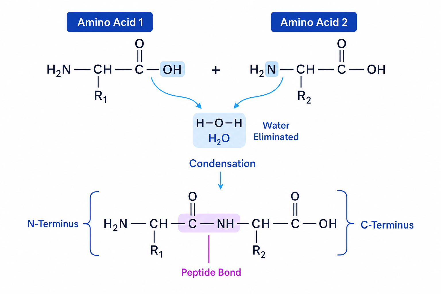
The left-hand end with a free amino group is called the N-terminus. The right-hand end with a free carboxyl group is the C-terminus.
Peptide formation is directional
Alanine-glycine and glycine-alanine are different dipeptides. The order of amino acids matters because the peptide chain has an N-terminus and a C-terminus.
Drawing two possible dipeptides from glycine and alanine
Glycine is H₂N–CH₂–COOH. Alanine is H₂N–CH(CH₃)–COOH.
-
First let alanine provide the carboxyl group and glycine provide the amino group. Remove OH from alanine’s –COOH and H from glycine’s –NH₂ to form water.
-
Join the remaining fragments through –CONH–. The product is H₂N–CH(CH₃)–CONH–CH₂–COOH, called Ala-Gly.
-
Now reverse the order: glycine provides the carboxyl group and alanine provides the amino group.
-
The second product is H₂N–CH₂–CONH–CH(CH₃)–COOH, called Gly-Ala.
-
These are different molecules because the amino acid sequence is different.
Removing the wrong atoms
In peptide formation, water comes from OH of the carboxyl group and H of the amino group. Do not remove the carbonyl oxygen from –COOH.
6. From dipeptides to polypeptides and proteins
A polypeptide is a long chain of amino acid residues joined by peptide bonds.
A residue is what is left of an amino acid after it has joined into a peptide chain by losing parts of water during condensation.
A protein is one or more polypeptide chains folded into a specific three-dimensional shape. Proteins are naturally occurring nitrogen compounds and are essential to living organisms.
The reverse of condensation is hydrolysis, where water is used to break peptide bonds. In the body, hydrolysis occurs during digestion of proteins. In the laboratory, proteins can be hydrolysed by heating with acid or alkali.
7. Protein structure
Protein function depends strongly on protein structure. You need to know the basic principles of primary, secondary and tertiary structure.
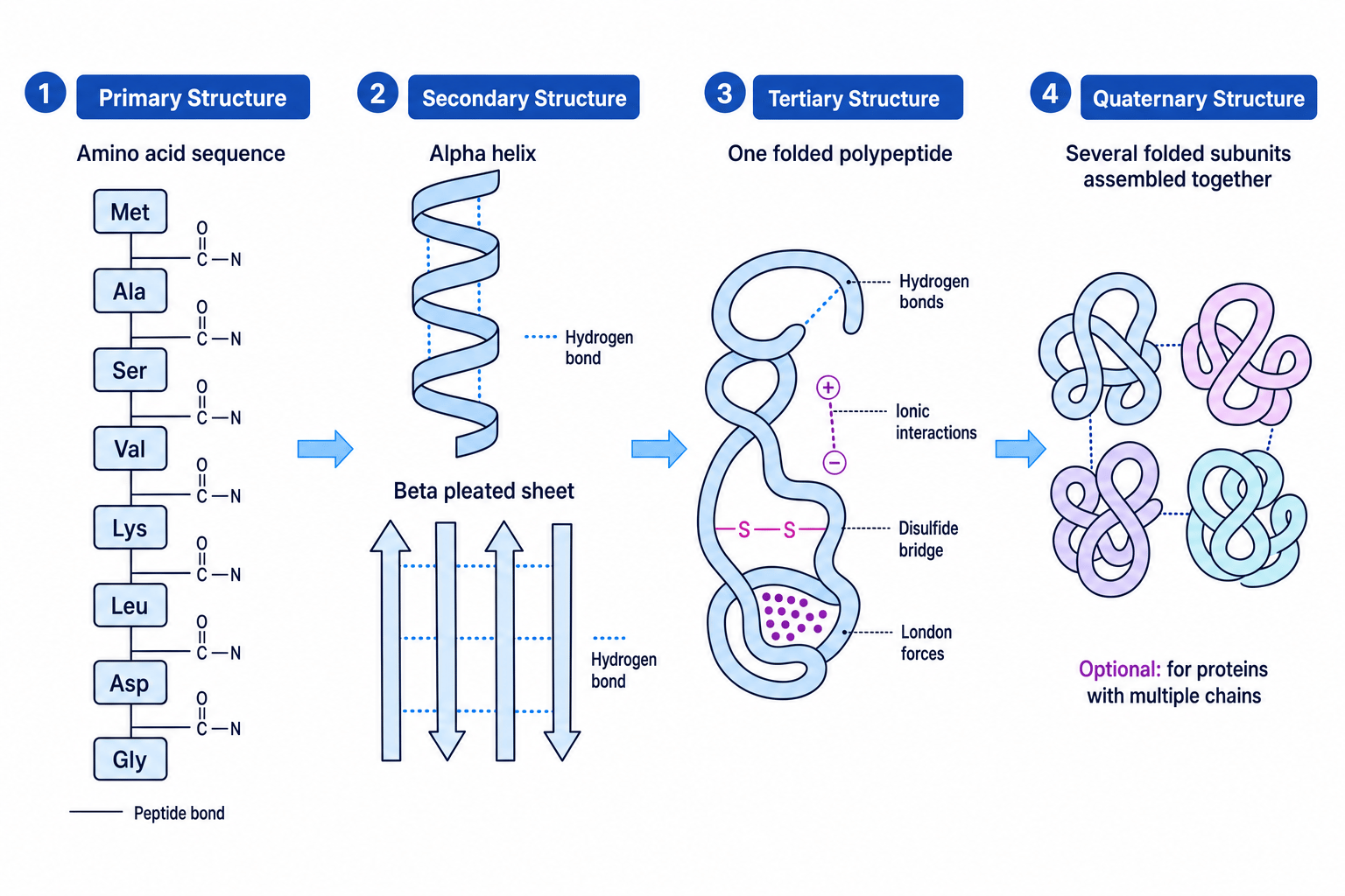
This overview shows the levels of protein structure, including quaternary structure for proteins made from several chains.
Primary structure
The primary structure is the sequence of amino acids in the polypeptide chain.
Example: Gly-Ala-Ser is not the same as Ser-Ala-Gly.
The primary structure is held together by covalent peptide bonds. It determines how the chain can fold, because different R groups form different interactions.
Secondary structure
The secondary structure is regular folding of the peptide backbone.
Common examples are:
- α-helix, a coiled shape
- β-pleated sheet, a folded sheet-like structure
These are stabilised by hydrogen bonds between C=O and N–H groups in the peptide backbone.
Tertiary structure
The tertiary structure is the overall three-dimensional shape of one polypeptide chain.
It is stabilised by interactions between R groups, including:
- hydrogen bonds between polar side chains
- ionic interactions between oppositely charged side chains
- disulfide bridges, covalent S–S bonds between cysteine residues
- London forces and hydrophobic interactions between non-polar side chains
Structure controls function
The primary structure determines the possible folding. The final tertiary structure gives the protein its specific shape and therefore its function.
8. Proteins in living systems: enzymes
Proteins have many essential roles in living systems. They act as:
- enzymes, which are biological catalysts
- structural materials, such as collagen
- transport molecules, such as haemoglobin
- antibodies in the immune system
- some hormones and cell-signalling molecules
For this topic, enzymes are especially important.
An enzyme has an active site, a region with a specific shape and arrangement of chemical groups. The substrate binds to the active site, forming an enzyme-substrate complex. The enzyme provides an alternative pathway with a lower activation energy, so the reaction is faster.
If the tertiary structure changes, the active site may change shape. The substrate may no longer fit, so enzyme activity decreases.
This loss of the functional three-dimensional shape is called denaturation.
Explaining loss of enzyme activity at low pH
An enzyme works best near pH 7 but loses activity in strongly acidic solution.
-
Strongly acidic solution contains excess H⁺ ions, which can protonate groups such as COO⁻ on amino acid side chains.
-
Changing the charges on side chains can break or alter ionic interactions and hydrogen bonding that help maintain the tertiary structure.
-
If the tertiary structure changes, the active site changes shape and the substrate is no longer complementary to it.
-
Fewer enzyme-substrate complexes form, so the reaction rate decreases.
Denaturation usually does not break peptide bonds
Denaturation changes secondary and tertiary structure. The primary structure, meaning the amino acid sequence joined by peptide bonds, usually remains intact unless hydrolysis occurs.
In the exam
-
For amino acid structure questions, always show both functional groups clearly: –NH₂ and –COOH, or their charged forms –NH₃⁺ and –COO⁻.
-
For peptide questions, circle or label the peptide bond as –CONH– and remember that water is eliminated during condensation.
-
For protein structure explanations, link each level to the correct bonding: peptide bonds for primary, backbone hydrogen bonds for secondary, and R-group interactions for tertiary.
Check yourself
- Why do amino acids usually have higher melting temperatures than similar-sized simple organic molecules?
- What two different dipeptides can be made from alanine and glycine?
- How can a change in pH reduce enzyme activity?

