

What you'll learn
- How amino acids join together by peptide links to form proteins.
- How to draw dipeptides, tripeptides, and the amino acids formed by hydrolysis.
- How primary, secondary and tertiary protein structures are maintained.
- How amino acids can be separated and identified using thin-layer chromatography and RfR_fRf values.
Starting point: amino acids
An amino acid contains two important functional groups:
- an amino group, –NH2
- a carboxyl group, –COOH
Most A-level protein questions use the general structure:
H2N–CH(R)–COOH
The R group is the side chain. It varies between different amino acids and gives each amino acid its particular properties.
Protein
A protein is a biological polymer made from a sequence of amino acid residues joined together by peptide links.
When an amino acid becomes part of a protein chain, it is often called an amino acid residue because part of the original molecule has been lost during condensation.
Peptide links: joining amino acids
Amino acids join by a condensation reaction. This means two molecules join together and a small molecule, usually water, is eliminated.
The –COOH group of one amino acid reacts with the –NH2 group of another amino acid. An –OH and an –H are removed to form H2O, and the remaining atoms join to make a peptide link.
Peptide link
A peptide link is the amide group –CO–NH– formed between two amino acid residues.
The same bond can be broken again by hydrolysis, which adds water across the peptide link.
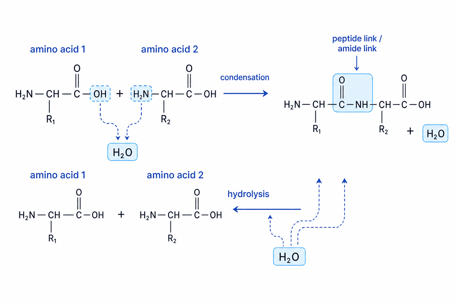
A short chain of amino acids has specific names:
- two amino acid residues: dipeptide
- three amino acid residues: tripeptide
- many amino acid residues: polypeptide
Order matters
Gly-Ala and Ala-Gly contain the same two amino acids, but they are different dipeptides because the sequence is reversed.
Drawing a dipeptide from glycine and alanine
Draw the dipeptide formed from glycine first, then alanine.
-
Choose the reacting groups. Glycine is H2N–CH2–COOH and alanine is H2N–CH(CH3)–COOH. The carboxyl group of glycine reacts with the amino group of alanine.
-
Remove water. Remove –OH from glycine’s –COOH group and remove H from alanine’s –NH2 group. These form H2O.
-
Join carbon to nitrogen. The resulting dipeptide is H2N–CH2–CO–NH–CH(CH3)–COOH. The peptide link is the –CO–NH– section.
Losing the wrong atoms
In peptide formation, the –OH comes from the carboxyl group and the H comes from the amino group. Do not remove the whole –COOH or –NH2 group.
Hydrolysis of peptide links
Hydrolysis means breaking a covalent bond by adding water. For peptides, hydrolysis breaks the peptide link and produces the original amino acids.
For a peptide containing nnn amino acid residues, there are n−1n - 1n−1 peptide links, so complete hydrolysis needs n−1n - 1n−1 water molecules.
Finding the amino acids from hydrolysis
A tripeptide has the structure:
H2N–CH(CH3)–CO–NH–CH2–CO–NH–CH(CH2OH)–COOH
-
Locate the peptide links. There are two –CO–NH– groups, so two bonds must be hydrolysed.
-
Break each C–N bond in the peptide link. Add –OH to the carbonyl carbon side, turning –CO– into –COOH.
-
Add H to the nitrogen side. This reforms amino groups, giving the constituent amino acids: alanine, glycine and serine.
Hydrolysis shortcut
For each –CO–NH– peptide link, break the C–N bond, put –OH on the C=O side, and put H on the N side.
Levels of protein structure
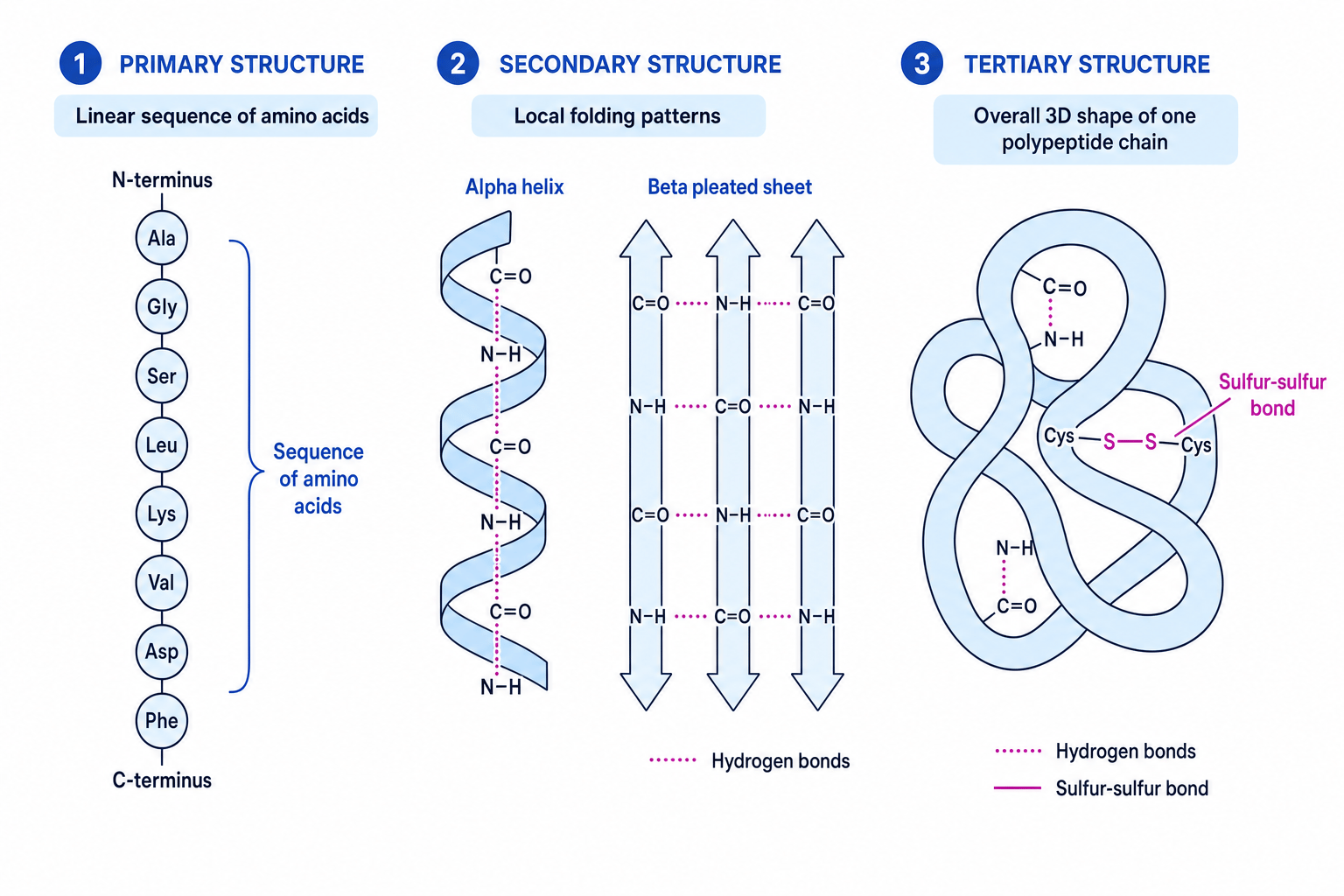
Proteins are not just random strings. Their function depends heavily on how the chain folds. This is described using different levels of structure.
Protein structure levels
- Primary structure is the sequence of amino acids in the polypeptide chain.
- Secondary structure is regular local folding, especially the α\alphaα-helix and β\betaβ-pleated sheet.
- Tertiary structure is the overall three-dimensional shape of one polypeptide chain.
Primary structure
The primary structure is held together by covalent peptide links. If the order of amino acids changes, the protein may fold differently and function differently.
Secondary structure
The two A-level secondary structures you need are:
- the α\alphaα-helix, a coiled shape
- the β\betaβ-pleated sheet, a folded sheet-like shape
These are maintained by hydrogen bonds between peptide backbone groups: the C=O group of one peptide link and the N–H group of another.
Hydrogen bond
A hydrogen bond is an attraction involving a partially positive hydrogen atom bonded to a very electronegative atom and a lone pair on another electronegative atom. In proteins, important hydrogen bonds form between C=O and N–H groups.
Tertiary structure
The tertiary structure is the full 3D folding of the polypeptide. It is maintained by interactions between parts of the chain, especially:
- hydrogen bonds involving polar groups
- sulfur–sulfur bonds, also called disulfide bonds
A sulfur–sulfur bond forms between sulfur atoms in two cysteine residues. It is a covalent bond, so it can strongly lock parts of the protein into position.
What holds the levels together
Primary structure is held by peptide links. Secondary structure is held mainly by hydrogen bonds in the backbone. Tertiary structure is held by side-chain interactions, including hydrogen bonds and sulfur–sulfur bonds.
Identifying structures in a protein diagram
A diagram shows a chain labelled Gly–Ser–Cys–Ala, a coiled section with dotted lines between C=O and N–H groups, and a folded region containing Cys–S–S–Cys.
-
Classify the sequence. Gly–Ser–Cys–Ala is the order of amino acids, so it shows primary structure.
-
Classify the regular coil. A coiled section with C=O···H–N hydrogen bonds is an α\alphaα-helix, so it shows secondary structure.
-
Classify the full fold. The overall folded shape with a Cys–S–S–Cys bond shows tertiary structure, because it involves the 3D arrangement of the chain.
Mixing up secondary and tertiary
An α\alphaα-helix or β\betaβ-pleated sheet is secondary structure, even if it is drawn inside a larger folded protein. The complete folded shape is tertiary structure.
Separating amino acids by TLC
Thin-layer chromatography, or TLC, is used to separate and identify amino acids.
In TLC:
- the stationary phase is a thin layer of solid, often silica or alumina, on a plate
- the mobile phase is a solvent that moves up the plate
- different amino acids travel different distances because they have different attractions to the stationary and mobile phases
Amino acids are often colourless, so the spots may need to be located using a developing agent such as ninhydrin, or by using ultraviolet light.
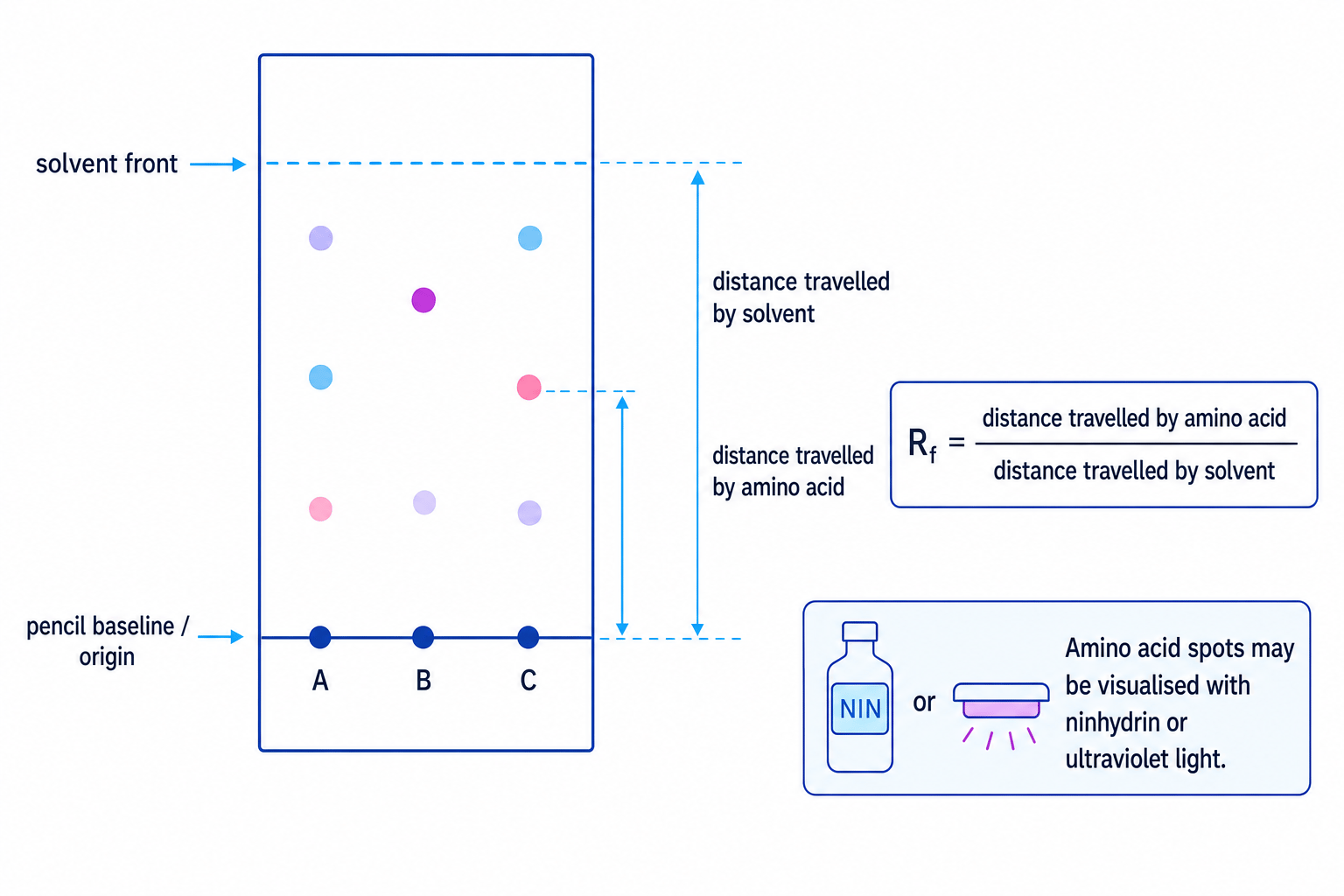
The RfR_fRf value is calculated using:
Rf=distance travelled by amino acid spotdistance travelled by solvent frontR_f = \frac{\text{distance travelled by amino acid spot}}{\text{distance travelled by solvent front}}Rf=distance travelled by solvent frontdistance travelled by amino acid spotBoth distances are measured from the pencil baseline. The value has no units because the units cancel.
Calculating and using an Rf value
An amino acid spot travels 4.8 cm from the baseline. The solvent front travels 8.0 cm from the baseline. Reference values in the same solvent are glycine 0.32, alanine 0.46 and leucine 0.60.
- Substitute the measured distances.
- Calculate the ratio.
- Compare with reference values. The unknown has the same RfR_fRf value as leucine under the same conditions, so it is identified as leucine.
Comparing Rf values from different conditions
Only compare RfR_fRf values if the stationary phase, solvent and temperature are the same. Changing the solvent can change how far each amino acid travels.
TLC measurement check
Measure to the centre of the spot and always measure from the baseline, not from the bottom of the plate.
In the exam
- When drawing peptides, keep the amino acid order exactly as given and show the peptide link as –CO–NH–.
- For hydrolysis questions, break every peptide link and add H and OH from water to regenerate the amino acids.
- For protein-structure explanations, link the structure named to the bonding involved: hydrogen bonds for secondary structure, and hydrogen bonds plus sulfur–sulfur bonds for tertiary structure.
- For TLC, show your measured distances, calculate RfR_fRf as a ratio, and state that comparison must be under the same conditions.
Check yourself
- Can you draw the tripeptide formed from three named amino acids in the order given?
- What type of bonding maintains an α\alphaα-helix?
- How would you calculate and use an RfR_fRf value from a chromatogram?
