

What you'll learn
- How ¹³C NMR and ¹H NMR give evidence about molecular structure.
- How to interpret chemical shift values on the δ scale.
- How integration and splitting patterns in ¹H NMR help you count protons.
- Why TMS and deuterated solvents are used when recording spectra.
Why chemists use NMR
Organic chemists rarely prove a structure using just one technique. Mass spectrometry can suggest a molecular mass or formula, infrared spectroscopy can identify functional groups, and nuclear magnetic resonance spectroscopy gives detailed information about the environments of atoms in a molecule.
This part is A-level only, and the emphasis is not on deriving the physics. You mainly need to use NMR data to solve structural puzzles.
Nuclear magnetic resonance spectroscopy
Nuclear magnetic resonance spectroscopy, usually shortened to NMR spectroscopy, is an analytical technique in which certain nuclei absorb energy in a magnetic field. The absorption pattern depends on the nuclei’s chemical environments, so it can be used to help deduce molecular structure.
For AQA A-level Chemistry, the two important types are:
- ¹³C NMR, which gives information about carbon environments.
- ¹H NMR, also called proton NMR, which gives information about hydrogen/proton environments.
Chemical environments
A chemical environment means the surroundings of an atom within a molecule. Atoms are in the same environment if they are equivalent by symmetry and are attached to the same types of neighbouring atoms.
Peaks show environments, not simply atoms
In NMR, the number of signals usually tells you the number of different chemical environments, not the total number of atoms.
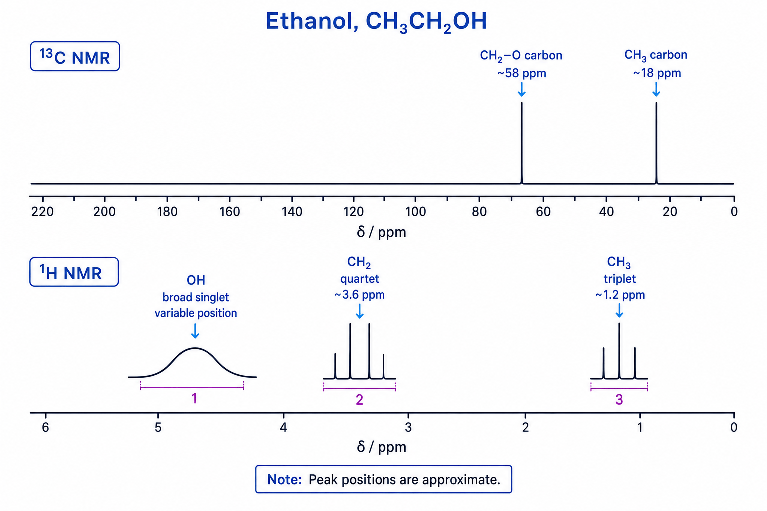
For example, in ethanol, CH₃CH₂OH, the three hydrogens in the CH₃ group are equivalent to each other, but they are not equivalent to the two hydrogens in the CH₂ group or the hydrogen in the OH group.
Counting carbon environments
Propanone has the structure CH₃COCH₃. Predict the number of peaks in its ¹³C NMR spectrum.
- Identify the carbon atoms: there are two methyl carbon atoms and one carbonyl carbon atom.
- Compare the two CH₃ groups: they are on opposite sides of the same C=O group, so they are equivalent by symmetry.
- Compare the methyl carbons with the carbonyl carbon: the carbonyl carbon is in a different environment because it is double-bonded to oxygen.
- Therefore, propanone has two carbon environments, so it gives two ¹³C NMR peaks.
The δ chemical shift scale
NMR peak positions are recorded using chemical shift, given the symbol δ and measured in ppm.
Chemical shift
Chemical shift is the position of an NMR signal on the δ scale, measured in parts per million, relative to a standard substance called TMS.
Chemical shift depends on molecular environment. A nucleus surrounded by more electron density is said to be shielded and tends to appear at a lower δ value. A nucleus near electronegative atoms, π bonds, or carbonyl groups is often deshielded and appears at a higher δ value.
NMR spectra are usually drawn with high δ values on the left and low δ values on the right, which is the opposite direction to many other graphs you meet.
The spectra below show the key ideas for ethanol: ¹³C NMR gives one peak per carbon environment, while ¹H NMR also includes integration and splitting information.
Use the Data Booklet
In structure questions, do not try to memorise every chemical shift. Learn the broad patterns, then compare the given δ values with the Chemistry Data Booklet.
¹³C NMR spectra
¹³C NMR spectra are usually simpler than ¹H NMR spectra. At A-level, you should treat a ¹³C NMR spectrum as showing one peak for each carbon environment.
Important points:
- The number of peaks tells you the number of carbon environments.
- The chemical shift helps identify the type of carbon environment.
- Peak heights are not normally used to count carbon atoms.
- You do not need to apply the n+1n+1n+1 splitting rule to ¹³C NMR.
Common ¹³C clues include:
- C–C carbon atoms: low δ values, often below about 50 ppm.
- C–O carbon atoms: higher, often around 50–90 ppm.
- C=O carbon atoms: much higher, often above 160 ppm.
Assuming ¹³C peak height shows carbon number
In ¹³C NMR, do not use peak height or peak area to decide how many carbons are in each environment. For A-level questions, focus on the number of peaks and their chemical shifts.
¹H NMR spectra: integration
¹H NMR gives more information than ¹³C NMR. One of the most useful features is integration.
Integration
Integration measures the relative area under each ¹H NMR signal. The areas are proportional to the relative numbers of protons in each environment.
So if a spectrum has integration ratio 3:2:1, the molecule has proton environments in the relative numbers 3H, 2H and 1H. If the molecular formula is known, you can often convert the ratio into actual numbers of hydrogens.
Converting integration to proton numbers
A compound has molecular formula C₂H₆O. Its ¹H NMR spectrum has three signals with integration ratio 3:2:1. Work out the number of hydrogens in each environment.
- Add the ratio parts: 3 + 2 + 1 = 6 parts.
- Compare this with the formula: C₂H₆O contains six hydrogens in total.
- Since six ratio parts correspond to six actual hydrogens, each ratio part represents one hydrogen.
- The three environments therefore contain 3H, 2H and 1H.
Using peak height instead of integration
For ¹H NMR, the relative number of protons comes from the integration trace or stated integration values, not from how tall the peaks look.
¹H NMR spectra: spin-spin splitting
Many ¹H NMR peaks are split into smaller peaks. This is called spin-spin splitting.
Spin-spin splitting
Spin-spin splitting occurs when protons in one environment are affected by nearby non-equivalent protons, causing their NMR signal to split into a pattern.
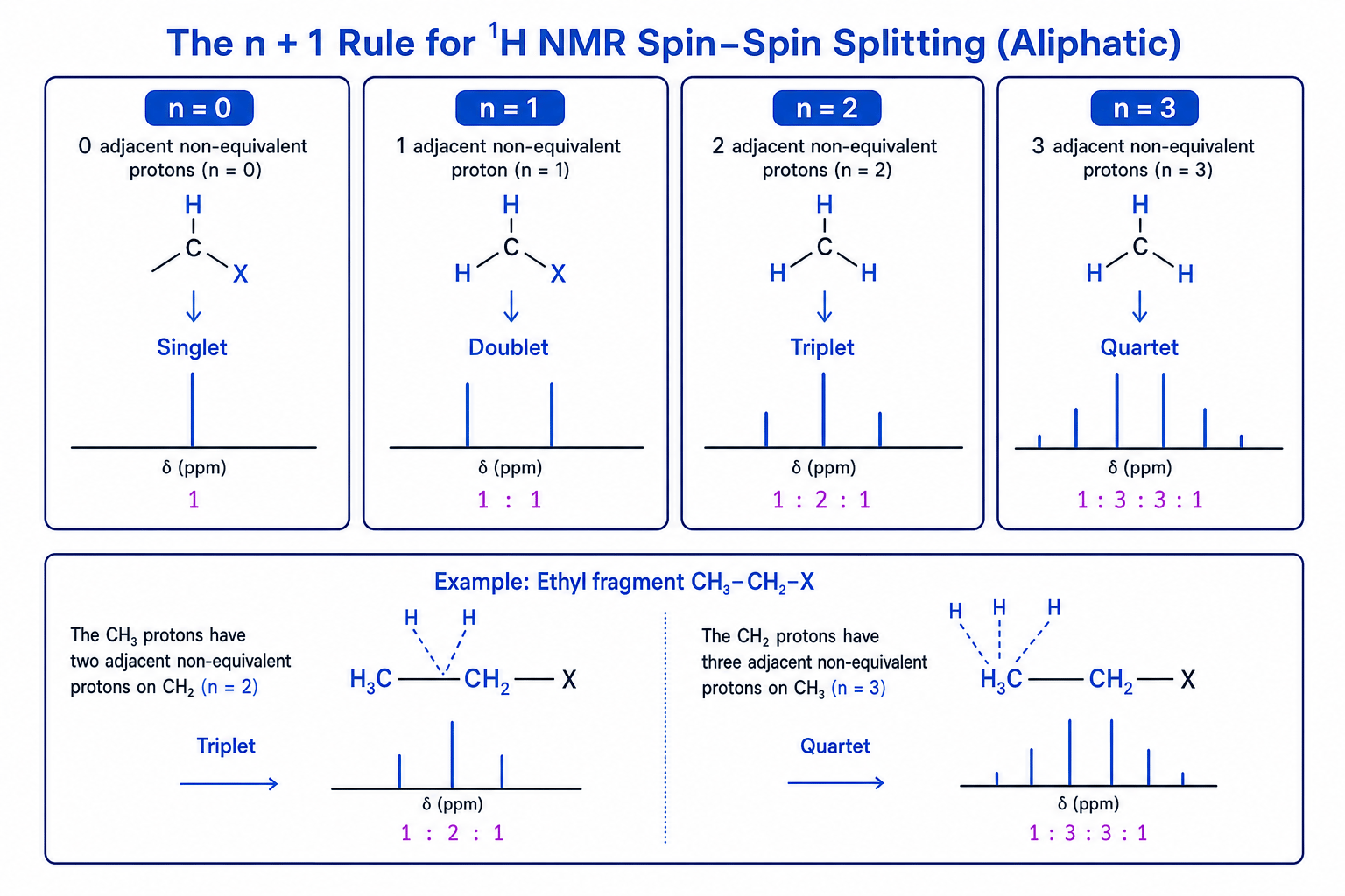
At A-level, use the n+1n+1n+1 rule for adjacent, non-equivalent protons in aliphatic compounds.
The n + 1 rule
If a proton environment has nnn adjacent non-equivalent protons, its signal is split into n+1n+1n+1 peaks.
The patterns you need are:
- 0 adjacent protons → singlet.
- 1 adjacent proton → doublet.
- 2 adjacent protons → triplet.
- 3 adjacent protons → quartet.
Predicting splitting in bromoethane
Bromoethane has the structure CH₃CH₂Br. Predict the splitting pattern for each proton environment.
- Identify the two proton environments: the CH₃ protons form one environment, and the CH₂ protons form another.
- For the CH₃ signal, count adjacent non-equivalent protons on the neighbouring carbon: the CH₂ group has two protons, so n=2n=2n=2.
- Apply the rule: n+1=2+1=3n+1=2+1=3n+1=2+1=3, so the CH₃ signal is a triplet.
- For the CH₂ signal, count adjacent non-equivalent protons on the neighbouring carbon: the CH₃ group has three protons, so n=3n=3n=3.
- Apply the rule: n+1=3+1=4n+1=3+1=4n+1=3+1=4, so the CH₂ signal is a quartet.
Spotting an ethyl group
A ¹H NMR spectrum with a 3H triplet and a 2H quartet often suggests an ethyl group, CH₃CH₂–.
Splitting by equivalent protons
Equivalent protons do not split each other. Only count adjacent non-equivalent protons when applying the n+1n+1n+1 rule.
OH and NH protons
OH and NH protons can give broad, variable signals and often do not show the splitting you might expect, because proton exchange can occur.
Solvents and TMS
¹H NMR samples are usually dissolved in a solvent before analysis. The solvent must not create confusing ¹H peaks.
Common choices include:
- Deuterated solvents, such as CDCl₃, where most ¹H atoms have been replaced by deuterium, ²H.
- CCl₄, which contains no hydrogen atoms.
TMS
Tetramethylsilane, TMS, has the formula Si(CH₃)₄ and is used as the standard reference compound for NMR chemical shifts.
TMS is suitable because:
- all 12 of its protons are equivalent, so it gives one sharp ¹H NMR peak;
- its peak is assigned δ = 0 ppm;
- it is chemically inert, so it does not react with the sample;
- it is volatile, so it can be removed easily;
- it is soluble in many organic solvents;
- its signal is away from most organic compound peaks, so it rarely overlaps.
Putting the evidence together
In exams, you are often given a mixture of data: molecular formula, IR peaks, ¹³C NMR and ¹H NMR. Treat it like building a jigsaw: each piece of evidence should fit the same structure.
Deducing ethyl ethanoate from NMR data
A compound has molecular formula C₄H₈O₂. Its IR spectrum shows a strong C=O absorption. Its ¹³C NMR spectrum has four peaks, including one near 171 ppm and one near 60 ppm. Its ¹H NMR spectrum has: a 3H triplet at 1.2 ppm, a 3H singlet at 2.1 ppm, and a 2H quartet at 4.1 ppm. Suggest a structure.
- Use the IR evidence: a strong C=O absorption means the molecule probably contains a carbonyl group.
- Use the ¹³C NMR evidence: a peak near 171 ppm supports a C=O carbon, and a peak near 60 ppm suggests a carbon attached to oxygen, such as C–O in an ester.
- Use the ¹H splitting pattern: the 3H triplet and 2H quartet together suggest an ethyl group, CH₃CH₂–.
- Use the chemical shift of the quartet: 4.1 ppm is high for CH₂, so this CH₂ is likely bonded to oxygen, giving –OCH₂CH₃.
- Use the remaining 3H singlet: a singlet at 2.1 ppm fits CH₃ next to C=O with no adjacent hydrogens, giving CH₃CO–.
- Combine the fragments: CH₃CO– and –OCH₂CH₃ give CH₃COOCH₂CH₃, ethyl ethanoate.
In the exam
- Start by counting environments: number of ¹³C peaks for carbon environments, number of ¹H signals for proton environments.
- Use chemical shifts to identify likely fragments, especially C=O, C–O, alkyl CH₃/CH₂, and aromatic environments.
- Use integration before splitting: first decide how many protons are in each environment, then apply the n+1n+1n+1 rule to connect neighbouring groups.
- Cross-check every proposed structure against all the data, including the molecular formula.
Check yourself
- Why does ethanol give two ¹³C NMR peaks but three main ¹H NMR environments?
- What splitting pattern would you expect for the CH₃ group in CH₃CH₂Cl?
- Why is TMS suitable as an NMR standard?
