

What you'll learn
- Why many transition metal ions are coloured.
- How d electrons absorb visible light and move to excited states.
- Why changing ligand, oxidation state or co-ordination number changes colour.
- How a colorimeter can be used to find the concentration of coloured ions.
Colour starts with visible light
Visible light is the part of the electromagnetic spectrum your eyes can detect. White light contains a mixture of wavelengths. A wavelength, given the symbol λ\lambdaλ, is the distance between matching points on neighbouring waves, such as crest to crest.
When light hits a substance, some wavelengths may be absorbed. The wavelengths not absorbed may be transmitted through a solution or reflected from a solid. These remaining wavelengths are what you see as the colour.
Seeing the wavelengths left over
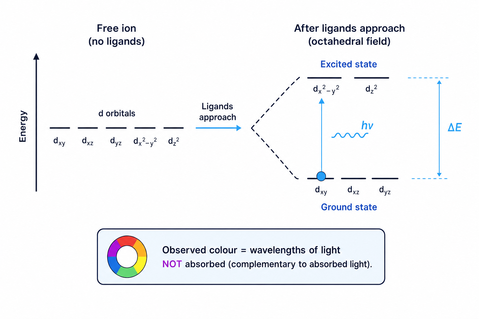
A coloured solution does not look the colour it absorbs. It looks the colour of the light that is transmitted or reflected after absorption.
For example, if a solution absorbs orange-red light strongly, the light reaching your eye may be mainly blue-green, so the solution appears blue-green.
Transition metal ions and d electrons
A transition metal ion often contains a partially filled d subshell. A d subshell contains five d orbitals, and d electrons are electrons in these orbitals.
Many transition metal ions are coloured because their d electrons can absorb visible light and move to a higher energy arrangement.
Ground state and excited state
The ground state is the lowest-energy arrangement of electrons. An excited state is a higher-energy arrangement formed when an electron absorbs energy.
Not every transition metal ion is coloured. Ions with no d electrons, such as d0d^0d0 ions, or a completely full d subshell, such as d10d^{10}d10 ions, are often colourless because the usual d-to-d electron promotion cannot occur.
Assuming every transition metal ion is coloured
Do not write “all transition metal ions are coloured”. For A-Level, remember that partially filled d orbitals are the important condition for the usual colour explanation.
Why d orbitals split in complex ions
Transition metal ions in solution usually exist as complex ions. A ligand is a species that donates a lone pair of electrons to a central metal ion, forming a co-ordinate bond. Water, ammonia and chloride ions are common ligands.
Complex ion and co-ordination number
A complex ion contains a central metal ion surrounded by ligands. The co-ordination number is the number of co-ordinate bonds from ligands to the central metal ion.
In an isolated gaseous ion, the five d orbitals have the same energy. We say they are degenerate, meaning equal in energy.
When ligands approach the metal ion, their lone pairs repel electrons in the d orbitals. Some d orbitals are repelled more than others, so the d orbitals split into two different energy levels. The energy gap between these levels is called $\Delta E`.
Absorbing light: the energy calculation
A d electron can move from the lower d energy level to the higher d energy level if it absorbs a photon with exactly the right energy.
A photon is a packet of electromagnetic radiation. The energy of the photon must match the energy gap $\Delta E`.
ΔE=hν=hcλ\Delta E = h\nu = \frac{hc}{\lambda}ΔE=hν=λhcwhere:
- ΔE\Delta EΔE is the energy difference for one electron transition, in J
- hhh is the Planck constant, 6.63×10⁻³⁴ J s6.63 \times 10^{-34}\ \text{J s}6.63×10⁻³⁴ J s
- ν\nuν is the frequency, in Hz
- ccc is the speed of light, 3.00×10⁸ m s−13.00 \times 10^8\ \text{m s}^{-1}3.00×10⁸ m s−1
- λ\lambdaλ is the wavelength, in m
Calculating the wavelength absorbed
A complex ion has an energy gap of ΔE=3.20×10⁻¹⁹ J\Delta E = 3.20 \times 10^{-19}\ \text{J}ΔE=3.20×10⁻¹⁹ J. Calculate the wavelength of light absorbed.
-
Choose the correct form of the equation because wavelength is required:
ΔE=hcλ\Delta E = \frac{hc}{\lambda}ΔE=λhc -
Rearrange for λ\lambdaλ:
λ=hcΔE\lambda = \frac{hc}{\Delta E}λ=ΔEhc -
Substitute the values with units:
λ=(6.63×10⁻³⁴ J s)(3.00×10⁸ m s−1)3.20×10⁻¹⁹ J\lambda = \frac{(6.63 \times 10^{-34}\ \text{J s})(3.00 \times 10^8\ \text{m s}^{-1})}{3.20 \times 10^{-19}\ \text{J}}λ=3.20×10⁻¹⁹ J(6.63×10⁻³⁴ J s)(3.00×10⁸ m s−1) -
Calculate and convert to nm:
λ=6.22×10⁻⁷ m\lambda = 6.22 \times 10^{-7}\ \text{m}λ=6.22×10⁻⁷ mThis is 622 nm, which is in the visible region.
If a substance absorbs visible light, the colour observed is linked to the wavelengths that are not absorbed.
Energy and wavelength move oppositely
A larger ΔE\Delta EΔE means a higher-energy photon is absorbed. Since ΔE=hcλ\Delta E = \frac{hc}{\lambda}ΔE=λhc, larger ΔE\Delta EΔE means a shorter wavelength.
Why the colour changes
The value of ΔE\Delta EΔE is not fixed for a metal ion. It depends on the surroundings of the metal.
Three important changes can alter ΔE\Delta EΔE:
- changing the oxidation state of the metal ion
- changing the ligand
- changing the co-ordination number
The oxidation state is the charge an atom would have if bonding electrons were assigned to the more electronegative atom. In transition metals, different oxidation states often have different colours. For example, vanadium ions in different oxidation states have noticeably different colours.
Changing the ligand also changes the repulsions around the d orbitals. For example, replacing water ligands with ammonia ligands around Cu2+\text{Cu}^{2+}Cu2+ changes the energy gap, so a different wavelength of visible light is absorbed.
Explaining a ligand-substitution colour change
A pale blue aqueous copper(II) complex forms a deep blue complex when excess ammonia is added. Explain why the colour changes.
-
Identify what has changed around the metal ion: some water ligands have been replaced by ammonia ligands.
-
Link the ligand change to the d orbitals: ammonia ligands interact differently with the copper(II) ion, so the splitting of the d orbitals changes.
-
Apply the energy equation: if ΔE\Delta EΔE changes, the absorbed wavelength changes because ΔE=hcλ\Delta E = \frac{hc}{\lambda}ΔE=λhc.
-
Connect absorption to observation: a different set of wavelengths is transmitted, so the observed colour changes from pale blue to deep blue.
Saying the ligand causes the colour directly
The ligand does not simply “give” the ion its colour. The ligand changes ΔE\Delta EΔE, which changes which wavelength is absorbed.
Absorption of visible light in spectroscopy
Spectroscopy is the study of how substances interact with electromagnetic radiation. In visible spectroscopy, you measure which wavelengths of visible light are absorbed.
For transition metal complexes, absorption in the visible region gives evidence about the size of ΔE\Delta EΔE. Different complexes absorb different wavelengths, so they can have different colours.
Real colours can be more complicated
At A-Level, explain transition metal colours using d electron transitions. Some very intense colours in real chemistry involve other processes, such as charge transfer, but that is beyond the core explanation here.
Using a colorimeter to find concentration
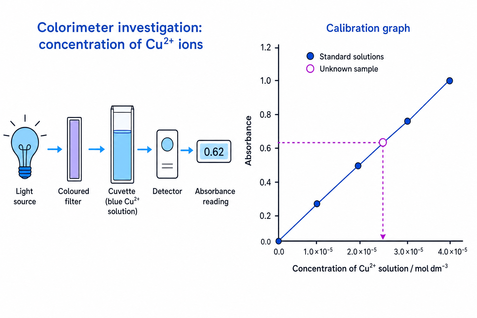
A colorimeter is an instrument that measures how much light is absorbed by a coloured solution.
The key idea is simple: a more concentrated coloured solution contains more coloured ions, so it usually absorbs more light. For dilute solutions, and with the same cuvette path length, absorbance is proportional to concentration.
Absorbance
Absorbance is a measure of how much light a sample absorbs. Higher absorbance means less light reaches the detector.
A typical method is:
- Prepare several standard solutions with known concentrations.
- Choose a filter colour that is strongly absorbed by the solution. This is often the complementary colour to the solution.
- Use a blank, usually the solvent or reagent mixture without the coloured ion, to zero the colorimeter.
- Measure the absorbance of each standard solution.
- Plot absorbance against concentration to make a calibration graph.
- Measure the absorbance of the unknown solution.
- Read the unknown concentration from the graph.
Good colorimetry technique
Wipe the outside of the cuvette, remove bubbles, keep the same orientation each time, and make sure the solution level is high enough for the light beam.
Finding a copper(II) concentration from absorbance
A calibration graph is a straight line through the origin. A 0.0400 mol dm⁻³ copper(II) standard has absorbance A=0.80A = 0.80A=0.80. An unknown copper(II) solution is made by diluting 10.0 cm³ of the original solution to 50.0 cm³. The diluted unknown has absorbance A=0.50A = 0.50A=0.50. Find the concentration of the original solution.
-
Calculate the gradient of the calibration graph:
gradient=0.800.0400 mol dm−3=20.0 dm3 mol−1\text{gradient} = \frac{0.80}{0.0400\ \text{mol dm}^{-3}} = 20.0\ \text{dm}^3\ \text{mol}^{-1}gradient=0.0400 mol dm−30.80=20.0 dm3 mol−1 -
Use the absorbance of the diluted unknown to find its concentration:
concentration=0.5020.0 dm3 mol−1=0.0250 mol dm−3\text{concentration} = \frac{0.50}{20.0\ \text{dm}^3\ \text{mol}^{-1}} = 0.0250\ \text{mol dm}^{-3}concentration=20.0 dm3 mol−10.50=0.0250 mol dm−3 -
Correct for the dilution. The dilution factor is:
50.0 cm310.0 cm3=5.00\frac{50.0\ \text{cm}^3}{10.0\ \text{cm}^3} = 5.0010.0 cm350.0 cm3=5.00 -
Multiply to find the original concentration:
0.0250 mol dm−3×5.00=0.125 mol dm−30.0250\ \text{mol dm}^{-3} \times 5.00 = 0.125\ \text{mol dm}^{-3}0.0250 mol dm−3×5.00=0.125 mol dm−3
Reading beyond the calibration range
Do not use a calibration graph to estimate concentrations far outside the range of your standards. Dilute the unknown if its absorbance is too high, then remember to apply the dilution factor.
In the exam
- Link colour to absorption of visible light, not just “presence of transition metals”.
- For colour changes, always mention that ligand, oxidation state or co-ordination number changes ΔE\Delta EΔE, so a different wavelength is absorbed.
- In colorimetry questions, describe standards, blanking, measuring absorbance, plotting a calibration graph, and reading off the unknown concentration.
Check yourself
- Why does a transition metal complex appear the colour of the light it does not absorb?
- What happens to the absorbed wavelength if ΔE\Delta EΔE becomes larger?
- How would you use a colorimeter to find the concentration of an unknown copper(II) solution?
