

As plants grow larger and more complex, they can no longer rely on simple diffusion to meet their metabolic needs. In these notes, we will look at how the vascular system of herbaceous dicotyledonous plants functions to transport water, mineral ions, and organic solutes.
What you'll learn
- Why multicellular plants require specialized transport systems based on surface area to volume ratio (SA:V), size, and metabolic rate.
- The structure, function, and distribution of xylem and phloem tissues.
- The pathways and mechanisms of water transport from roots to leaves (transpiration).
- How plants adapt to extreme water availability (xerophytes and hydrophytes).
- The energy-requiring mechanism of translocation in the phloem.
The need for transport systems
In small, single-celled organisms, the surface area to volume ratio (SA:V) is large enough that simple diffusion across the cell surface membrane can supply all necessary nutrients and remove metabolic waste. However, multicellular plants require a specialized vascular transport system due to three primary factors:
Size
Large plants (such as mature trees) have a long pathway between the roots (which absorb water and mineral ions) and the leaves (which produce sugars via photosynthesis). Diffusion is too slow over distances greater than a few millimetres because diffusion time is proportional to the square of the distance.
Surface Area to Volume Ratio (SA:V)
As an organism increases in size, its volume increases much faster than its surface area (volume scales by x3x^3x3 while surface area scales by x2x^2x2). This means that the outer surface area of a large plant is far too small relative to its total volume to supply all internal cells with nutrients by diffusion alone.
Metabolic Rate
While plants are less active than mammals and have a lower overall metabolic rate, parts of the plant (such as growing meristems, roots, and leaves) still require high rates of transport for water, minerals, and sugars. Leaves produce large quantities of organic assimilates (sugars) that must be distributed, and roots need these sugars for respiration to actively transport minerals from the soil.
Assimilates
Substances made by the plant itself using inorganic nutrients and light energy, such as sucrose and amino acids.
Calculating the surface area to volume ratio of a plant stem model
A student models a section of a young herbaceous plant stem as a simple cylinder with a radius (rrr) of 4 mm and a height (hhh) of 80 mm. Calculate the surface area to volume ratio (SA:V) of this stem model.
- Recall the geometric formulas for a cylinder. The total surface area (AAA) including both circular ends and the curved surface is:
The volume (VVV) of a cylinder is:
V=πr2h V = \pi r^2 h V=πr2h- Substitute the values into the surface area equation. Keep units consistent in millimetres (mm):
- Substitute the values into the volume equation:
- Divide the surface area by the volume to find the ratio:
Expressing this as a simplified ratio gives approximately 0.53:10.53 : 10.53:1. A ratio this low demonstrates that internal tissues cannot be supported by diffusion from the outer surface alone, highlighting the necessity of a transport system.
Structure and function of the vascular system
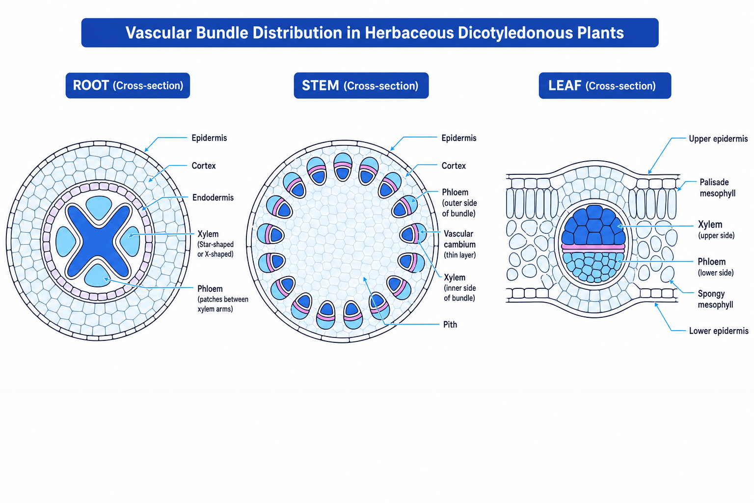
The vascular system of herbaceous dicotyledonous (dicot) plants is composed of two main tissues: xylem and phloem. These are arranged in vascular bundles throughout the plant. Their distribution varies depending on the organ (root, stem, or leaf) to suit its mechanical and physiological demands.
Distribution of vascular tissue
- In the root: The vascular bundle is located in the centre. The xylem forms an 'X' or star-shaped core, with phloem patches located between the arms. This central arrangement helps the root resist pulling forces (tug of wind/gravity) and anchor the plant in the soil.
- In the stem: Vascular bundles are arranged in a neat ring near the outer edge. The xylem is positioned on the inside of each bundle, and the phloem is on the outside, separated by a thin layer of meristematic cells called the cambium. This peripheral arrangement provides scaffold-like support against bending forces.
- In the leaf: The vascular bundles form the midrib and veins. The xylem is located on the upper side of the bundle, and the phloem is on the lower side. This gives mechanical support to hold the leaf flat for maximum light interception.
Xylem structure and function
Xylem vessels transport water and dissolved mineral ions upwards from the roots to the leaves. They are adapted for this function in several key ways:
- Dead cells: Xylem vessels are formed from column-like cells fused end-to-end. As they mature, the cell contents die and break down, leaving a hollow, continuous tube.
- No end walls: The lack of horizontal end walls between cells minimizes resistance to the upward flow of water.
- Lignification: The cell walls are thickened with lignin, a tough, waterproof polymer. Lignin prevents the vessel walls from collapsing inward under the high negative pressures (tension) created by transpiration. It also waterproofs the walls to prevent water from escaping.
- Pits: Small unlignified areas in the cell walls, called pits, allow water to move laterally between adjacent vessels or into surrounding living tissue.
Phloem structure and function
Phloem tissue transports organic solutes (mainly sucrose) up and down the plant. It is a living tissue composed of two main cell types:
-
Sieve tube elements:
- Long, thin cells joined end-to-end to form a continuous tube.
- Unlike xylem, they are living cells, but they have no nucleus, very little cytoplasm, and few organelles (no vacuole, no ribosomes) to allow maximum space for the flow of sap.
- The end walls are perforated to form sieve plates, which let the liquid phloem sap pass through easily.
-
Companion cells:
- Located alongside sieve tube elements and connected to them via microscopic cytoplasmic channels called plasmodesmata.
- Companion cells are highly metabolically active; they contain a large nucleus, abundant mitochondria (to produce ATP for active transport), and ribosomes.
- They carry out the metabolic functions required to maintain and support the sieve tube elements.
Sieve tubes vs Companion cells
A common mistake is thinking sieve tubes are dead because they lack a nucleus. They are living cells but lack the metabolic machinery to survive independently; they rely entirely on their adjacent companion cells.
Practical skill: Dissection and drawing of plant tissues (PAG 1 and PAG 2)
To study the distribution of xylem and phloem in the laboratory, you will perform stem dissections and examine prepared slides.
- Transverse section (TS): Cutting across the stem at a right angle to its length. This shows the ring of vascular bundles.
- Longitudinal section (LS): Cutting along the length of the stem. This shows the long, continuous nature of the xylem vessels.
When producing biological drawings from your sections:
- Use a sharp pencil and single, continuous lines (no shading or sketching).
- Ensure your drawing is large enough to show detail (occupying at least half the page).
- Draw only the outlines of the tissues (a low-power plan diagram) rather than individual cells, unless specifically instructed.
- Use a ruler to draw straight, horizontal label lines that touch the target tissue without crossing over each other.
Transpiration
Transpiration
The loss of water vapour from the aerial parts of a plant (mainly the leaves) by evaporation from cell surfaces followed by diffusion out through the stomata.
Transpiration as a consequence of gas exchange
Plants must open their stomata to allow carbon dioxide (CO2CO_2CO2) to diffuse into the leaf for photosynthesis. Because the inside of the leaf is moist and has a high water potential, water evaporates from the wet cell walls of the mesophyll cells into the air spaces.
When the stomata are open, water vapour diffuses down its water potential gradient into the drier air outside. Thus, transpiration is an unavoidable consequence of gas exchange required for photosynthesis.
Environmental factors affecting transpiration rate
Any factor that alters the concentration gradient of water vapour between the inside of the leaf and the external air, or changes the kinetic energy of water molecules, will affect the rate of transpiration:
| Factor | Effect on Transpiration Rate | Explanation |
|---|---|---|
| Light Intensity | Increases | Light triggers stomata to open for photosynthesis, providing a wider pathway for water vapour to escape. |
| Temperature | Increases | Increases the kinetic energy of water molecules, increasing the rate of evaporation from mesophyll walls. It also decreases the relative humidity of the external air, steepening the water potential gradient. |
| Wind/Air Movement | Increases | Blows away the saturated "boundary layer" of water vapour accumulated around the stomatal pore, maintaining a steep water potential gradient. |
| Relative Humidity | Decreases | High external humidity reduces the water potential gradient between the leaf interior and the outside air, slowing diffusion. |
Practical investigation of transpiration using a potometer (PAG 5)
A potometer measures the rate of water uptake by a cut shoot. Since more than 99% of water taken up by a plant is lost through transpiration, water uptake is a very close estimate of transpiration rate.
       Cut plant shoot
            │
            ▼
      [ Rubber tubing ]
            │
    ┌───────┴───────┐
    │  Capillary    │   ◄─── [ Water reservoir with tap ]
    │    tube       │
    │  (with scale) │
    └───────┬───────┘
            │
            ▼
      [ Air bubble ]
Uptake does not equal loss exactly
In the exam, do not state that a potometer measures transpiration rate directly. It measures water uptake. A small fraction of the water absorbed is kept by the cells for photosynthesis or to maintain turgor pressure.
To set up a potometer successfully and prevent errors:
- Cut the shoot underwater: This prevents air from entering the xylem vessels, which would break the continuous column of water (cohesion).
- Assemble the apparatus underwater: Ensures there are no air bubbles inside the capillary tube or connections.
- Seal all joints with waterproof jelly (e.g., petroleum jelly): Ensures the apparatus is completely airtight.
- Dry the leaves: Wet leaves will artificially reduce transpiration by raising the humidity immediately around the stomata.
- Introduce a single air bubble: Open the reservoir tap briefly or lift the capillary tube out of the water to let a small bubble in, then place it back in the water.
- Record the distance moved by the bubble per unit time.
Calculating transpiration rate using potometer data
A student uses a potometer with a capillary tube of internal diameter d=0.8 mmd = 0.8\text{ mm}d=0.8 mm to measure water uptake. Over a period of 15 minutes, the air bubble moves a distance of 45 mm. Calculate the rate of water uptake in mm3 min−1\text{mm}^3\text{ min}^{-1}mm3 min−1.
- Find the radius (rrr) of the capillary tube in millimetres:
- Calculate the volume (VVV) of water taken up using the volume of a cylinder formula (V=πr2hV = \pi r^2 hV=πr2h), where hhh is the distance the bubble moved (45 mm45\text{ mm}45 mm):
- Calculate the rate of water uptake per minute by dividing the volume by the time taken (15 minutes15\text{ minutes}15 minutes):
Water transport pathways and mechanisms
Water moves down a gradient of water potential (ψ\psiψ), measured in kilopascals (kPa\text{kPa}kPa), from the soil (highest ψ\psiψ, close to 0 kPa0\text{ kPa}0 kPa) to the air outside the leaf (lowest ψ\psiψ, highly negative).
Pathways through the root cortex
Once water is absorbed by the root hair cells, it travels across the root cortex toward the xylem via two distinct pathways:
- The Apoplast Pathway: Water travels through the non-living parts of the plant — specifically, the mesh of cellulose fibers in the cell walls and the intercellular spaces. Water moves by mass flow rather than osmosis, as it does not cross any cell membranes.
- The Symplast Pathway: Water enters the cytoplasm of a cell by osmosis across the plasma membrane. It then travels from cell to cell through the plasmodesmata (cytoplasmic connections).
The Casparian Strip is the gatekeeper
At the endodermis (the cell layer surrounding the vascular bundle), the cell walls are blocked by a waterproof, waxy band of suberin called the Casparian strip.
This forces all water traveling via the apoplast pathway to cross the cell membrane and enter the symplast pathway. This allows the plant's cell membranes to selectively filter and control which mineral ions enter the xylem.
Mechanism of water movement up the stem
Water moves up the xylem vessels in a continuous column driven by the cohesion-tension theory:
- Transpiration pull: Evaporation of water from the leaves pulls the water column upwards.
- Cohesion: Water molecules are polar and form hydrogen bonds with one another. This cohesion holds the water molecules together in a continuous, unbroken column.
- Adhesion: Water molecules form hydrogen bonds with the hydrophilic components of the xylem vessel walls (such as cellulose and lignin). This adhesion prevents the column from pulling away from the walls and helps support the weight of the water.
- Tension: The pulling force from the top of the column creates a negative pressure (tension) within the xylem.
Adaptations to water availability
Plants are adapted to survive in different environmental conditions depending on water availability.
1. Xerophytes
Plants adapted to dry habitats where water is scarce (e.g., Ammophila arenaria / Marram grass, and cacti).
- Rolled leaves: Traps a layer of humid air inside the roll, reducing the water potential gradient between the inside and outside of the leaf.
- Sunken stomata: Stomata are located in pits or grooves, sheltering them from wind and trapping moist air.
- Hairs on leaves: Microscopic hairs trap a boundary layer of water vapour, reducing diffusion.
- Thick waxy cuticle: A thick layer of cutin on the epidermis prevents water loss directly through the leaf surface.
- Reduced leaf surface area: Leaves may be modified into spines (e.g., cacti), minimizing the surface area available for transpiration.
2. Hydrophytes
Plants adapted to live in aquatic environments or waterlogged soils (e.g., Nymphaea / Water lilies).
- Very thin or absent waxy cuticle: There is no need to conserve water.
- Stomata on the upper epidermis only: Leaves float on water; having stomata on the upper surface allows gas exchange with the atmosphere.
- Large air spaces (Aerenchyma): Highly porous tissue that provides buoyancy to keep the leaves afloat and allows oxygen to diffuse quickly to submerged roots.
- Reduced structural support: Water supports the plant's weight, so they do not need heavy lignified tissues.
Translocation
Translocation
The transport of organic assimilates (predominantly sucrose) through the phloem from sources (where they are loaded) to sinks (where they are removed and used).
- Sources include photosynthesising leaves and green stems, or storage organs (like tubers) when they are breaking down starch at the start of the growing season.
- Sinks include growing meristems (roots, shoots, flowers), developing fruits, and storage organs storing starch.
Active loading at the source
To move sucrose into the phloem, companion cells must actively load sucrose against its concentration gradient. This is an energy-requiring process.
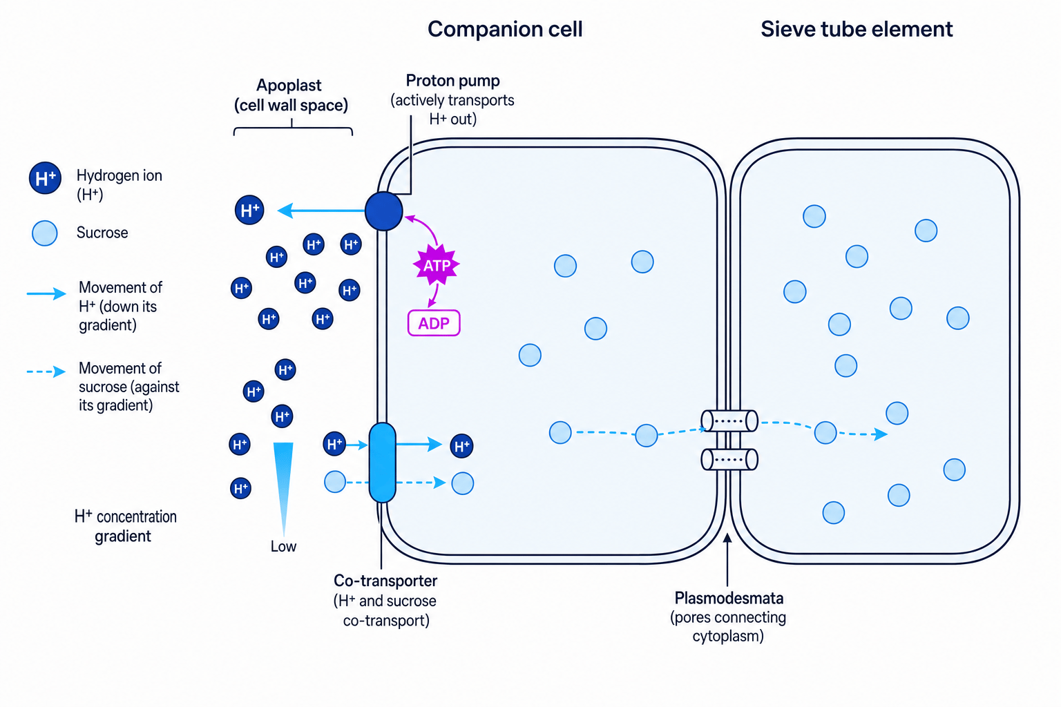
- Proton pumping: Companion cells use ATP to actively transport hydrogen ions (H+H^+H+) out of their cytoplasm, across the plasma membrane, and into the surrounding apoplast (cell wall space). This creates a steep electrochemical gradient of H+H^+H+ ions.
- Co-transport: The H+H^+H+ ions diffuse back into the companion cell down their concentration gradient through a specialized co-transporter protein.
- Sucrose entry: As the H+H^+H+ ions pass through the co-transporter, they bring a sucrose molecule along with them against its concentration gradient (facilitated co-transport).
- Diffusion to sieve tubes: The high concentration of sucrose in the companion cell causes it to diffuse into the sieve tube element through the plasmodesmata.
Mass flow in the phloem
The movement of phloem sap is driven by hydrostatic pressure gradients:
    [ Source ]                                                 [ Sink ]
  Active loading                                            Sucrose unloaded
   of sucrose                                                 at the sink
        │                                                          │
        ▼                                                          ▼
  Water potential                                           Water potential
     decreases                                                 increases
        │                                                          │
        ▼                                                          ▼
  Water enters by                                            Water leaves by
  osmosis from xylem                                         osmosis into xylem
        │                                                          │
        ▼                                                          ▼
 High hydrostatic pressure  ───► [ MASS FLOW OF SAP ] ───►  Low hydrostatic pressure
- At the source: Active loading of sucrose into the sieve tube lowers its water potential (ψ\psiψ). Water enters the sieve tube element from the adjacent xylem by osmosis down a water potential gradient. This increases the hydrostatic pressure inside the sieve tube at the source.
- At the sink: Sucrose is unloaded from the sieve tube (for respiration or storage as starch), which increases the water potential inside the sieve tube. Water leaves the phloem by osmosis, lowering the hydrostatic pressure at the sink.
- Mass flow: The pressure difference forces the phloem sap to flow from the high-pressure region (source) to the low-pressure region (sink).
In the exam
- When describing active loading, always state that hydrogen ions (H+H^+H+) are pumped out first. Do not say sucrose is pumped out directly.
- Be precise with terms: water moves by osmosis, but phloem sap moves by mass flow down a hydrostatic pressure gradient.
- Remember that translocation is two-way (bidirectional), whereas transpiration is one-way (unidirectional, upwards only).
Check yourself
- Why does cutting a shoot underwater matter when setting up a potometer?
- What are the structural differences between a xylem vessel element and a phloem sieve tube element?
- How does the Casparian strip affect the pathway of water through the root?
- Describe the step-by-step process of active loading of sucrose in the companion cell.
