

As animals become larger and more active, simple diffusion becomes entirely inadequate to meet their metabolic demands. Controlling the supply of nutrients and the removal of waste requires the highly coordinated activity of a specialised circulatory system.
In these study notes, we will break down the mechanics of animal transport, from the cellular level to the organ system, focusing on the concepts, calculations, and practical skills you need to master for your OCR A-Level Biology exams.
What you'll learn
- Why multicellular organisms require specialised transport systems, using surface area to volume ratio (SA:VSA:VSA:V) calculations.
- How open, closed, single, and double circulatory systems compare across different animal groups.
- The structure and functions of arteries, arterioles, capillaries, venules, and veins.
- How hydrostatic and oncotic pressures balance to form and reabsorb tissue fluid.
- The mechanics of the cardiac cycle, heart initiation, ECG interpretation, and the biochemistry of oxygen transport.
The Need for Transport Systems
Why can an amoeba rely on simple diffusion across its body surface, while a mammal requires a complex network of blood vessels and a muscular pump? The answer lies in three key factors:
- Size: In larger organisms, the distance between the external environment and the innermost cells is too great. Diffusion would take far too long to deliver oxygen and nutrients.
- Metabolic Rate: Multicellular animals are highly active. They undergo aerobic respiration at high rates, demanding a rapid, constant supply of oxygen and glucose, and producing toxic waste products (like carbon dioxide) that must be removed immediately.
- Surface Area to Volume Ratio (SA:VSA:VSA:V): As an organism increases in size, its volume increases much faster than its surface area.
Surface Area to Volume Ratio (SA:V)
The ratio of the area of the external surface of an organism to its internal volume. It determines the efficiency of exchange across the body surface.
The SA:V Rule of Scale
As size increases, the SA:VSA:VSA:V ratio decreases. Cells deep within a large organism are too far from the exchange surface for diffusion to be effective. Consequently, large organisms must develop specialised exchange surfaces (such as alveoli or gills) and mass flow transport systems (the circulatory system) to move substances over long distances.
Calculating surface area to volume ratio
Compare the surface area to volume ratio (SA:VSA:VSA:V) of a single-celled amoeba, modelled as a cube of side length 10 μm10\,\mu\text{m}10μm, with a multicellular organism modelled as a cube of side length 1000 μm1000\,\mu\text{m}1000μm (1 mm1\,\text{mm}1mm).
- Calculate the surface area (SA=6l2SA = 6l^2SA=6l2) and volume (V=l3V = l^3V=l3) of the single-celled model:
- Calculate the surface area and volume of the multicellular model:
-
Simplify the SA:VSA:VSA:V ratios for both to compare them:
- Amoeba: 6001000=0.6 μm−1\frac{600}{1000} = 0.6\,\mu\text{m}^{-1}1000600=0.6μm−1 (or a ratio of 3:53:53:5)
- Multicellular organism: 6,000,0001,000,000,000=0.006 μm−1\frac{6,000,000}{1,000,000,000} = 0.006\,\mu\text{m}^{-1}1,000,000,0006,000,000=0.006μm−1 (or a ratio of 3:5003:5003:500)
The single-celled organism has an SA:VSA:VSA:V ratio 100 times larger than the multicellular model, illustrating why the larger organism cannot rely on diffusion.
Types of Circulatory Systems
Different animal groups have evolved different ways to transport substances. Circulatory systems are classified based on whether the blood is always contained within vessels, and how many times it passes through the heart during a complete circuit of the body.
Open vs. Closed Circulatory Systems
- Open Circulatory Systems (e.g., Insects): The transport medium (called haemolymph) is not enclosed in blood vessels. Instead, it is pumped straight from a tube-like heart into a large open body cavity called the haemocoel. Under low pressure, the haemolymph directly bathes the organs. It returns to the heart through small valves called ostia.
Insect Gas Exchange is Separate!
Insects do not use their circulatory system to transport oxygen or carbon dioxide! Their haemolymph only transports nutrients and nitrogenous waste. Gas exchange is handled entirely by their tracheal system.
- Closed Circulatory Systems (e.g., Fish and Mammals): The blood is entirely enclosed within blood vessels. It does not come into direct contact with the cells. This allows the blood to travel under high pressure and at high velocity, making transport extremely rapid and highly directed.
Single vs. Double Circulatory Systems
- Single Circulatory Systems (e.g., Fish): Blood passes through the heart once for each complete circuit of the body.
As blood passes through the delicate capillaries of the gills, its pressure drops significantly. This means it flows through the rest of the body at low pressure, limiting the metabolic rate of the fish.
- Double Circulatory Systems (e.g., Mammals):
Blood passes through the heart twice for each complete circuit. It has two distinct loops:
- Pulmonary Circulation: The right side of the heart pumps deoxygenated blood to the lungs to pick up oxygen and lose carbon dioxide. The pressure is kept relatively low to avoid damaging the delicate lung capillaries.
- Systemic Circulation: Oxygenated blood returns to the left side of the heart, which pumps it out to the rest of the body at high pressure, ensuring rapid delivery to active tissues.
Structure and Function of Blood Vessels
To withstand and maintain pressure, and to control blood flow, different blood vessels contain specialized distributions of tissues.
Endothelium
A single layer of flattened, smooth epithelial cells lining the inside of all blood vessels, reducing friction to maximize blood flow rate.
The walls of major vessels (except capillaries) contain varying proportions of:
- Elastic fibres: Made of elastin, these allow vessels to stretch when blood is pumped in and recoil to maintain high blood pressure between heartbeats.
- Smooth muscle: Contracts or relaxes to narrow (vasoconstrict) or widen (vasodilate) the vessel lumen, controlling blood flow distribution.
- Collagen: A tough structural protein that provides strength to prevent the vessel from bursting under high pressure.
Comparing Blood Vessel Walls
| Vessel | Elastic Fibres | Smooth Muscle | Collagen | Endothelium / Lumen Characteristics |
|---|---|---|---|---|
| Artery | Very thick layer | Thick layer | Thick layer | Smooth, folded endothelium (allows stretching); narrow lumen to maintain high pressure. |
| Arteriole | Thin layer | Very thick layer | Thin layer | Smooth endothelium; highly muscular to constrict and control blood flow into capillary beds. |
| Capillary | None | None | None | Extremely thin wall consisting of only a single layer of endothelial cells (minimal diffusion distance). |
| Venule | None | Extremely thin layer | Thin layer | Thin wall, collecting blood from capillaries. |
| Vein | Thin layer | Thin layer | Thick layer | Thin walls with a very wide lumen to reduce resistance; contains valves to prevent backflow of low-pressure blood. |
PAG 2: Blood Vessel Microscopy
When drawing blood vessels under a light microscope, look for the highly folded, wavy inner lining (endothelium) of the artery compared to the collapsed, thin wall of the vein. Always use a sharp pencil, do not shade, and label the tissue layers (tunica intima, media, and externa) clearly.
The Formation of Tissue Fluid
Blood consists of cells suspended in a straw-coloured liquid called plasma. Cells cannot directly access the plasma; instead, they are bathed in tissue fluid, which acts as the exchange medium.
Tissue fluid is formed at the capillary beds by the interaction of two opposing pressures:
- Hydrostatic Pressure: The physical pressure exerted by the fluid (blood) against the capillary walls, generated by the contraction of the heart. It forces fluid out of the capillaries through tiny gaps between the endothelial cells.
- Oncotic Pressure: The osmotic pressure generated by plasma proteins (such as albumin) trapped inside the capillary. Because these proteins are too large to pass through the capillary wall, they lower the water potential (ψ\psiψ) of the blood, drawing water into the capillaries by osmosis.
The Balance of Pressures
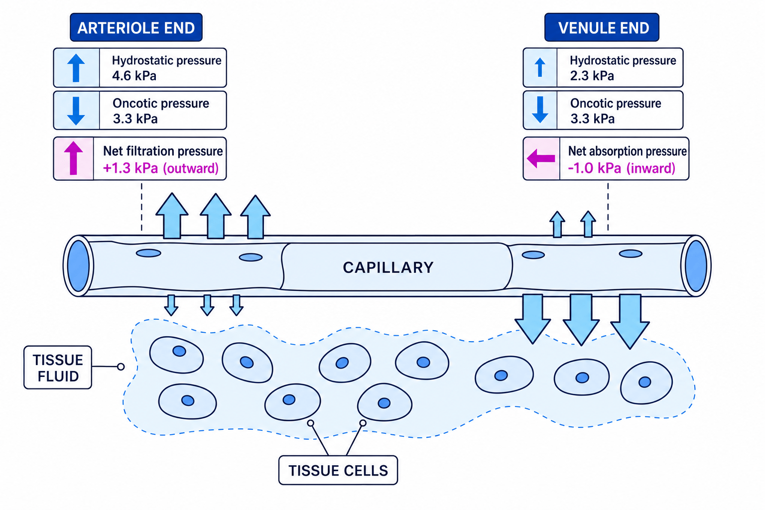
- At the Arteriole End: The hydrostatic pressure of the blood is high (approx. 4.6 kPa4.6\,\text{kPa}4.6kPa), which is much greater than the inward-acting oncotic pressure (approx. 3.3 kPa3.3\,\text{kPa}3.3kPa). This creates a net outward filtration pressure of +1.3 kPa+1.3\,\text{kPa}+1.3kPa, forcing plasma water, glucose, amino acids, and oxygen out into the spaces between the cells.
- At the Venule End: As fluid leaves, the hydrostatic pressure within the capillary drops significantly (to approx. 2.3 kPa2.3\,\text{kPa}2.3kPa). The oncotic pressure remains constant at 3.3 kPa3.3\,\text{kPa}3.3kPa because the plasma proteins remain behind. This creates a net inward absorption pressure of −1.0 kPa-1.0\,\text{kPa}−1.0kPa, drawing water and dissolved metabolic wastes (like carbon dioxide) back into the blood by osmosis.
Lymphatic System
Not all the fluid that leaves at the arteriole end returns at the venule end. The excess fluid (about 10%) drains into blind-ended vessels called lymph capillaries. Once inside, this fluid is called lymph. It is eventually returned to the blood system via the subclavian veins in the chest.
Differences in Composition
| Component | Blood Plasma | Tissue Fluid | Lymph |
|---|---|---|---|
| Red Blood Cells | Yes | No | No |
| White Blood Cells | Yes | Very few (only during immune response) | Yes (mainly lymphocytes released from lymph nodes) |
| Platelets | Yes | No | No |
| Large Proteins | Yes | No (too large to pass capillary wall) | No |
| Glucose & Oxygen | High concentration | High (absorbed by body cells) | Low concentration |
Mammalian Heart Structure and Dissection
The mammalian heart is a muscular double-pump designed to keep blood flowing in one direction under precise pressure control.
Internal Structure of the Heart
- Atria: Thin-walled chambers that receive blood returning from the veins (vena cava on the right, pulmonary vein on the left). They only need to pump blood a short distance into the ventricles.
- Ventricles: Thick-walled chambers. The left ventricle has a muscular wall approximately three times thicker than the right ventricle.
Why is the left ventricle thicker?
Never say the left ventricle pumps blood "further". The correct explanation is that the left ventricle must generate higher pressure to overcome the high resistance of the systemic circulation (the entire body), whereas the right ventricle only pumps blood to the lungs, which are nearby and have delicate capillaries.
- Atrioventricular (AV) Valves: Located between the atria and ventricles (tricuspid on the right, bicuspid/mitral on the left). They prevent blood from flowing back into the atria during ventricular contraction. They are anchored by tough, fibrous strings called tendinous cords (heartstrings) to prevent the valves from turning inside out.
- Semilunar Valves: Located at the entrance to the major arteries leaving the heart (pulmonary artery on the right, aorta on the left). They prevent blood from flowing back into the ventricles during diastole.
- Septum: A thick wall of muscle separating the right and left sides of the heart, preventing oxygenated and deoxygenated blood from mixing.
Dissection Skills (PAG 2)
During a heart dissection (typically a sheep or pig heart):
- Safety: Wear a lab coat, gloves, and safety goggles. Perform all cutting on a clean dissecting board using a sharp scalpel.
- External Examination: Identify the front (anterior) of the heart by locating the coronary arteries running diagonally across the ventricles. These supply oxygenated blood directly to the cardiac muscle. Note the difference in compressibility between the thin-walled atria and the firm, thick ventricles.
- Internal Cuts: Use a scalpel to make a clean, longitudinal cut down through the left ventricle wall to observe its thickness, then repeat on the right side. Identify the AV valves, semilunar valves, and the fibrous tendinous cords.
The Cardiac Cycle
The cardiac cycle describes the sequence of events in a single heartbeat. It is divided into three main stages:
- Diastole (Relaxation): Both the atria and ventricles relax. Blood flows into the atria from the vena cava and pulmonary vein. As the atria fill, pressure rises above that of the ventricles, causing the AV valves to open passively so blood begins to trickle into the ventricles.
- Atrial Systole (Atrial Contraction): The atria contract, forcing the remaining blood through the AV valves into the ventricles.
- Ventricular Systole (Ventricular Contraction): The ventricles contract from the apex (bottom) upwards. This dramatically increases the pressure inside the ventricles. The high pressure forces the AV valves shut (making the first "lub" sound of the heartbeat). Once ventricular pressure exceeds the pressure in the aorta and pulmonary artery, the semilunar valves are forced open, and blood is ejected.
Analyzing Pressure Changes
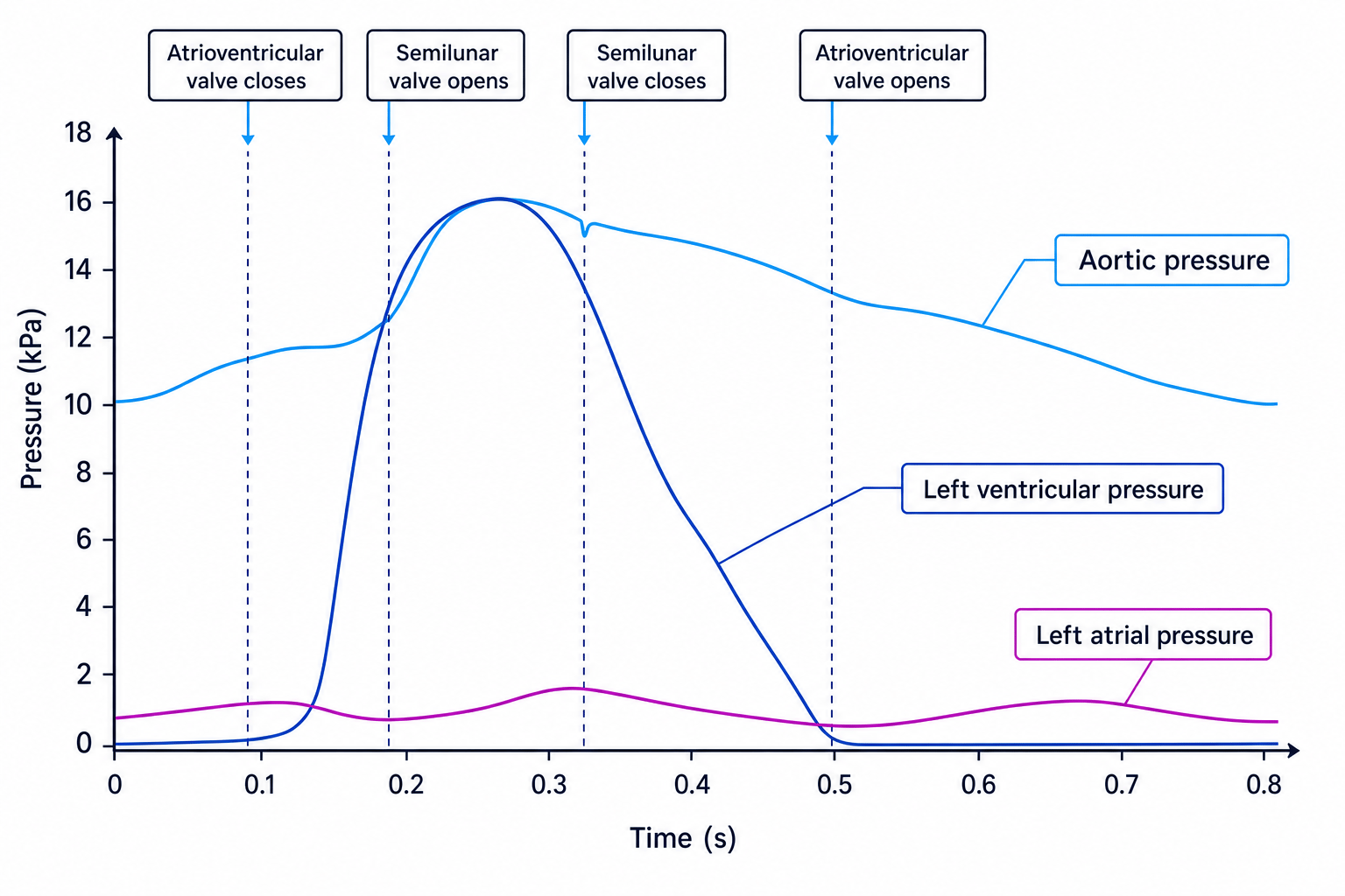
By tracking the pressures of the left atrium, left ventricle, and aorta, you can determine exactly when valves open and close.
The Golden Rule of Valves
Valves open when the pressure behind them is greater than the pressure in front of them. They snap shut when the pressure in front exceeds the pressure behind.
Using the graph above, we can identify key pressure cross-over points:
- AV valve closes: When ventricular pressure rises above atrial pressure (at approx. 0.1 s0.1\,\text{s}0.1s).
- Semilunar valve opens: When ventricular pressure rises above aortic pressure (at approx. 0.18 s0.18\,\text{s}0.18s).
- Semilunar valve closes: When ventricular pressure falls below aortic pressure, causing blood to attempt to flow backwards and fill the valve pockets (at approx. 0.32 s0.32\,\text{s}0.32s).
- AV valve opens: When ventricular pressure falls below atrial pressure (at approx. 0.5 s0.5\,\text{s}0.5s).
Calculating Cardiac Output
Cardiac Output (CO)
The total volume of blood pumped by one ventricle of the heart per minute. It is calculated using the formula:
Cardiac Output=Heart Rate×Stroke Volume \text{Cardiac Output} = \text{Heart Rate} \times \text{Stroke Volume} Cardiac Output=Heart Rate×Stroke VolumeWhere:
- Heart Rate (HR) is measured in beats per minute (bpm\text{bpm}bpm).
- Stroke Volume (SV) is the volume of blood pumped out of a ventricle in a single beat, measured in cm3\text{cm}^3cm3 or dm3\text{dm}^3dm3.
Calculating cardiac output
A student calculates from their ECG that their resting heart rate is 75 bpm75\,\text{bpm}75bpm. Their stroke volume is measured at 72 cm372\,\text{cm}^372cm3. Calculate their cardiac output in dm3 min−1\text{dm}^3\,\text{min}^{-1}dm3min−1.
- Convert stroke volume from cm3\text{cm}^3cm3 to dm3\text{dm}^3dm3 (since 1 dm3=1000 cm31\,\text{dm}^3 = 1000\,\text{cm}^31dm3=1000cm3):
- Substitute the values into the formula:
- Compute the final answer and apply the correct units:
Coordination of Heart Action
Cardiac muscle is myogenic, meaning it can initiate its own contraction without requiring external nerve impulses. To ensure the chambers contract in a coordinated sequence, the heart uses a specialized electrical conduction pathway:
- The Sino-Atrial Node (SAN): Located in the wall of the right atrium, the SAN acts as the natural pacemaker. It generates a wave of electrical excitation (depolarisation) that spreads rapidly across the walls of both atria, causing them to contract in unison (atrial systole).
- Non-conducting Collagen Tissue: A band of non-conducting tissue at the base of the atria acts as an electrical barrier, preventing the wave of excitation from passing directly down into the ventricles.
- The Atrio-Ventricular Node (AVN): The excitation wave can only pass to the ventricles via the AVN. The AVN introduces a vital delay (approx. 0.1 s0.1\,\text{s}0.1s) before transmitting the impulse down.
Why must the AVN delay the electrical impulse?
The delay introduced by the AVN is essential because it gives the atria enough time to contract fully and empty all their blood into the ventricles before the ventricles begin to contract.
- Bundle of His and Purkyne Tissue: After the delay, the electrical impulse is conducted rapidly down the septum via the Bundle of His to the apex of the heart. The impulse then spreads upwards through the Purkyne tissue in the walls of the ventricles, causing the ventricles to contract from the apex upwards. This squeezes blood efficiently up and out into the major arteries.
Interpreting Electrocardiograms (ECGs)
An ECG measures the electrical activity of the heart over time using electrodes placed on the skin. A normal trace shows a characteristic pattern of waves:
- P wave: Represents the depolarisation (electrical excitation) of the atria, leading to atrial contraction.
- QRS complex: Represents the depolarisation of the ventricles, leading to ventricular contraction (the larger wave reflects the larger muscle mass of the ventricles).
- T wave: Represents the repolarisation (recovery) of the ventricles.
Diagnostic ECG Anomalies
You must be able to recognize four common heart abnormalities from an ECG strip:
- Tachycardia: An abnormally fast resting heart rate (typically over 100 bpm100\,\text{bpm}100bpm). The waves are normal in shape but packed very closely together.
- Bradycardia: An abnormally slow resting heart rate (typically below 60 bpm60\,\text{bpm}60bpm). The waves are normal but spaced very far apart.
- Ectopic Heartbeat: An early or "skipped" beat that disrupts the normal rhythm, often followed by a brief pause. It is caused by an out-of-sequence depolarisation.
- Fibrillation: A chaotic, irregular, and rapid electrical signal with no clear P, QRS, or T waves. The heart muscle simply shudders (quivers) instead of contracting effectively.
Oxygen Transport and Haemoglobin
Red blood cells are packed with haemoglobin (Hb), a globular protein with a quaternary structure composed of four polypeptide chains. Each chain contains a prosthetic haem group containing an iron ion (Fe2+\text{Fe}^{2+}Fe2+), which can bind reversibly to one oxygen molecule (O2\text{O}_2O2).
Hb+4O2⇌HbO8 \text{Hb} + 4\text{O}_2 \rightleftharpoons \text{HbO}_8 Hb+4O2⇌HbO8The Oxygen Dissociation Curve
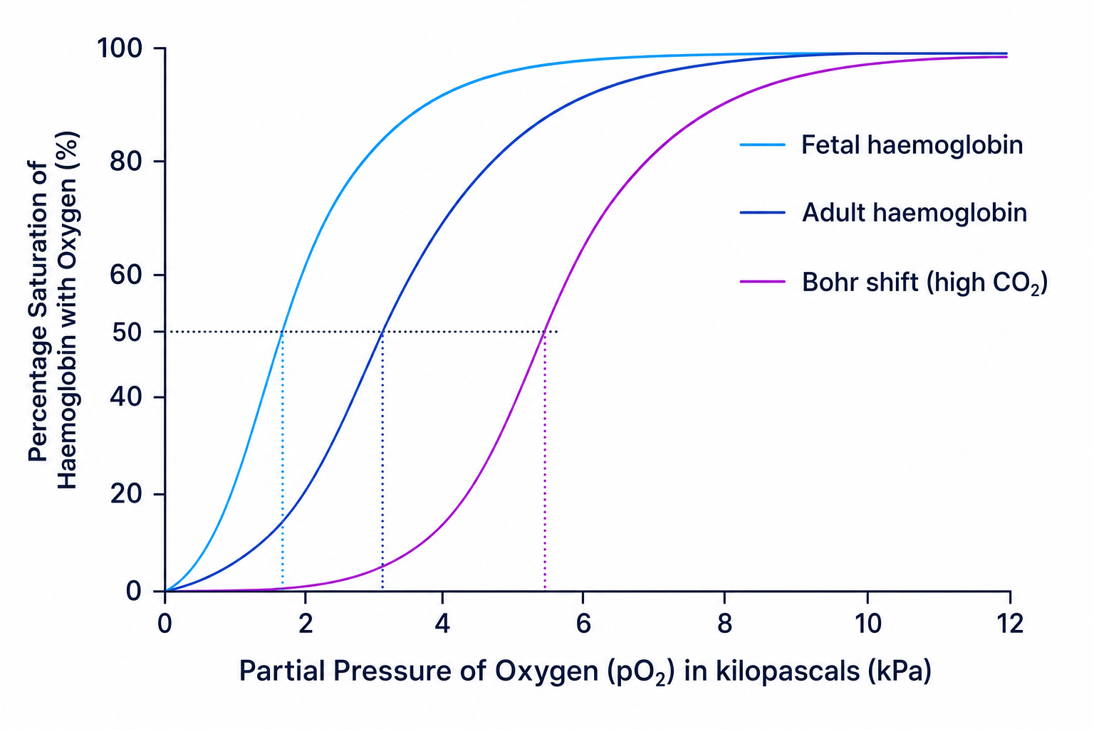
The affinity (binding strength) of haemoglobin for oxygen changes depending on the partial pressure of oxygen (pO2\text{pO}_2pO2). When represented on a graph, this relationship produces an S-shaped (sigmoid) curve.
The S-shape is caused by cooperative binding:
- At very low pO2\text{pO}_2pO2, the shape of the haemoglobin molecule makes it difficult for the first oxygen molecule to bind.
- Once the first oxygen molecule binds, it induces a conformational change (shape change) in the quaternary structure of the haemoglobin.
- This conformational change makes it much easier for the second and third oxygen molecules to bind, causing the curve to steepen rapidly.
- As the haemoglobin becomes highly saturated, it becomes harder for the fourth oxygen molecule to find an empty binding site, causing the curve to plateau.
Fetal Haemoglobin vs. Adult Haemoglobin
A fetus relies entirely on obtaining oxygen from its mother's blood across the placenta. By the time maternal blood reaches the placenta, its oxygen saturation has decreased.
To survive, fetal haemoglobin has a higher affinity for oxygen than adult haemoglobin. This shifts the fetal oxygen dissociation curve to the left. At any given partial pressure of oxygen, fetal haemoglobin will bind oxygen more readily, allowing it to load oxygen from the mother's oxyhaemoglobin.
The Bohr Effect (Carbon Dioxide Transport)
In highly active tissues (like exercising muscle), respiration produces high levels of carbon dioxide, resulting in a high partial pressure of carbon dioxide (pCO2\text{pCO}_2pCO2).
When pCO2\text{pCO}_2pCO2 is high, the affinity of haemoglobin for oxygen decreases. This causes the oxygen dissociation curve to shift to the right (the Bohr shift). This shift is highly beneficial: at respiring tissues, haemoglobin releases (unloads) its oxygen much more easily to sustain aerobic respiration.
Carbon Dioxide Transport in the Blood
Carbon dioxide produced by respiring cells is transported back to the lungs in three ways:
- Dissolved directly in the plasma (approx. 5%).
- Bound directly to haemoglobin to form carbaminohaemoglobin (approx. 10%).
- Converted into soluble hydrogencarbonate ions (HCO3−\text{HCO}_3^-HCO3−) in the cytoplasm of red blood cells (approx. 85%).
The Hydrogencarbonate Pathway and the Chloride Shift
Inside the red blood cell, the following biochemical sequence takes place:
- Carbon dioxide diffuses into the red blood cell and reacts with water to form weak carbonic acid (H2CO3\text{H}_2\text{CO}_3H2CO3). This reaction is catalyzed by the enzyme carbonic anhydrase:
- Carbonic acid spontaneously dissociates into hydrogen ions (H+\text{H}^+H+) and hydrogencarbonate ions (HCO3−\text{HCO}_3^-HCO3−):
- The negatively charged hydrogencarbonate ions (HCO3−\text{HCO}_3^-HCO3−) diffuse out of the red blood cell into the plasma down their concentration gradient.
- To maintain electrical neutrality inside the cell, chloride ions (Cl−\text{Cl}^-Cl−) diffuse from the plasma into the red blood cell. This movement of ions is known as the chloride shift.
- The accumulation of hydrogen ions (H+\text{H}^+H+) lowers the pH inside the cell, which could denature proteins. To prevent this, haemoglobin acts as a buffer by binding to the free hydrogen ions to form haemoglobinic acid (HHb\text{HHb}HHb):
This binding of hydrogen ions alters the shape of the haemoglobin, reducing its affinity for oxygen, which further explains the molecular mechanism behind the Bohr effect.
In the exam
- Explain the shape of the oxygen dissociation curve: Always use the term cooperative binding and describe the conformational change in haemoglobin's shape that occurs when the first oxygen molecule binds.
- Distinguish left and right shifts: Remember: Left = Linked to Loading (higher affinity, e.g., fetal haemoglobin or organisms living at high altitudes); Right = Rapid Release (lower affinity, e.g., during the Bohr shift to dump oxygen at working muscles).
- Be quantitative with tissue fluid questions: If asked to explain tissue fluid formation, quote values for hydrostatic and oncotic pressures, calculate net filtration/absorption pressures, and describe how the balance changes from the arteriole end to the venule end.
- Identify the exact roles of cardiac nodes: Do not confuse the SAN and AVN. The SAN initiates the wave of excitation. The AVN delays the wave.
Check yourself
- A patient has a resting heart rate of 60 bpm60\,\text{bpm}60bpm and a cardiac output of 4.8 dm3 min−14.8\,\text{dm}^3\,\text{min}^{-1}4.8dm3min−1. What is their stroke volume in cm3\text{cm}^3cm3?
- Why is it critical that the wave of electrical excitation cannot pass directly from the atria to the ventricles across the entire boundary layer?
- Explain how the binding of hydrogen ions (H+\text{H}^+H+) to haemoglobin inside red blood cells leads to increased oxygen delivery to respiring tissues.
- Contrast the structural tissue layers of a large vein with those of a large artery.
