

Living organisms must detect and respond to changes in their internal and external environments to survive. In plants, responses are coordinated by plant growth regulators (hormones), while in animals, responses are coordinated by both the nervous and endocrine (hormonal) systems.
This topic covers how these response systems operate at a cellular, physiological, and whole-organism level.
What you'll learn
- How plants respond to abiotic stress, herbivory, and directional stimuli using plant hormones.
- The organisation of the mammalian nervous system and the anatomy of the human brain.
- The coordination of the "fight-or-flight" response and the homeostatic regulation of heart rate.
- The structure of mammalian muscle and the molecular mechanism of skeletal muscle contraction.
1. Plant Responses and Tropisms
Plants do not have nervous systems, but they are highly responsive to their environments. They coordinate responses to abiotic stress (such as drought or freezing) and biotic stress (such as herbivory) using chemical messengers.
Types of Plant Responses
Plants exhibit tropisms, which are growth responses to directional external stimuli.
Tropism
A directional growth response of a plant where the direction of the response is determined by the direction of the external stimulus.
- Phototropism: Growth response to unilateral light (shoots are positively phototropic; roots are negatively phototropic).
- Geotropism (Gravitropism): Growth response to gravity (shoots are negatively geotropic; roots are positively geotropic).
- Chemotropism: Growth response to chemicals (e.g., pollen tubes growing towards chemicals secreted by the ovary).
- Thigmotropism: Growth response to touch (e.g., climbing plants winding around support structures).
Responses to Herbivory
Herbivory is the consumption of plant material by animals. Plants have evolved sophisticated chemical and physical mechanisms to defend themselves:
- Alkaloids: Nitrogen-containing organic compounds that taste extremely bitter and act as toxins to herbivores. Examples include nicotine, caffeine, and morphine. They can inhibit protein synthesis or disrupt crucial metabolic pathways in the herbivore.
- Pheromones: Volatile chemical signals released by a plant that affect the physiology or behaviour of other organisms. For instance, when attacked, some plants release volatile organic compounds (VOCs) that signal neighbouring plants to start synthesising defensive chemicals, or attract predatory insects that feed on the herbivores.
- Folding in response to touch: Mimosa pudica exhibits a rapid, non-directional movement (a thigmonastic response). If touched, its leaflets fold inward within seconds. This sudden movement startles herbivores and exposes sharp stems or makes the leaves appear smaller and less appetizing. This is driven by rapid electrochemical signals causing water to leave vacuoles in specialized cells at the leaf joint (pulvinule), leading to a loss of turgor pressure.
2. Roles of Plant Hormones
Plant responses are coordinated by plant hormones (often called plant growth regulators). Unlike animal hormones, plant hormones are not produced by specialized endocrine glands but are synthesised by cells in active regions (like meristems) and diffuse or are actively transported to target cells.
Key Plant Hormones and Their Roles
| Hormone | Primary Functions |
|---|---|
| Auxins (e.g., IAA) | Controls cell elongation, maintains apical dominance, inhibits leaf abscission, and stimulates root growth. |
| Gibberellins | Stimulates stem elongation (internode growth) and triggers seed germination. |
| Abscisic Acid (ABA) | Coordinates stomatal closure during water stress and maintains seed dormancy. |
| Ethene | Promotes fruit ripening and stimulates leaf abscission. |
Control of Leaf Loss (Abscission)
Deciduous plants shed their leaves in autumn to prevent water loss and damage from frost.
- As a leaf ages, the level of auxin in the leaf decreases.
- This drop in auxin makes cells in the abscission zone (at the base of the leaf stalk) more sensitive to ethene.
- Ethene activates genes that code for cellulase and pectinase enzymes.
- These enzymes digest the cell walls in the abscission layer, weakening the connection until the leaf falls off under its own weight or wind.
Control of Seed Germination
Gibberellins are essential for initiating seed germination:
- The seed absorbs water (imbibition), activating the embryo.
- The active embryo secretes gibberellin.
- Gibberellin diffuses to the aleurone layer (a protein-rich layer surrounding the endosperm).
- In the aleurone layer, gibberellin triggers the transcription of genes coding for amylase and protease enzymes.
- Amylase hydrolyses stored starch in the endosperm into maltose and glucose.
- This glucose is transported to the embryo, providing a respiratory substrate to produce ATP for growth.
Hormonal Antagonism
Abscisic acid (ABA) acts as an antagonist to gibberellin. High concentrations of ABA maintain seed dormancy and prevent premature germination. Germination only occurs when the ratio of gibberellin to ABA becomes high enough.
Stomatal Closure under Water Stress
When water is scarce, roots synthesise ABA, which is transported to the leaves where it causes rapid stomatal closure:
- ABA binds to specific receptors on the cell-surface membranes of the guard cells.
- This binding triggers an influx of calcium ions (Ca2+Ca^{2+}Ca2+) into the cytosol from vacuoles.
- The elevated cytosolic Ca2+Ca^{2+}Ca2+ concentration acts as a second messenger, opening ion channels that allow potassium ions (K+K^+K+) and anions (like Cl−Cl^-Cl−) to exit the guard cells.
- The loss of these ions raises the water potential (ψ\psiψ) inside the guard cells.
- Water leaves the guard cells by osmosis down a water potential gradient into neighbouring epidermal cells.
- The guard cells lose turgor pressure, become flaccid, and close the stomatal pore.
3. Experimental Evidence for Auxins and Gibberellins
Apical Dominance
Apical dominance is the phenomenon where the growing apical bud (the tip of the main shoot) suppresses the growth of lateral buds lower down the stem. This ensures the plant grows vertically to access light.
- The Auxin Hypothesis: Auxins (specifically Indoleacetic Acid, IAA) are produced in the shoot tip and transported basipetally (downwards). High concentrations of auxin in the stem inhibit the growth of lateral buds.
- Experimental Evidence:
- Decapitation: If the apical bud is cut off, lateral buds begin to grow because the source of auxin is removed.
- Auxin Replacement: If the apical bud is removed and an agar block containing auxin is placed on the cut stump, the lateral buds remain dormant. This confirms that auxin is the chemical messenger inhibiting lateral growth.
- Transport Inhibitors: Applying an auxin transport inhibitor (such as TIBA) below an intact apical bud leads to lateral bud growth, showing that active downward transport of auxin is required for inhibition.
Confusing Concentrations
Do not assume auxin always stimulates growth. Auxin has different effects depending on its concentration and the tissue type. High concentrations of auxin stimulate stem elongation but inhibit root elongation and lateral bud growth.
Stem Elongation
Gibberellins control stem elongation by stimulating both cell division and cell elongation.
- Experimental Evidence:
- Dwarf varieties of plants (like dwarf peas or dwarf maize) often have a genetic mutation that prevents them from producing active gibberellins (they lack the functional enzyme to convert inactive precursors into active GA1GA_1GA1).
- When active gibberellin is applied externally to these dwarf plants, they grow to the same height as wild-type tall plants.
- Applying gibberellin to wild-type tall plants has little to no additional effect, demonstrating that gibberellin concentration is the limiting factor in dwarf plant growth.
4. Practical Investigations and Commercial Uses
Serial Dilutions in Plant Hormone Experiments
To investigate the effect of auxin or gibberellin concentration on plant tissue growth (e.g., oat coleoptile length), you must prepare a range of hormone concentrations. This is done using a serial dilution.
Preparing a serial dilution of auxin
You are provided with a stock solution of auxin with a concentration of 10.0 g dm−310.0\text{ g dm}^{-3}10.0 g dm−3. You need to prepare 10 cm310\text{ cm}^310 cm3 of five solutions ranging from 1.0 g dm−31.0\text{ g dm}^{-3}1.0 g dm−3 down to 1.0×10⁻⁴ g dm−31.0 \times 10^{-4}\text{ g dm}^{-3}1.0×10⁻⁴ g dm−3 using a 101010-fold (1:101:101:10) serial dilution series.
Calculate the volumes of solution and distilled water required for each step.
-
Calculate the dilution factor: To perform a 101010-fold dilution, each subsequent solution must be 110\frac{1}{10}101 of the concentration of the previous one. The dilution ratio is 111 part stock solution to 999 parts distilled water.
-
Determine the volume for the first dilution (1.0 g dm−31.0\text{ g dm}^{-3}1.0 g dm−3): To make 10 cm310\text{ cm}^310 cm3 of a 1.0 g dm−31.0\text{ g dm}^{-3}1.0 g dm−3 solution from a 10.0 g dm−310.0\text{ g dm}^{-3}10.0 g dm−3 stock:
Add 1.0 cm31.0\text{ cm}^31.0 cm3 of stock solution to 9.0 cm39.0\text{ cm}^39.0 cm3 of distilled water. Thoroughly mix this solution.
-
Perform the subsequent steps systematically:
- To make 1.0×10⁻¹ g dm−31.0 \times 10^{-1}\text{ g dm}^{-3}1.0×10⁻¹ g dm−3 (0.1 g dm−30.1\text{ g dm}^{-3}0.1 g dm−3): Take 1.0 cm31.0\text{ cm}^31.0 cm3 of the 1.0 g dm−31.0\text{ g dm}^{-3}1.0 g dm−3 solution and add it to 9.0 cm39.0\text{ cm}^39.0 cm3 of distilled water.
- To make 1.0×10⁻² g dm−31.0 \times 10^{-2}\text{ g dm}^{-3}1.0×10⁻² g dm−3: Take 1.0 cm31.0\text{ cm}^31.0 cm3 of the 1.0×10⁻¹ g dm−31.0 \times 10^{-1}\text{ g dm}^{-3}1.0×10⁻¹ g dm−3 solution and add it to 9.0 cm39.0\text{ cm}^39.0 cm3 of distilled water.
- To make 1.0×10⁻³ g dm−31.0 \times 10^{-3}\text{ g dm}^{-3}1.0×10⁻³ g dm−3: Take 1.0 cm31.0\text{ cm}^31.0 cm3 of the 1.0×10⁻² g dm−31.0 \times 10^{-2}\text{ g dm}^{-3}1.0×10⁻² g dm−3 solution and add it to 9.0 cm39.0\text{ cm}^39.0 cm3 of distilled water.
- To make 1.0×10⁻⁴ g dm−31.0 \times 10^{-4}\text{ g dm}^{-3}1.0×10⁻⁴ g dm−3: Take 1.0 cm31.0\text{ cm}^31.0 cm3 of the 1.0×10⁻³ g dm−31.0 \times 10^{-3}\text{ g dm}^{-3}1.0×10⁻³ g dm−3 solution and add it to 9.0 cm39.0\text{ cm}^39.0 cm3 of distilled water.
-
Note final volumes for the experiment: Each tube will contain 9.0 cm39.0\text{ cm}^39.0 cm3 of solution (after 1.0 cm31.0\text{ cm}^31.0 cm3 is transferred to the next tube), except the final tube which will contain 10.0 cm310.0\text{ cm}^310.0 cm3 unless 1.0 cm31.0\text{ cm}^31.0 cm3 is discarded.
Commercial Uses of Plant Hormones
Plant hormones are widely exploited in agriculture and horticulture:
- Auxins:
- Rooting powders: Applying auxin to the cut end of a plant cutting stimulates rapid root development, allowing clonal propagation.
- Hormonal weedkillers: Synthetic auxins (like 2,4-D) are selective for broad-leaved dicot plants (weeds). They cause rapid, distorted growth that the plant's transport systems cannot sustain, leading to exhaustion and death.
- Ethene:
- Controlled ripening: Climacteric fruits (such as bananas and tomatoes) are harvested green and unripe because they are firmer and less easily damaged during transport. Before retail, they are exposed to ethene gas to trigger synchronous ripening.
- Gibberellins:
- Fruit production: Gibberellins make grape stalks elongate, giving individual grapes more room to grow larger. They also stimulate the growth of seedless fruit.
- Brewing: Gibberellins are applied to barley seeds to speed up the synthesis of amylase during the malting process.
5. Organisation of the Mammalian Nervous System
The mammalian nervous system is structurally and functionally organised to rapidly transmit electrical impulses.
Structural Organisation
- Central Nervous System (CNS): Consists of the brain and the spinal cord. It processes sensory information and coordinates responses.
- Peripheral Nervous System (PNS): Consists of all the neurones that lie outside the CNS. It connects the CNS to the rest of the body.
Functional Organisation
The PNS is functionally divided into:
- Somatic Nervous System: Coordinates voluntary responses. It consists of motor neurones that carry impulses from the CNS to skeletal muscles under conscious control.
- Autonomic Nervous System (ANS): Coordinates involuntary/unconscious responses. It connects the CNS to cardiac muscle, smooth muscle, and glands.
The Autonomic Nervous System is further divided into two antagonistic branches:
| Feature | Sympathetic Nervous System | Parasympathetic Nervous System |
|---|---|---|
| General Role | "Fight or flight" (prepares body for activity) | "Rest and digest" (conserves energy, promotes recovery) |
| Neurotransmitter | Noradrenaline | Acetylcholine |
| Heart Rate | Increases | Decreases |
| Airways | Dilates bronchioles | Constricts bronchioles |
| Digestive System | Inhibits peristalsis and gland secretion | Stimulates peristalsis and gland secretion |
6. Structure and Function of the Human Brain
The brain is the primary processing centre of the mammalian nervous system. It consists of specialized regions that handle distinct physiological and cognitive tasks.
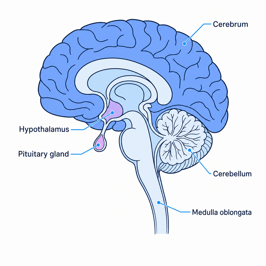
Brain Structures and Their Roles
1. Cerebrum
- The largest part of the brain, divided into left and right cerebral hemispheres.
- Coordinates conscious thought, intelligence, memory, learning, language, and emotional responses.
- It receives sensory inputs (sensory areas), integrates them (association areas), and sends out voluntary motor impulses (motor areas).
2. Cerebellum
- Located at the back of the brain, highly folded.
- Coordinates motor control, balance, posture, and fine movement. It does not initiate muscle movement but coordinates complex movements (like writing or riding a bicycle) based on sensory feedback from eyes and joints.
3. Medulla Oblongata
- Located at the base of the brain stem.
- Controls critical autonomic (involuntary) functions. It contains centres regulating heart rate (cardiovascular centre), breathing rate (ventilation centre), and smooth muscle contraction in blood vessel walls.
4. Hypothalamus
- Located below the cerebrum.
- The master control centre for homeostasis. It monitors blood temperature (thermoregulation) and blood water potential (osmoregulation). It links the nervous and endocrine systems by controlling the secretory activity of the pituitary gland.
5. Pituitary Gland
- Located at the base of the hypothalamus, split into two lobes:
- Anterior Pituitary: Produces and releases its own hormones (e.g., FSH, LH, growth hormone) under the control of releasing factors from the hypothalamus.
- Posterior Pituitary: Stores and releases hormones produced by the hypothalamus (such as ADH).
7. Reflex Actions
A reflex action is a rapid, involuntary, and automatic response to a sensory stimulus. It bypasses the conscious areas of the brain, which minimizes the time taken to respond.
Survival Value of Reflexes
Reflexes are essential for survival because:
- They are extremely fast because they involve very few synapses and do not require conscious processing by the brain.
- They are involuntary, meaning the response occurs automatically without decision-making, reducing delay.
- They protect the body from tissue damage (e.g., the withdrawal reflex from hot objects, or the pupil reflex protecting the retina from bright light).
The Knee-Jerk Reflex
The knee-jerk reflex is a spinal reflex that helps maintain balance and posture.
- Stimulus: Tapping the patellar tendon (just below the kneecap) stretches the quadriceps muscle.
- Receptors: Stretch receptors (muscle spindles) in the quadriceps detect the stretch.
- Sensory Neurone: Sends action potentials directly to the spinal cord.
- Synapse: In the spinal cord, the sensory neurone synapses directly with a motor neurone (this is a monosynaptic reflex arc, as there is no relay neurone in the primary pathway).
- Motor Neurone: Carries action potentials back to the effector.
- Effector: The quadriceps muscle contracts, causing the lower leg to kick forward.
Reciprocal Inhibition
While the motor neurone stimulates the quadriceps to contract, the sensory neurone also synapses with an inhibitory interneurone in the spinal cord. This interneurone inhibits the motor neurone of the antagonistic hamstring muscle, preventing it from contracting and allowing the knee to extend smoothly.
8. The Fight-or-Flight Response and Cell Signalling
When a mammal perceives a threat, its nervous and endocrine systems coordinate a rapid response to prepare the animal for physical exertion (either fighting the threat or running away).
Coordination of the Response
- Sensory inputs (visual or auditory) are processed by the cerebral cortex, which activates the hypothalamus.
- The hypothalamus activates both the sympathetic nervous system and the endocrine system:
- Nervous Pathway: Sympathetic nerves directly stimulate target organs (e.g., increasing heart rate) and stimulate the adrenal medulla to release adrenaline and noradrenaline into the blood.
- Hormonal Pathway: The hypothalamus releases CRH (corticotropin-releasing hormone), causing the anterior pituitary to secrete ACTH (adrenocorticotropic hormone), which stimulates the adrenal cortex to release glucocorticoids (such as cortisol) to increase blood glucose.
Adrenaline Action: The Second Messenger Model
Adrenaline is a hydrophilic hormone; it cannot pass through the phospholipid bilayer of target cells. It must trigger its intracellular effect using a transmembrane receptor and a second messenger:
- Adrenaline (the first messenger) binds to a specific complementary transmembrane receptor on the target cell (e.g., a hepatocyte).
- The binding changes the tertiary structure of the receptor, activating a G-protein on the cytoplasmic side of the membrane.
- The active G-protein stimulates the membrane-bound enzyme adenylyl cyclase.
- Adenylyl cyclase catalyses the conversion of ATP into cyclic AMP (cAMP), which acts as the second messenger.
- cAMP binds to and activates protein kinase enzymes inside the cell.
- The active protein kinases trigger a cascade of phosphorylation reactions that activate glycogen phosphorylase. This enzyme catalyses glycogenolysis (the hydrolysis of glycogen to glucose), raising blood glucose levels for respiration.
9. Coordination of Heart Rate
The resting heart rate is determined by the intrinsic rhythm of the Sinoatrial Node (SAN) but is continuously modified by the cardiovascular centre in the medulla oblongata.
Autonomic Control of the SAN
Two antagonistic autonomic nerves run from the medulla oblongata to the SAN:
- The Accelerans (Sympathetic) Nerve: Releases noradrenaline at the SAN, which increases the frequency of electrical impulses generated by the SAN, thereby increasing heart rate.
- The Vagus (Parasympathetic) Nerve: Releases acetylcholine at the SAN, which decreases the frequency of electrical impulses, thereby decreasing heart rate.
Sensory Inputs to the Cardiovascular Centre
The medulla oblongata processes inputs from two types of receptors located in the walls of the carotid sinus and the aortic arch:
-
Chemoreceptors:
- Detect changes in blood pHpHpH, CO2CO_2CO2 tension, and O2O_2O2 levels.
- During exercise, increased aerobic respiration increases the partial pressure of carbon dioxide (pCO2pCO_2pCO2) in the blood.
- CO2CO_2CO2 dissolves in blood plasma to form carbonic acid, which dissociates into H+H^+H+ and HCO3−HCO_3^-HCO3−, lowering blood pHpHpH.
- Chemoreceptors detect the drop in pHpHpH and send a higher frequency of nerve impulses to the cardiovascular centre.
- The medulla sends more impulses down the accelerans nerve, increasing heart rate to flush out CO2CO_2CO2 via the lungs and supply O2O_2O2 to respiring tissues.
-
Baroreceptors (Pressure Receptors):
- Detect changes in blood pressure.
- If blood pressure rises too high, baroreceptors are stretched and send a higher frequency of impulses to the medulla.
- The medulla sends more impulses down the vagus nerve to decrease heart rate, preventing damage to arterial walls.
- If blood pressure drops (e.g., during sudden blood loss or standing up quickly), baroreceptors send fewer impulses. The medulla increases sympathetic output via the accelerans nerve to increase heart rate and restore blood pressure.
10. Mammalian Muscle Structure and Contraction
Mammalian movement is achieved by the contraction of muscle tissue. There are three structurally and functionally distinct types of muscle:
| Feature | Skeletal Muscle | Involuntary (Smooth) Muscle | Cardiac Muscle |
|---|---|---|---|
| Structure | Striated (banded), multinucleated, unbranched fibres | Non-striated, spindle-shaped single cells, single nucleus | Striated, branched fibres joined by intercalated discs |
| Control | Somatic (voluntary) | Autonomic (involuntary) | Autonomic (involuntary/myogenic) |
| Contraction Speed | Very fast, fatigues quickly | Slow, sustained, does not fatigue | Moderate, rhythmic, does not fatigue |
| Location | Attached to skeleton | Walls of gut, blood vessels, airways | Heart wall (myocardium) |
Structure of Skeletal Muscle
Skeletal muscle tissue is highly organised:
- Muscle is composed of bundles of muscle fibres (muscle cells).
- Each muscle fibre is surrounded by a cell-surface membrane called the sarcolemma and contains cytoplasm called sarcoplasm.
- The sarcolemma folds deep into the sarcoplasm to form T-tubules (transverse tubules), which conduct electrical impulses rapidly into the cell.
- Muscle fibres are multinucleated because they are formed from the fusion of many embryonic cells (myoblasts).
- The sarcoplasm contains many mitochondria and a specialised smooth endoplasmic reticulum called the sarcoplasmic reticulum (SR), which stores and releases Ca2+Ca^{2+}Ca2+.
- Each muscle fibre contains many parallel, longitudinal organelles called myofibrils, which are composed of contractile proteins arranged in repeating functional units called sarcomeres.
The Structure of the Sarcomere
A sarcomere is the functional unit of myofibril contraction, bounded by two Z-lines.
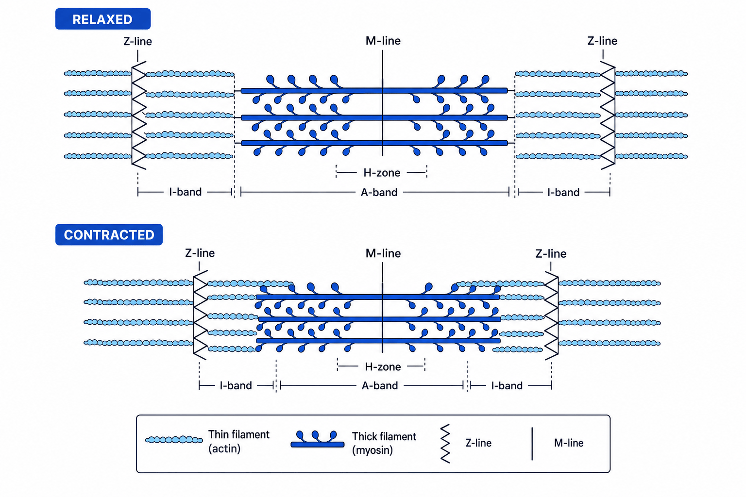
- Thin Filaments: Composed of the protein actin anchored to the Z-lines.
- Thick Filaments: Composed of the protein myosin anchored at the M-line.
- I-band: Contains only thin (actin) filaments. It appears light under a microscope.
- A-band: The entire length of the thick (myosin) filament. It appears dark.
- H-zone: The region in the centre of the A-band containing only thick (myosin) filaments.
The Sliding Filament Hypothesis
During muscle contraction, the actin and myosin filaments do not shorten. Instead, they slide past each other, pulling the Z-lines closer together.
- The I-band narrows.
- The H-zone narrows (and may disappear).
- The A-band remains exactly the same length.
Neuromuscular Junctions
A neuromuscular junction is a specialized synapse between a motor neurone and a muscle fibre:
- An action potential arrives at the presynaptic membrane of the motor neurone, opening voltage-gated Ca2+Ca^{2+}Ca2+ channels.
- Ca2+Ca^{2+}Ca2+ enters the synaptic knob, causing synaptic vesicles containing acetylcholine (ACh) to fuse with the presynaptic membrane.
- ACh is released by exocytosis, diffuses across the synaptic cleft, and binds to ligand-gated nicotinic receptors on the sarcolemma.
- This opens sodium ion channels, causing Na+Na^+Na+ influx, which depolarises the sarcolemma.
- This depolarisation generates an action potential that spreads rapidly down the T-tubules.
The Sliding Filament Mechanism
Once the action potential reaches the T-tubules, contraction is initiated:
- Calcium Release: The action potential in the T-tubules triggers the opening of voltage-gated Ca2+Ca^{2+}Ca2+ channels in the membrane of the sarcoplasmic reticulum. Ca2+Ca^{2+}Ca2+ diffuses rapidly down its concentration gradient out of the SR into the sarcoplasm.
- Exposing the Binding Site: Ca2+Ca^{2+}Ca2+ binds to troponin on the actin filament. This causes a conformational change in troponin, which pulls the attached tropomyosin filament out of the myosin-binding site on the actin.
- Cross-Bridge Formation: Myosin heads bind to the exposed binding sites on actin, forming actin-myosin cross-bridges.
- The Power Stroke: The myosin head releases ADPADPADP and inorganic phosphate (PiP_iPi). This causes the myosin head to pivot/bend, pulling the actin filament towards the centre of the sarcomere (M-line).
- Cross-Bridge Detachment: A new molecule of ATP binds to the myosin head, causing a conformational change that forces the myosin head to detach from the actin filament.
- Cocked Position: The enzyme ATPase (on the myosin head) hydrolyses the bound ATP into ADP and PiP_iPi. The energy released is used to return the myosin head to its original "cocked" (high-energy) position, ready to bind to another actin site further along.
- Relaxation: When stimulation stops, Ca2+Ca^{2+}Ca2+ is actively transported back into the sarcoplasmic reticulum using ATP-driven calcium pumps. Troponin returns to its original shape, and tropomyosin blocks the binding sites again.
Maintaining the ATP Supply in Muscles
Muscle contraction consumes massive quantities of ATP. Active muscle fibres maintain their ATP supply using three pathways:
- Aerobic Respiration: The primary source of ATP during moderate, long-term exercise. It takes place in mitochondria, requiring oxygen and respiratory substrates (like glucose and fatty acids).
- Anaerobic Respiration (Lactate Fermentation): Provides a rapid, short-term supply of ATP in the sarcoplasm when oxygen delivery is insufficient. It has a low yield of ATP per glucose molecule and produces lactate, which can lower cellular pHpHpH and lead to fatigue.
- The Creatine Phosphate System:
- Creatine phosphate is an organic compound stored in the sarcoplasm that acts as an immediate reserve of phosphate groups.
- It directly phosphorylates ADP to form ATP in a reversible reaction catalysed by the enzyme creatine kinase:
- This pathway requires no oxygen, is incredibly rapid, but can only sustain maximum contraction for approximately 10 seconds10\text{ seconds}10 seconds before creatine phosphate stores are depleted.
In the exam
- Explain the structural changes in the sarcomere during contraction: Ensure you state clearly that actin and myosin filaments do not shorten. Instead, describe how actin slides over myosin, causing the light I-band and H-zone to narrow, while the dark A-band remains constant.
- Be precise with hormonal vs. electrical pathways: When discussing the "fight-or-flight" response, make sure you distinguish between the rapid, short-term pathway (sympathetic nerves directly stimulating the adrenal medulla to secrete adrenaline) and the slower, longer-term endocrine pathway (hypothalamus releasing CRH, pituitary releasing ACTH, and adrenal cortex releasing cortisol).
- Master the second messenger steps: When explaining adrenaline's action, use the exact terminology: first messenger (adrenaline), G-protein, adenylyl cyclase, second messenger (cyclic AMP / cAMP), and enzyme cascade/protein kinase. Do not miss any of these sequential terms.
- Be comfortable with serial dilution calculations: Practice using the formula C1V1=C2V2C_1 V_1 = C_2 V_2C1V1=C2V2 for dilution calculations, and always state the units clearly in your working.
Check yourself
- How does the concentration of auxin affect the growth of apical shoots compared to lateral buds?
- Detail the exact step-by-step mechanism of how a rise in carbon dioxide concentration during exercise results in an increased heart rate.
- Describe the structural differences visible under a light microscope between skeletal muscle and involuntary (smooth) muscle.
- Explain the role of ATP in both the contraction and relaxation phases of muscle activity.
