What you'll learn
- How sensory receptors, such as the Pacinian corpuscle, act as transducers.
- How sensory, relay and motor neurones are adapted for their roles.
- How resting potentials, action potentials and saltatory conduction work.
- How cholinergic synapses transmit, excite, inhibit and summate signals.
The big picture
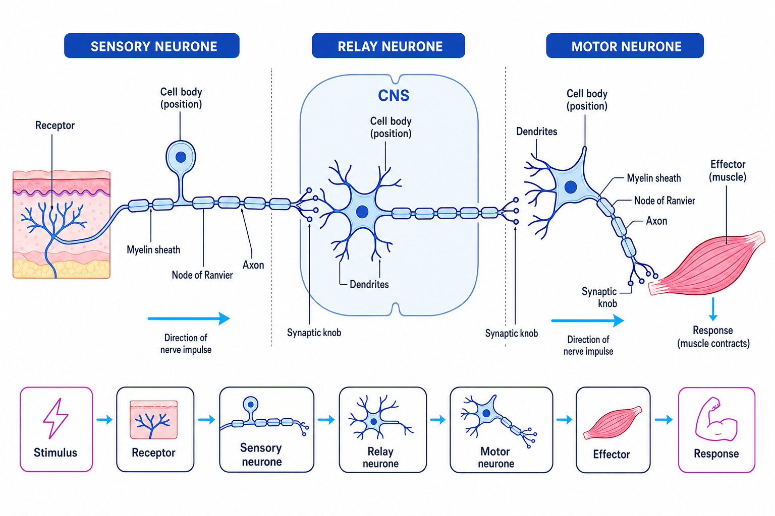
A stimulus is a detectable change in the internal or external environment. In mammals, many stimuli are detected by sensory receptors, passed along neurones as electrical signals, processed in the central nervous system, or CNS, and then sent to effectors, such as muscles or glands.
Pathway of communication
A typical response pathway is: stimulus → receptor → sensory neurone → CNS → relay neurone → motor neurone → effector → response.
Sensory receptors as transducers
Transducer
A transducer converts one form of energy into another. A sensory receptor transduces stimulus energy, such as pressure, light or chemicals, into an electrical signal in a neurone.
Different receptor types respond to different stimuli. For example, mechanoreceptors respond to mechanical pressure, chemoreceptors respond to chemicals, and photoreceptors respond to light.
The Pacinian corpuscle is a mammalian mechanoreceptor found in places such as the skin. It responds to pressure and vibration. When pressure deforms the corpuscle, stretch-mediated sodium ion channels in the sensory neurone membrane open. Sodium ions diffuse in, making the membrane less negative. This produces a generator potential, which is a local change in membrane potential caused by a stimulus.
If the generator potential reaches the threshold potential, it triggers an action potential.
Deciding whether a stimulus reaches threshold
A receptor has a resting potential of -70 mV and a threshold of -55 mV.
- A weak stimulus causing a 10 mV depolarisation changes the membrane potential to −70 mV+10 mV=−60 mV-70\ \text{mV} + 10\ \text{mV} = -60\ \text{mV}−70 mV+10 mV=−60 mV.
- Since −60 mV-60\ \text{mV}−60 mV is still below the threshold of −55 mV-55\ \text{mV}−55 mV, no action potential is generated.
- A stronger stimulus causing an 18 mV depolarisation changes the membrane potential to −70 mV+18 mV=−52 mV-70\ \text{mV} + 18\ \text{mV} = -52\ \text{mV}−70 mV+18 mV=−52 mV, which exceeds threshold, so action potentials are produced.
Types of neurone
A neurone is a specialised cell that transmits electrical impulses. Most neurones have dendrites, which receive impulses, an axon, which carries impulses away from the cell body, and synaptic knobs, which communicate with other cells.
Sensory neurones
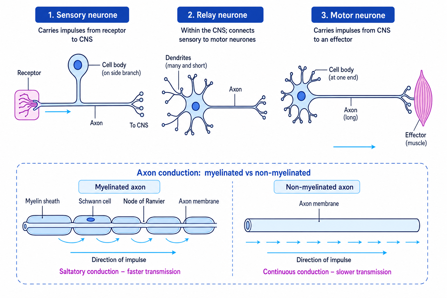
A sensory neurone carries impulses from a receptor to the CNS. Its cell body is usually on a side branch, and it has a long fibre carrying information towards the spinal cord or brain.
Relay neurones
A relay neurone is found inside the CNS. It connects sensory neurones to motor neurones and helps process information. Relay neurones often have many short dendrites so they can receive signals from several neurones.
Motor neurones
A motor neurone carries impulses from the CNS to an effector, such as a muscle or gland. Its cell body is in the CNS, and it has a long axon leading to the effector.
Myelinated and non-myelinated neurones
Myelin is a lipid-rich insulating layer around some axons. In mammals, it is produced by Schwann cells. The small gaps between myelin sheath sections are called nodes of Ranvier.
In a myelinated neurone, action potentials occur mainly at the nodes of Ranvier. This makes transmission faster because the impulse effectively jumps from node to node. This is called saltatory conduction.
In a non-myelinated neurone, the action potential is regenerated along the whole axon membrane, so transmission is slower.
Myelin does not carry the impulse
Myelin is an electrical insulator. The action potential is regenerated at the exposed membrane of the nodes of Ranvier, not through the myelin itself.
Resting potential
Resting potential
The resting potential is the voltage across a neurone membrane when it is not transmitting an impulse. In many mammalian neurones it is about -70 mV, meaning the inside is more negative than the outside.
A resting potential is established and maintained by ion movement:
- The sodium-potassium pump uses ATP to move 3 sodium ions out of the neurone and 2 potassium ions in.
- The membrane is more permeable to potassium ions than sodium ions at rest, so potassium ions diffuse out through leak channels.
- Large negatively charged proteins and other anions remain inside the neurone.
- The inside becomes negative relative to the outside.
Generating an action potential
Action potential
An action potential is a rapid, temporary reversal of membrane potential across a neurone membrane. It is the electrical event that forms a nerve impulse.
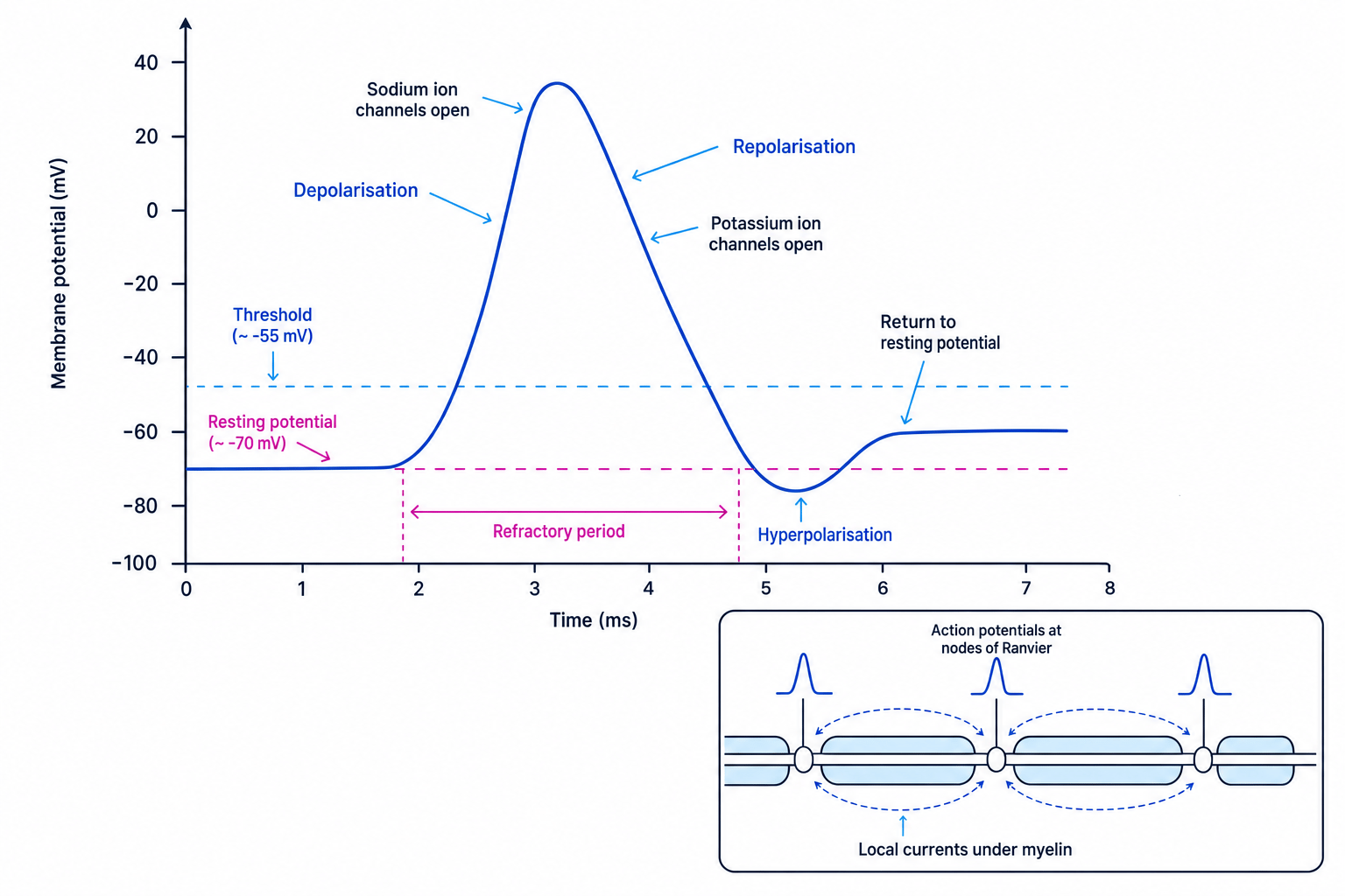
An action potential follows a set sequence:
- A stimulus causes depolarisation. Depolarisation means the membrane potential becomes less negative.
- If threshold is reached, voltage-gated sodium ion channels open. Sodium ions diffuse into the neurone.
- Sodium ion entry causes more depolarisation, opening more sodium ion channels. This is positive feedback.
- The inside becomes positive relative to the outside.
- Sodium ion channels inactivate, and voltage-gated potassium ion channels open.
- Potassium ions diffuse out, making the inside negative again. This is repolarisation.
- Potassium channels close slowly, so too many potassium ions may leave. This causes hyperpolarisation, where the membrane becomes more negative than the resting potential.
- The resting potential is restored.
The refractory period is the short time after an action potential when another action potential cannot be generated in that part of the membrane. This ensures impulses travel in one direction and limits the maximum frequency of impulses.
Stronger stimuli do not make bigger action potentials
Action potentials are all-or-nothing. Once threshold is reached, the amplitude is roughly the same. A stronger stimulus is represented by a higher frequency of action potentials, not larger action potentials.
Transmission along a myelinated neurone
When one region of the axon depolarises, local electrical currents depolarise the next region. In a myelinated axon, these local currents pass under the myelin sheath to the next node of Ranvier, where another action potential is generated.
This makes transmission fast and energy-efficient, because ions cross the membrane mainly at the nodes.
Calculating conduction velocity
A nerve impulse travels 0.80 m along a myelinated axon in 12 ms.
-
Convert milliseconds into seconds: 12 ms=12×10−3 s=0.012 s12\ \text{ms} = 12 \times 10^{-3}\ \text{s} = 0.012\ \text{s}12 ms=12×10−3 s=0.012 s.
-
Use the rate formula:
speed=distancetime=0.80 m0.012 s\begin{aligned} \text{speed} &= \frac{\text{distance}}{\text{time}} \\ &= \frac{0.80\ \text{m}}{0.012\ \text{s}} \end{aligned}speed=timedistance=0.012 s0.80 m -
Calculate the conduction velocity: speed=66.7 m s−1\text{speed} = 66.7\ \text{m s}^{-1}speed=66.7 m s−1, so to two significant figures this is 67 m s−167\ \text{m s}^{-1}67 m s−1.
Synapses and neurotransmission
Synapse
A synapse is a junction between two neurones, or between a neurone and an effector. The tiny gap between cells is the synaptic cleft.
A cholinergic synapse uses the neurotransmitter acetylcholine, often abbreviated to ACh. A neurotransmitter is a chemical messenger released by a neurone.
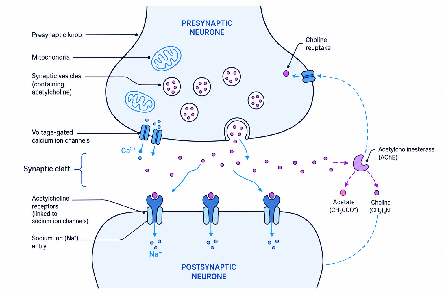
Sequence at a cholinergic synapse
- An action potential arrives at the presynaptic knob.
- Voltage-gated calcium ion channels open, so calcium ions diffuse into the presynaptic neurone.
- Calcium ions cause vesicles containing acetylcholine to fuse with the presynaptic membrane.
- Acetylcholine is released by exocytosis and diffuses across the synaptic cleft.
- Acetylcholine binds to receptors on the postsynaptic membrane.
- Sodium ion channels open, sodium ions enter, and the postsynaptic membrane depolarises.
- If threshold is reached, an action potential is generated in the postsynaptic neurone.
- Acetylcholinesterase breaks down acetylcholine into acetate and choline, preventing continuous stimulation.
Synapses make transmission one-way because neurotransmitter vesicles are on the presynaptic side, while receptors are on the postsynaptic side.
Excitation, inhibition and summation
An excitatory synapse makes an action potential more likely. It produces an excitatory postsynaptic potential, or EPSP, which is a small depolarisation.
An inhibitory synapse makes an action potential less likely. It produces an inhibitory postsynaptic potential, or IPSP, often by making the postsynaptic membrane more negative.
Summation is the adding together of EPSPs and IPSPs at a postsynaptic neurone. Temporal summation occurs when one presynaptic neurone releases neurotransmitter repeatedly in a short time. Spatial summation occurs when several presynaptic neurones act on the same postsynaptic neurone.
Predicting summation at a postsynaptic neurone
A postsynaptic neurone starts at -70 mV and has a threshold of -55 mV. It receives EPSPs of +6 mV and +5 mV, plus an IPSP of -4 mV.
- Add the effects on membrane potential: +6 mV+5 mV−4 mV=+7 mV+6\ \text{mV} + 5\ \text{mV} - 4\ \text{mV} = +7\ \text{mV}+6 mV+5 mV−4 mV=+7 mV.
- Apply this net change to the resting potential: −70 mV+7 mV=−63 mV-70\ \text{mV} + 7\ \text{mV} = -63\ \text{mV}−70 mV+7 mV=−63 mV.
- Compare with threshold: −63 mV-63\ \text{mV}−63 mV is below −55 mV-55\ \text{mV}−55 mV, so no action potential is generated.
Synapse wording shortcut
For a cholinergic synapse, keep the order clear: calcium ions enter → acetylcholine released → acetylcholine binds receptors → sodium ions enter → depolarisation.
In the exam
- Link each ion movement to the correct stage: sodium ions enter during depolarisation; potassium ions leave during repolarisation.
- When explaining stimulus intensity, write about frequency of impulses, not the size of each action potential.
- For synapse questions, include both release and removal of neurotransmitter; acetylcholinesterase prevents continuous stimulation.
Check yourself
- Why does a stronger stimulus cause more frequent action potentials rather than larger ones?
- How does myelination increase the speed of transmission in a mammalian neurone?
- What is the difference between temporal summation and spatial summation at a synapse?