

What you'll learn
- What excretion is, and why removing carbon dioxide and nitrogenous waste matters.
- How the liver and kidneys are structured for their excretory and homeostatic roles.
- How nephrons form urine by ultrafiltration and selective reabsorption.
- How ADH, dialysis, transplants and urine tests link excretion to health.
Why excretion matters
Living cells constantly carry out metabolism, meaning all the enzyme-controlled chemical reactions in the body. Some reactions produce useful products, but others produce waste substances that would become toxic if they accumulated.
Excretion
Excretion is the removal of toxic products of metabolism and substances in excess of requirements from the body. It helps maintain homeostasis, the maintenance of a stable internal environment.
The main excretory organs in this topic are:
- Lungs — excrete carbon dioxide from respiration. Carbon dioxide can form carbonic acid in solution, lowering pH and affecting enzyme activity.
- Liver — detoxifies some ingested toxins and converts toxic ammonia into less toxic urea.
- Kidneys — remove urea, excess ions and excess water in urine, helping control blood water potential.
Excretion is not egestion
Egestion is the removal of undigested food as faeces. Excretion is the removal of metabolic waste, such as carbon dioxide and urea.
The liver: structure, histology and functions
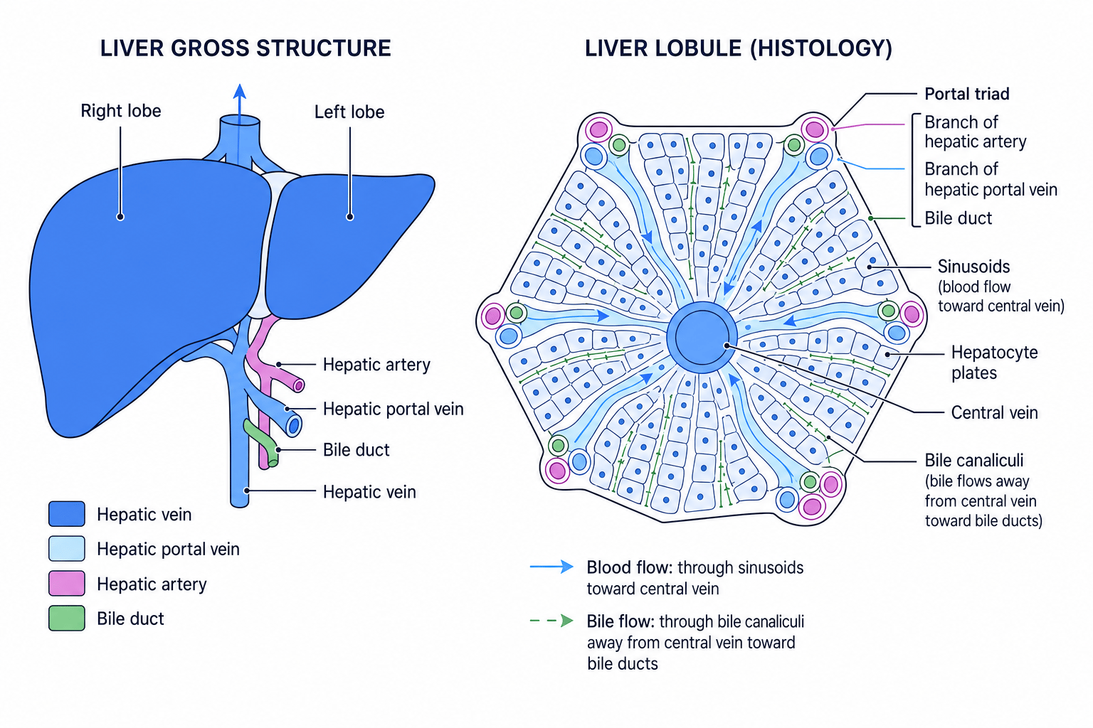
The mammalian liver is a large organ with right and left lobes. It receives oxygenated blood from the hepatic artery and nutrient-rich blood from the gut via the hepatic portal vein. Blood leaves through the hepatic vein. Bile leaves through the bile duct.
Liver histology
A stained liver section shows repeating units called lobules. In a lobule:
- Hepatocytes are liver cells arranged in plates.
- Sinusoids are blood-filled spaces between hepatocyte plates.
- Blood flows from branches of the hepatic artery and hepatic portal vein towards the central vein.
- Bile canaliculi carry bile in the opposite direction, towards bile ducts in the portal triads.
Liver functions you need here
The liver has many roles, but for this specification focus on three:
- Storage of glycogen — hepatocytes convert glucose into glycogen for storage, then break it down when blood glucose falls.
- Detoxification — hepatocytes metabolise toxins such as alcohol and some drugs, making them less harmful.
- Formation of urea — excess amino acids are deaminated, producing ammonia. Ammonia is very toxic, so the liver converts ammonia and carbon dioxide into urea as part of the ornithine cycle.
You do not need the detailed steps of the ornithine cycle; know its purpose and where it happens.
Where urea is made
The liver makes urea. The kidneys remove urea from the blood and excrete it in urine.
Drawing liver tissue
For a biological drawing of stained liver tissue, draw clear outlines only: central vein, hepatocyte plates, sinusoids and portal triads. Avoid sketchy shading; use labels and a title or magnification if given.
The kidney: gross structure and nephron structure
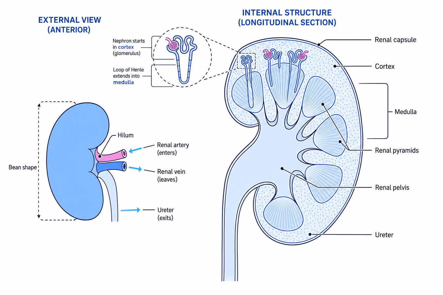
Each kidney has a tough renal capsule, an outer cortex, an inner medulla, several renal pyramids, a central renal pelvis, and a ureter that carries urine to the bladder. Blood enters through the renal artery and leaves through the renal vein.
Nephron
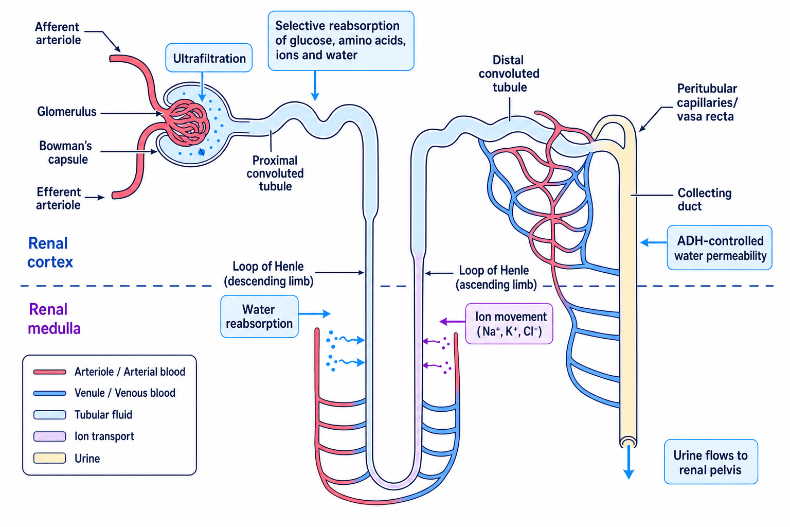
A nephron is the functional unit of the kidney: a microscopic tubule and associated blood vessels that filter blood and modify the filtrate to produce urine.
A nephron includes the glomerulus, Bowman’s capsule, proximal convoluted tubule, loop of Henle, distal convoluted tubule and collecting duct. The glomerulus is supplied by an afferent arteriole and drained by an efferent arteriole, which leads to capillaries around the tubule.
In stained kidney sections, the cortex contains many renal corpuscles: glomeruli inside Bowman’s capsules. The medulla contains loops of Henle and collecting ducts.
How urine is formed
Ultrafiltration
Ultrafiltration happens in the renal corpuscle: the glomerulus and Bowman’s capsule. Blood in the glomerulus is under high hydrostatic pressure, partly because the efferent arteriole is narrower than the afferent arteriole.
Small molecules are forced out of the blood into Bowman’s capsule, including water, glucose, amino acids, ions and urea. Blood cells and large plasma proteins remain in the blood because they are too large to pass through the filtration barrier.
Glomerular filtration rate
Glomerular filtration rate, or GFR, is the volume of filtrate formed by the kidneys per unit time. A reduced GFR means less filtration, so wastes such as urea may accumulate.
Calculating glomerular filtration rate
-
Use the rate relationship:
GFR=volume of filtrate formedtime\text{GFR}=\frac{\text{volume of filtrate formed}}{\text{time}}GFR=timevolume of filtrate formed -
If 0.75 dm³ of filtrate is formed in 6.0 min, substitute with units:
GFR=0.75 dm36.0 min\text{GFR}=\frac{0.75\,\text{dm}^{3}}{6.0\,\text{min}}GFR=6.0min0.75dm3 -
Calculate and state the result:
GFR=0.125 dm3 min−1\text{GFR}=0.125\,\text{dm}^{3}\,\text{min}^{-1}GFR=0.125dm3min−1
Selective reabsorption
Selective reabsorption is the movement of useful substances from the filtrate back into the blood. It mainly happens in the proximal convoluted tubule.
The proximal convoluted tubule is adapted with many mitochondria for active transport and microvilli for a large surface area. Sodium ions are actively transported out of tubule cells, helping glucose and amino acids enter by co-transport. Water then follows by osmosis.
The loop of Henle helps create a low water potential in the medulla. The descending limb loses water, while the ascending limb moves ions out and is relatively impermeable to water. This gradient is important for water reabsorption from collecting ducts.
Urine is modified filtrate
Filtrate begins as blood plasma without cells or large proteins. Urine is what remains after useful substances and variable amounts of water have been reabsorbed.
Following molecules through the nephron
-
Compare molecule size at ultrafiltration: glucose and urea are small enough to enter Bowman’s capsule, but plasma proteins are too large and remain in the blood.
-
Apply selective reabsorption: glucose is normally reabsorbed from the proximal convoluted tubule back into the blood, while urea is not fully reabsorbed.
-
Predict final urine composition in a healthy person: urea should be present, but glucose and plasma proteins should usually be absent.
Control of blood water potential
Water potential and ADH
Water potential describes the tendency of water to move by osmosis. ADH, or antidiuretic hormone, is a hormone that increases water reabsorption by making collecting duct walls more permeable to water.
The control system works by negative feedback:
- Osmoreceptors in the hypothalamus detect changes in blood water potential.
- The posterior pituitary gland releases more or less ADH into the blood.
- ADH acts on the collecting ducts of nephrons.
- Water reabsorption changes, helping return blood water potential towards normal.
If blood water potential is too low, such as after sweating, more ADH is released. More water is reabsorbed, producing a smaller volume of more concentrated urine. If blood water potential is too high, less ADH is released, so less water is reabsorbed and a larger volume of dilute urine is produced.
Predicting the ADH response after dehydration
-
Dehydration removes water from the blood, so blood water potential falls.
-
Osmoreceptors in the hypothalamus detect this change, causing increased ADH release from the posterior pituitary gland.
-
ADH makes collecting ducts more permeable to water, so more water enters the blood by osmosis and urine becomes more concentrated.
Kidney failure and treatments
In kidney failure, nephrons cannot maintain normal filtration and reabsorption. Problems include:
- reduced GFR, causing accumulation of urea and other wastes
- disturbed electrolyte balance, especially ions such as potassium and sodium
- poor control of blood water potential, causing fluid imbalance and high blood pressure risks
Haemodialysis
In haemodialysis, blood is passed through a dialysis machine, separated from dialysis fluid by a partially permeable membrane. Urea diffuses from the blood into the dialysis fluid. The dialysis fluid has carefully controlled concentrations of glucose and ions so that useful substances are not lost in large amounts.
Setting dialysis-fluid concentrations
-
To remove urea, the dialysis fluid should contain no urea, creating a diffusion gradient from blood to fluid.
-
To avoid losing glucose, the dialysis fluid should contain a normal blood concentration of glucose, so there is little or no net diffusion.
-
To correct excess ions, ion concentrations in dialysis fluid are controlled so excess ions diffuse out while essential ions are maintained.
Dialysis fluid is not pure water
Pure water would cause useful ions and glucose to diffuse out of the blood too rapidly and could cause dangerous osmotic effects.
Kidney transplants
A kidney transplant can restore kidney function more continuously than dialysis. However, it requires a suitable donor, tissue matching, surgery, and immunosuppressant drugs to reduce rejection risk. Transplants can greatly improve quality of life, but donor organs are limited.
Excretory products in medical diagnosis
Urine is useful for diagnosis because it is easy to collect and contains excreted substances or metabolites from the blood.
Monoclonal antibodies
Monoclonal antibodies are identical antibodies produced from one clone of cells. They bind specifically to one antigen, so they can be used in diagnostic tests.
Pregnancy tests detect hCG, a hormone present in urine during pregnancy. Monoclonal antibodies bind hCG and produce a visible line on a test strip.
Urine tests can also detect anabolic steroids and drugs, often by detecting the drug itself or metabolites produced when the body breaks it down. Results must be interpreted carefully because concentration depends on timing, hydration and test sensitivity.
In the exam
-
Link each waste to its source and organ: carbon dioxide from respiration via lungs, urea made in liver and removed by kidneys.
-
For nephron questions, separate ultrafiltration from selective reabsorption; they happen in different places and use different mechanisms.
-
For ADH questions, always include the stimulus, osmoreceptors, posterior pituitary gland, collecting duct permeability and urine concentration.
Check yourself
- Why are plasma proteins not normally found in Bowman’s capsule filtrate?
- How does ADH change the volume and concentration of urine?
- Why must dialysis fluid contain controlled concentrations of glucose and ions?
