What you'll learn
- How the fluid-mosaic model explains membrane structure and selective permeability.
- How simple diffusion, facilitated diffusion, osmosis, active transport and co-transport differ.
- How gradients, surface area and membrane proteins affect the rate of transport.
- How to approach the two required practicals on water potential and membrane permeability.
Why membranes matter
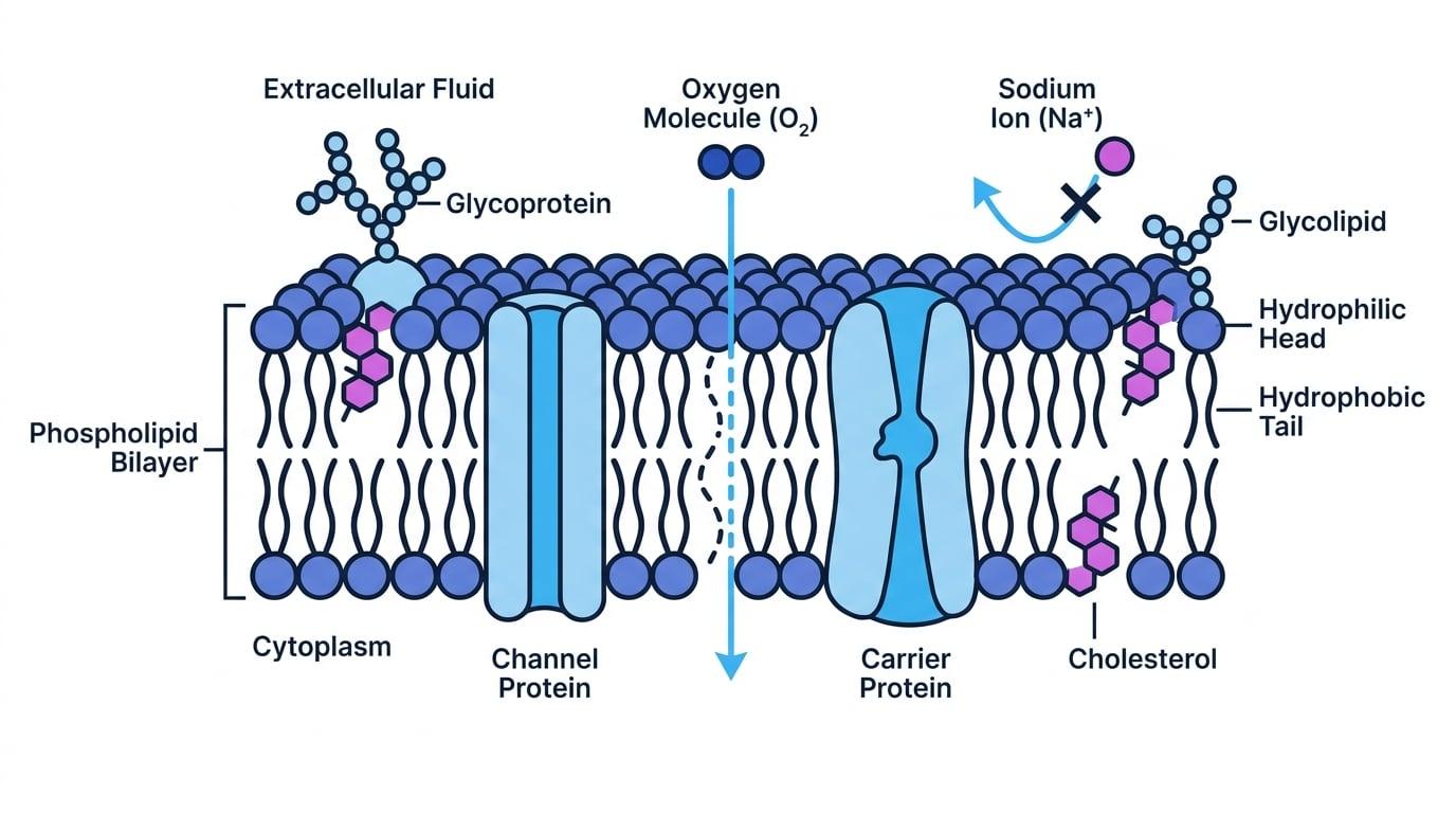
A cell-surface membrane surrounds the whole cell. Eukaryotic cells also have membranes around organelles, such as mitochondria, chloroplasts and the nucleus. The basic structure of all these membranes is the same: a phospholipid bilayer with proteins and other molecules embedded in it.
A membrane is partially permeable: it lets some substances cross more easily than others. Whether a substance crosses depends on its size, charge, lipid solubility, and whether the membrane contains suitable transport proteins.
The fluid-mosaic model
A phospholipid has a hydrophilic phosphate head, meaning it is attracted to water, and two hydrophobic fatty acid tails, meaning they are repelled by water. In water, phospholipids form a bilayer: heads face the watery cytoplasm or extracellular fluid, while tails point inwards away from water.
The fluid-mosaic model describes the membrane as fluid because phospholipids and many proteins can move sideways, and mosaic because different proteins, glycoproteins and glycolipids are scattered through it.
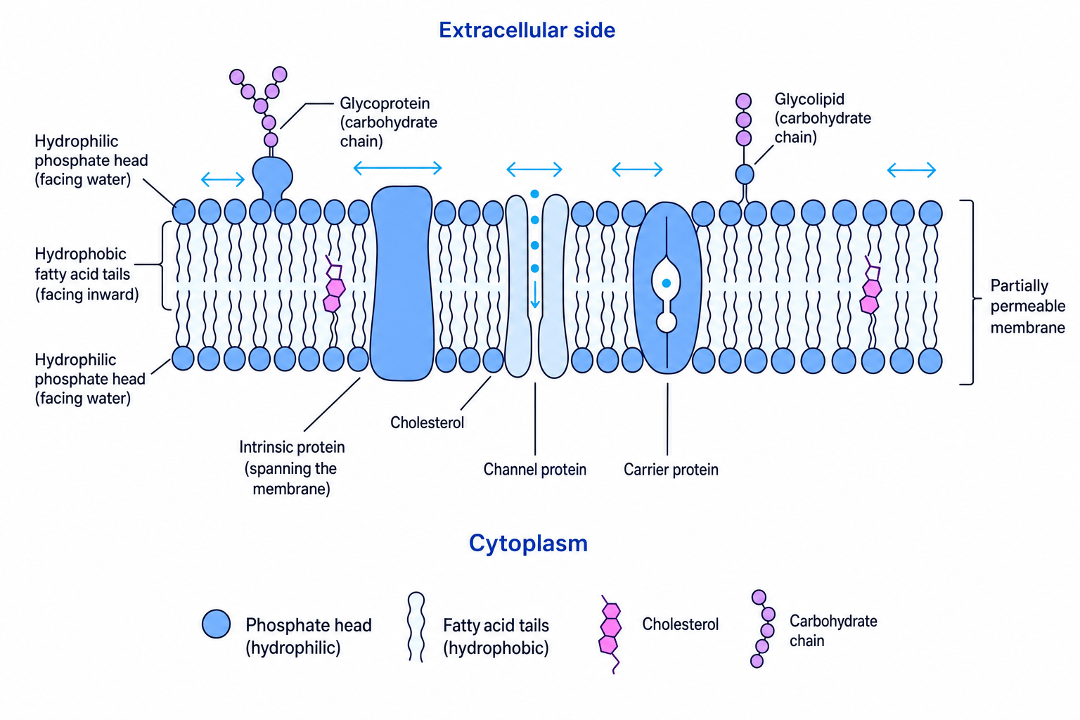
Proteins have different roles. Channel proteins form pores for specific ions or polar substances. Carrier proteins bind to a molecule and change shape to move it across. Glycoproteins and glycolipids have carbohydrate chains, often involved in cell recognition and receptors. Cholesterol may be present between phospholipids, where it restricts movement of membrane molecules and helps stabilise the membrane.
Fluid mosaic
Membrane function depends on both the phospholipid bilayer and the specific proteins present. Two membranes may have the same basic structure but very different transport abilities.
Concentration gradients and passive movement
A concentration gradient is a difference in concentration between two regions. If particles move from a higher concentration to a lower concentration, they move down the gradient. This does not require ATP, so it is called passive transport.
Simple diffusion
Simple diffusion is the net movement of particles from a higher concentration to a lower concentration, due to their random movement.
Small, non-polar molecules such as oxygen and carbon dioxide can diffuse directly through the phospholipid bilayer. Charged ions and most large polar molecules cannot easily pass through the hydrophobic centre of the bilayer.
Facilitated diffusion
Facilitated diffusion is passive movement down a concentration gradient through membrane proteins. It does not use ATP.
Channel proteins allow specific ions or polar molecules to pass through a hydrophilic pore. Carrier proteins bind to a specific molecule, change shape, then release it on the other side.
Explaining a facilitated diffusion plateau
-
At low external glucose concentration, increasing the concentration makes the gradient steeper, so more glucose molecules bind to carrier proteins per second and the rate increases.
-
At high external glucose concentration, all carrier proteins are occupied or changing shape, so the rate reaches a maximum.
-
Adding more carrier proteins would increase the maximum rate, but simply increasing glucose concentration further would not help much.
Protein does not always mean active
Facilitated diffusion uses proteins, but it is still passive because particles move down their concentration gradient and ATP is not hydrolysed.
Osmosis and water potential
Osmosis is the net movement of water molecules across a partially permeable membrane from a region of higher water potential to a region of lower water potential.
Water potential
Water potential is a measure of the tendency of water molecules to move. It is represented by Ψ\PsiΨ and measured in kilopascals, kPa. Pure water has Ψ=0 kPa\Psi = 0\ \text{kPa}Ψ=0 kPa; adding solute makes water potential more negative.
Water moves from less negative to more negative water potential. For example, water would move from -200 kPa to -700 kPa if a partially permeable membrane separated the two solutions.
More negative means lower
0 kPa is higher than -500 kPa. In osmosis questions, compare the values carefully before deciding the direction of water movement.
In plant cells, water entering by osmosis increases turgor pressure and makes the cell turgid. Water leaving can cause the cytoplasm and cell-surface membrane to pull away from the cell wall; this is plasmolysis.
Required practical 3: finding water potential of plant tissue
A dilution series is a set of solutions made by diluting a solute, such as sucrose, to different concentrations. A calibration curve is a graph made from known values that is then used to estimate an unknown value.
For plant tissue, you place equal-sized samples, such as potato cylinders, into solutions with known water potentials. Measure initial mass, leave for the same time, blot dry in the same way, then measure final mass.
Use:
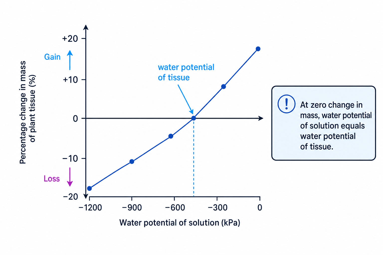
percentage change in mass=final mass−initial massinitial mass×100\text{percentage change in mass}=\frac{\text{final mass}-\text{initial mass}}{\text{initial mass}}\times 100percentage change in mass=initial massfinal mass−initial mass×100Plot mean percentage change in mass against water potential of the solution. Where the curve crosses 0% change in mass, there is no net osmosis, so the solution water potential equals the tissue water potential.
Finding water potential from an intercept
A graph has two points either side of 0% change: -700 kPa gives -4.0%, and -500 kPa gives +2.0%.
-
The change from -4.0% to +2.0% is 6.0 percentage points, so 0% is 4.0 percentage points above the lower point.
-
Interpolate between the two water potentials:
- Add this to the lower water potential:
So the tissue water potential is about -570 kPa.
Active transport
Active transport is the movement of substances across a membrane against their concentration gradient, from lower concentration to higher concentration. It requires carrier proteins and energy from ATP.
Hydrolysis means breaking a chemical bond using water. ATP hydrolysis releases energy:
ATP+H2O→ADP+Pi\text{ATP}+\text{H}_2\text{O}\to \text{ADP}+\text{P}_{\text{i}}ATP+H2O→ADP+Pi
The energy allows a carrier protein to change shape and move the substance across the membrane. Root hair cells use active transport to take up mineral ions from soil when the ion concentration is already higher inside the cell.
Identifying active transport
A root hair cell absorbs nitrate ions from soil where the nitrate ion concentration is lower than inside the cell.
-
The ions are moving from a lower concentration in the soil to a higher concentration inside the cell, so they are moving against the concentration gradient.
-
Nitrate ions are charged, so they cannot simply pass through the hydrophobic part of the phospholipid bilayer.
-
Movement therefore needs a carrier protein and ATP hydrolysis, so the process is active transport.
Co-transport in the ileum
Co-transport is when the movement of one substance down its concentration gradient helps move another substance across the same membrane. In the mammalian ileum, this is how glucose is absorbed after digestion.
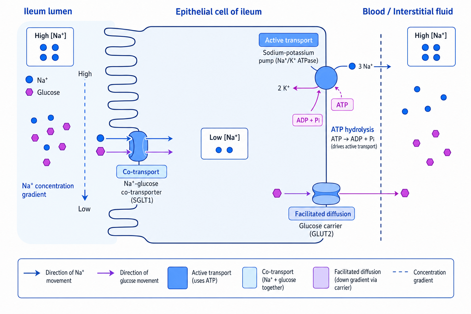
The ileum has epithelial cells lining its lumen, which is the space inside the gut.
- The sodium-potassium pump in the membrane facing the blood actively transports Na⁺ out of the epithelial cell, using ATP.
- This keeps the Na⁺ concentration low inside the epithelial cell.
- Na⁺ then moves from the ileum lumen into the epithelial cell down its concentration gradient through a sodium-glucose co-transporter.
- Glucose is carried into the cell at the same time, even if glucose is moving against its own concentration gradient.
- Glucose then leaves the epithelial cell into the blood by facilitated diffusion through a carrier protein.
Co-transport depends on ATP indirectly
The sodium-glucose co-transporter itself does not hydrolyse ATP, but it relies on the Na⁺ gradient created by ATP-driven sodium-potassium pumps.
What affects the rate of transport?
The rate of movement across membranes depends on:
- Surface area: larger membrane surface area allows more particles to cross per second.
- Number of channel or carrier proteins: more transport proteins increase the maximum rate of facilitated diffusion or active transport.
- Steepness of the gradient: a larger concentration gradient, or larger difference in water potential, increases the rate.
- ATP supply: active transport and the pumps maintaining co-transport gradients need ATP, so many mitochondria can help.
Specialised cells often show these adaptations. Ileum epithelial cells have microvilli to increase surface area, many co-transport proteins, and many mitochondria. Root hair cells have long extensions for large surface area and carrier proteins for mineral ion uptake. Internal membranes can also be adapted: folded membranes provide more area for embedded proteins.
Required practical 4: membrane permeability
A common investigation uses beetroot cells because their red pigment leaks out when membranes become more permeable. A named variable could be temperature, ethanol concentration or pH.
For temperature:
- Cut equal-sized beetroot pieces and rinse them to remove pigment from damaged cells.
- Place pieces in tubes at different temperatures for the same time.
- Keep tissue size, volume of water, time, beetroot source and rinsing method the same.
- Use a colorimeter to measure absorbance of the surrounding solution. Higher absorbance means more pigment has leaked out, so membrane permeability is higher.
Interpreting beetroot permeability data
At 20 °C the absorbance is 0.08. At 60 °C the absorbance is 0.64.
-
Higher absorbance at 60 °C means more pigment has diffused out of the beetroot cells.
-
The relative increase is:
0.64÷0.08=8.00.64 \div 0.08 = 8.00.64÷0.08=8.0
So the solution is 8 times more absorbent at 60 °C.
- This suggests the cell-surface membranes are more permeable at 60 °C, because high temperature increases phospholipid movement and can denature membrane proteins.
In the exam
-
Name the transport process, then justify it using three clues: direction of gradient, whether a protein is involved, and whether ATP is needed.
-
For osmosis, always compare water potentials numerically: water moves from higher Ψ\PsiΨ to lower Ψ\PsiΨ, and solutes make Ψ\PsiΨ more negative.
-
In practical questions, mention repeats, control variables, an appropriate graph, and the intercept or trend used to support your conclusion.
Check yourself
- Why can oxygen cross the phospholipid bilayer by simple diffusion, but Na⁺ needs a protein?
- A plant tissue gains mass in -300 kPa solution but loses mass in -700 kPa solution. What does this tell you about its water potential?
- How does the sodium-potassium pump allow glucose to be absorbed from the ileum?