

What you'll learn:
- How cells recognise "self" vs "non-self" using antigens.
- The difference between non-specific (phagocytosis) and specific (cellular and humoral) immune responses.
- How vaccines and monoclonal antibodies work, and their ethical implications.
- The structure of HIV and how it leads to AIDS.
Identifying cells: The role of antigens
Every cell has specific molecules on its surface. These are primarily proteins, and because proteins have highly specific 3D tertiary structures, they allow the immune system to identify:
- Pathogens (disease-causing organisms like bacteria or viruses).
- Cells from other organisms of the same species (e.g. transplanted organs).
- Abnormal body cells (e.g. cancer cells).
- Toxins (poisons released by some bacteria, like cholera).
Antigen
An antigen is a foreign protein that stimulates an immune response.
Pathogens can undergo mutations that change the tertiary structure of their surface proteins. This is called antigen variability. When the antigen changes, your memory cells no longer recognise it, so you can catch the same disease again (e.g. influenza). This variability is also what makes developing a permanent vaccine for some viruses so difficult.
Non-specific defence: Phagocytosis
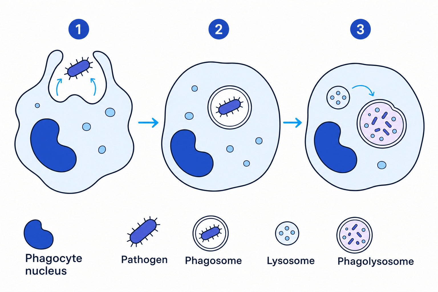
The immune response starts immediately with a non-specific mechanism: phagocytosis. Phagocytes (a type of white blood cell) engulf pathogens.
- The phagocyte is attracted to the pathogen by chemical products (chemoattractants).
- The phagocyte's receptors bind to the pathogen's surface antigens.
- The phagocyte changes shape to engulf the pathogen, enclosing it in a vesicle called a phagosome.
- Lysosomes migrate towards the phagosome and fuse with it.
- The lysosomes release lysozymes (digestive enzymes) into the phagosome, which hydrolyse and destroy the pathogen.
- The soluble products of the breakdown are absorbed into the phagocyte's cytoplasm.
The Specific Immune Response
If a pathogen survives phagocytosis, the specific immune response takes over. It involves white blood cells called lymphocytes. This response takes longer to ramp up, but it is highly targeted and produces long-term immunity. There are two main branches: the cellular response (T cells) and the humoral response (B cells).
The Cellular Response (T Lymphocytes)
T lymphocytes (T cells) only respond to antigens that are physically presented on the surface of a body cell (rather than floating free in the blood). Cells that display foreign antigens on their surface are called antigen-presenting cells (APCs). For example, once a phagocyte has broken down a pathogen, it presents the pathogen's antigens on its own cell-surface membrane.
- Receptors on a specific Helper T cell (THT_HTH cell) fit exactly onto these presented antigens.
- This attachment activates the THT_HTH cell to divide rapidly by mitosis, forming clones.
- These cloned THT_HTH cells do several jobs:
- They develop into memory cells for a rapid response in the future.
- They stimulate phagocytes to engulf more pathogens.
- They stimulate B cells to divide and secrete their antibodies.
- They activate Cytotoxic T cells (TCT_CTC cells), which destroy infected body cells by making holes in their cell-surface membrane.
The Humoral Response (B Lymphocytes)
The humoral response deals with antigens circulating freely in the blood and tissue fluid, relying on B cells.
- A B cell takes up a foreign circulating antigen, processes it, and presents it on its own surface.
- A Helper T cell (THT_HTH) binds to these processed antigens, activating the B cell.
- The activated B cell divides rapidly by mitosis to give clones of plasma cells. This process is known as clonal selection.
- The plasma cells secrete highly specific monoclonal antibodies into the blood. This initial production of antibodies is the primary immune response.
- Some B cells develop into memory cells. If the exact same antigen is encountered again months or years later, memory cells divide rapidly into plasma cells to produce a vast number of antibodies very quickly (the secondary immune response).
Primary vs Secondary
The secondary immune response is much faster and produces a far higher concentration of antibodies than the primary response. It often destroys the pathogen before you even show any symptoms.
Antibodies
Antibody
An antibody is a protein produced by B cells (specifically plasma cells) that is complementary to a specific antigen.
Antibodies are Y-shaped proteins made of four polypeptide chains: two heavy chains and two light chains. The specific antigen-binding site at the top forms the variable region. Because the variable region has a highly specific 3D tertiary structure, it will only bind to one complementary antigen, forming an antigen-antibody complex. The rest of the molecule is known as the constant region.
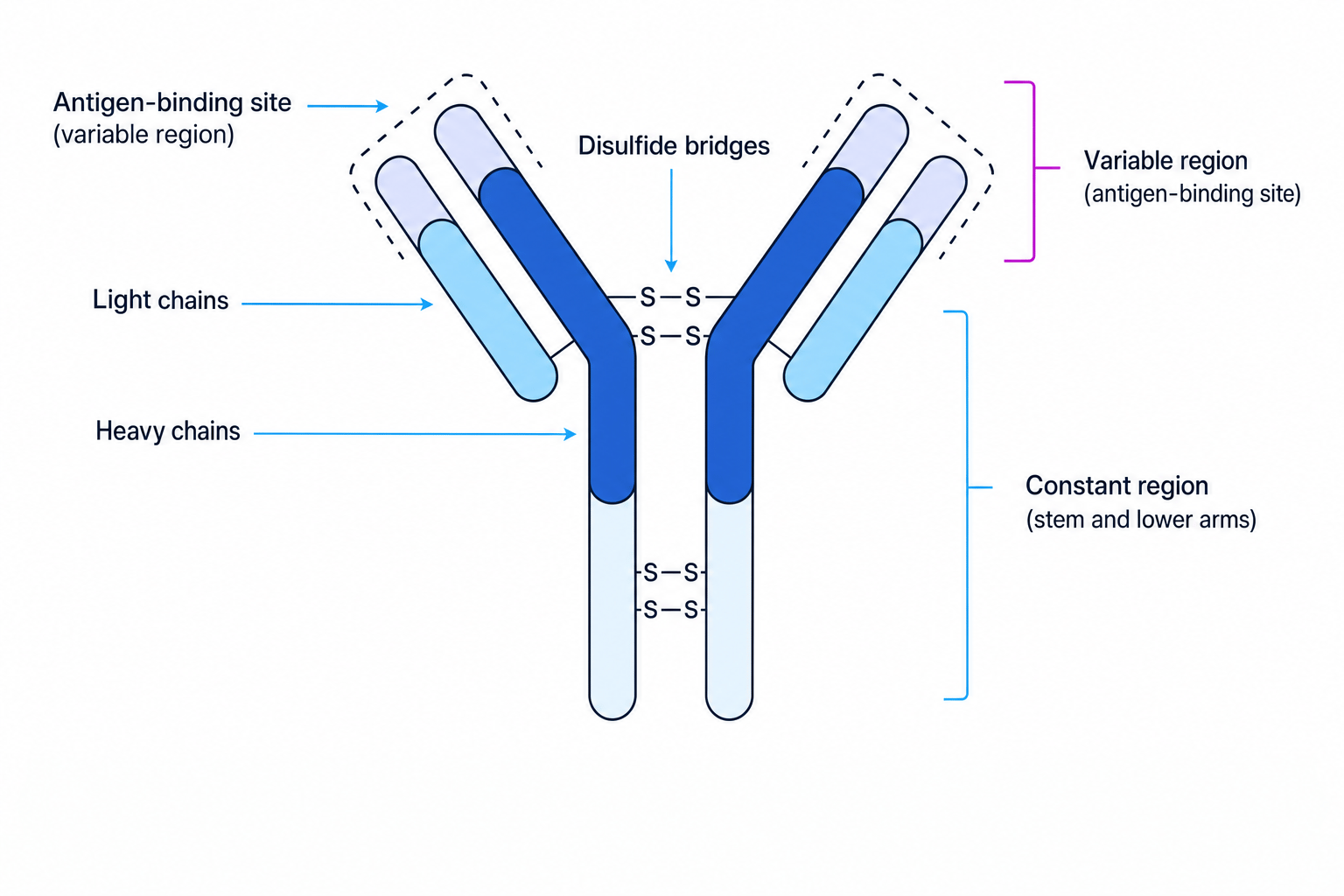
Antibodies do not destroy pathogens directly; they prepare them for destruction. Because each antibody has two binding sites, a single antibody can bind to two pathogens at once, clumping them together. This clumping is called agglutination. Agglutination makes it much easier for phagocytes to locate and engulf multiple bacterial cells at once.
Immunity and Vaccines
- Passive immunity is acquired by receiving antibodies from an outside source (e.g. antivenom given for a snakebite, or antibodies passing across the placenta). It provides immediate protection, but because no memory cells are formed, it does not offer long-term immunity.
- Active immunity is created when your own immune system produces antibodies and memory cells following exposure to an antigen. It takes longer to develop but is long-lasting. It can be natural (catching a disease) or artificial (vaccination).
Vaccines and Herd Immunity
A vaccine contains antigens (often isolated from a dead or weakened pathogen) that stimulate the production of memory cells against a particular disease without causing the disease itself.
Herd Immunity
Herd immunity arises when a sufficiently large proportion of a population has been vaccinated. This makes it highly unlikely that a susceptible individual will come into contact with an infected person, indirectly protecting those who cannot be vaccinated (e.g. due to medical conditions or compromised immune systems).
HIV and AIDS
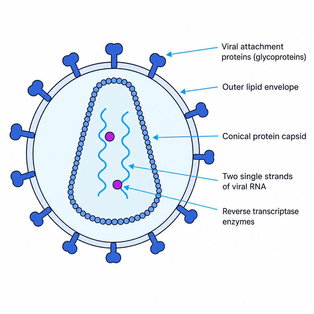
The Human Immunodeficiency Virus (HIV) is a retrovirus that ultimately causes Acquired Immune Deficiency Syndrome (AIDS).
HIV Replication
Like all viruses, HIV cannot replicate on its own; it must hijack a host cell. Crucially, HIV specifically targets and infects Helper T cells (THT_HTH cells).
- The attachment proteins on the surface of HIV bind to a receptor on the THT_HTH cell.
- The viral capsid fuses with the cell membrane, allowing HIV's RNA and enzymes to enter the THT_HTH cell.
- The viral enzyme reverse transcriptase copies the single-stranded viral RNA into double-stranded DNA.
- The viral DNA is inserted into the host cell's chromosomes.
- The host cell's machinery is hijacked to create viral proteins and new viral RNA.
- New HIV particles are assembled and bud off from the THT_HTH cell, destroying it in the process.
How HIV causes AIDS
As HIV continuously replicates, the number of THT_HTH cells in the blood drops drastically. Without enough THT_HTH cells, the body cannot stimulate B cells to produce antibodies or activate cytotoxic T cells to kill infected cells. At this point, the patient has developed AIDS and becomes highly susceptible to opportunistic infections (like pneumonia or tuberculosis), which are ultimately what cause death.
Antibiotics and viruses
Antibiotics target specific bacterial structures (like murein cell walls) or bacterial metabolic pathways (like bacterial ribosomes). Viruses do not have cell walls or their own metabolic machinery; they hide inside your own host cells. Therefore, antibiotics are completely ineffective against viruses.
Monoclonal Antibodies
Monoclonal antibodies are artificially produced antibodies isolated from a single clone of B cells. They are identical in structure and bind to one specific antigen.
Their powerful specificity has two major medical uses:
- Targeted medication: By attaching a therapeutic drug to a monoclonal antibody that is complementary to a cancer cell's antigens, the drug is delivered directly to the tumour. This destroys the cancer cells while minimising toxic side effects on healthy cells.
- Medical diagnosis: They can be used to detect the presence of specific proteins or pathogens, such as in pregnancy tests (detecting the hormone hCG) or tests for HIV.
The ELISA Test
The Enzyme-Linked Immunosorbent Assay (ELISA) uses antibodies to detect both the presence and the quantity of a protein in a sample. A standard setup (the indirect ELISA) works like this:
- Apply the test sample to a surface (like a plastic well) so any antigens present will attach.
- Wash the surface to remove unattached antigens.
- Add a primary antibody that is specific to the antigen we are testing for. Leave them to bind, then wash away any unattached antibodies.
- Add a second antibody, which has an enzyme attached to it. This second antibody binds to the first antibody. Wash the surface again.
- Add the colourless substrate of the enzyme. If the enzyme is present, it will act on the substrate to produce a coloured product.
- The intensity of the colour is proportional to the amount of antigen present in the original sample.
Why do we wash the well?
In an ELISA test, the washing steps are crucial. If unattached antibodies are not washed away, the enzyme will remain in the well even if the antigen was completely absent from the sample. This would lead to the enzyme acting on the substrate anyway, giving a false-positive result.
Ethical Issues
You need to be able to discuss the ethics surrounding the use of these technologies:
- Vaccines: Testing often involves animals, which some consider unethical. Vaccines can also have rare side effects. When facing an epidemic, there is a complex debate about whether vaccines should be made compulsory to achieve herd immunity, balancing public health against individual freedom.
- Monoclonal antibodies: Producing them historically involved deliberately inducing cancer in mice. Furthermore, there have been instances where human trials of new monoclonal antibodies have caused severe, life-threatening side effects (such as multiple organ failure) in volunteers.
In the exam
- When describing the specific immune response, be precise with cell names: always state "Helper T cells" (THT_HTH) and "Cytotoxic T cells" (TCT_CTC) rather than just "T cells".
- Remember that antibodies do not directly destroy pathogens. They bind to antigens to form an antigen-antibody complex, causing agglutination, which makes it easier for phagocytes to destroy them.
- If a past-paper question asks why a vaccine failed or a person got sick twice, look for the keyword "antigen variability" or "a mutation changing the tertiary structure of the antigen".
- When explaining why antibiotics don't work on viruses, explicitly state that viruses lack the target sites (like a murein cell wall) and rely entirely on the host cell's internal machinery.
Check yourself
- What is an antigen, and how does antigen variability affect disease prevention?
- Describe the process of phagocytosis and the specific role of lysozymes.
- How do Helper T cells coordinate the specific immune response?
- Describe the structure of an antibody and explain how it leads to agglutination.
- Why are the washing steps so vital in ensuring an accurate ELISA test result?
