

Welcome to the topic of cell division! Within multicellular organisms, not all cells keep the ability to divide (for example, human nerve cells and muscle cells generally do not). However, the cells that can divide are responsible for growth, tissue repair, and reproduction.
What you'll learn:
- How eukaryotic cells progress through the cell cycle and the specific stages of mitosis.
- How to prepare a root tip squash and calculate actual cell sizes and mitotic indices.
- How uncontrolled cell division leads to cancer.
- The differences between eukaryotic mitosis, prokaryotic binary fission, and viral replication.
The Eukaryotic Cell Cycle
Eukaryotic cells that retain the ability to divide undergo a continuous sequence of events known as the cell cycle.
The Cell Cycle
The regular pattern of growth and cell division that occurs in eukaryotic cells. It consists of three main stages: interphase, nuclear division (mitosis or meiosis), and cytokinesis.
The longest part of the cell cycle is interphase. During this time, the cell is incredibly active. It grows, produces new organelles, synthesizes proteins, and—most importantly—replicates its DNA. By the end of interphase, the cell has two identical copies of its genetic material, ready to be split into two new cells.
DNA replication and mitosis
A frequent exam error is stating that DNA replicates during mitosis. It does not! DNA replication strictly occurs during interphase, before mitosis even begins. Mitosis is simply the process of separating the DNA that has already been copied.
Mitosis and its Stages
Mitosis is the part of the cell cycle where a eukaryotic parent cell divides to produce two genetically identical daughter cells. Before we look at the stages, we need to clarify some terminology about how DNA is packaged.
Chromatids and Centromeres
- Chromatid: One of the two identical strands of DNA that make up a replicated chromosome. Because there are two identical copies joined together, they are called sister chromatids.
- Centromere: The central region of a chromosome where the two sister chromatids are held together, and where spindle fibres attach during division.
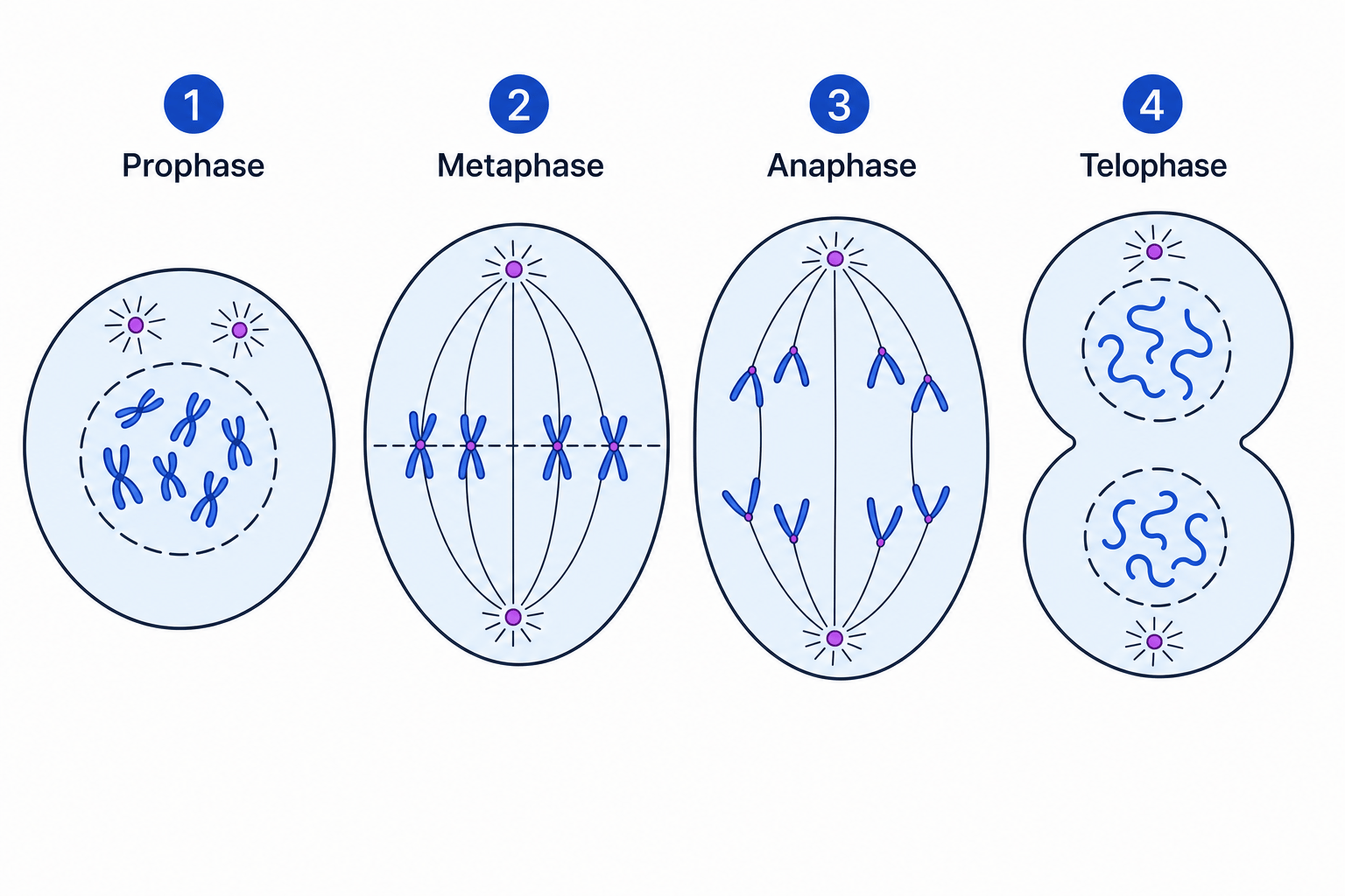
Mitosis is a continuous process, but biologists divide it into four main stages to make it easier to study: Prophase, Metaphase, Anaphase, and Telophase.
Remembering the stages
You can remember the order of the stages using the mnemonic PMAT:
- Prophase (Prepare)
- Metaphase (Middle)
- Anaphase (Apart)
- Telophase (Two)
1. Prophase
The long, tangled threads of DNA condense (shorten and thicken) to become visible chromosomes. Because the DNA was replicated in interphase, each chromosome appears as two sister chromatids joined at a centromere. The nuclear envelope begins to break down, leaving the chromosomes free in the cytoplasm.
2. Metaphase
The chromosomes line up neatly along the equator (the middle) of the cell. Protein structures called spindle fibres form across the cell and attach to the centromere of each chromosome.
3. Anaphase
The centromeres divide in two, separating the sister chromatids. The spindle fibres contract, actively pulling the separated chromatids to opposite poles (ends) of the cell. (Once separated, we refer to the chromatids as individual chromosomes again).
4. Telophase
The chromosomes reach the opposite poles and begin to uncoil, becoming long and thin again. A new nuclear envelope forms around each set of chromosomes, creating two distinct nuclei.
Cytokinesis
Finally, the cytoplasm divides. This is called cytokinesis. In animal cells, the cell membrane pinches inwards to split the cell in two. In plant cells, a new cell wall forms down the middle. This produces two separate daughter cells.
Cancer and Tumours
Mitosis is a highly controlled process, regulated by specific genes. If these genes mutate, the cell may start dividing uncontrollably. This uncontrolled cell division leads to the formation of tumours and, potentially, cancers.
Because cancer is essentially cell division out of control, many cancer treatments are specifically directed at disrupting the cell cycle. For example, chemotherapy drugs might prevent DNA from replicating during interphase, or stop spindle fibres from forming during metaphase. By doing this, they force the cancer cells to destroy themselves. (Note: These treatments can also affect normal, rapidly dividing cells, like hair follicles, which causes the side effects of chemotherapy).
Required Practical 2: Root Tip Squashes
To observe the stages of mitosis, you will prepare and examine stained squashes of plant root tips under an optical microscope.
Why do we use the tip of a root? Plant growth happens at localized regions called meristems. The very tip of a root is a meristem where cells are actively and rapidly undergoing mitosis to lengthen the root.
You must squash the tissue on the microscope slide. This is crucial because it spreads the tissue out into a single layer of cells, allowing light from the microscope to pass through so you can actually see the internal structures. You will also add a stain (such as toluidine blue) to bind to the DNA, making the chromosomes clearly visible.
The Mitotic Index
When you look at your slide, you can calculate the mitotic index. This is simply the proportion of cells in your field of view that are currently undergoing mitosis (i.e., any cells where chromosomes are visible).
Mitotic index=number of cells in mitosistotal number of cells \text{Mitotic index} = \frac{\text{number of cells in mitosis}}{\text{total number of cells}} Mitotic index=total number of cellsnumber of cells in mitosisCalculating magnification and mitotic index
A student observes a root tip squash containing 150 cells. 24 of these cells have visible condensed chromosomes. The student takes a micrograph photograph of the slide. On the printed photograph, one of the cells in metaphase has a length of 36 mm36\text{ mm}36 mm. The overall magnification of the micrograph is ×1200\times 1200×1200. Calculate the mitotic index of the tissue and the actual length of the metaphase cell in micrometres (μm\mu\text{m}μm).
- Calculate the mitotic index: Use the formula by dividing the number of cells actively dividing by the total number. Substitute the values:
- Convert the image measurement to the correct units: The image length is given as 36 mm36\text{ mm}36 mm. To convert millimetres to micrometres, multiply by 100010001000.
- Calculate the actual size: Apply the magnification equation:
Substitute the converted values:
Division in Prokaryotic Cells
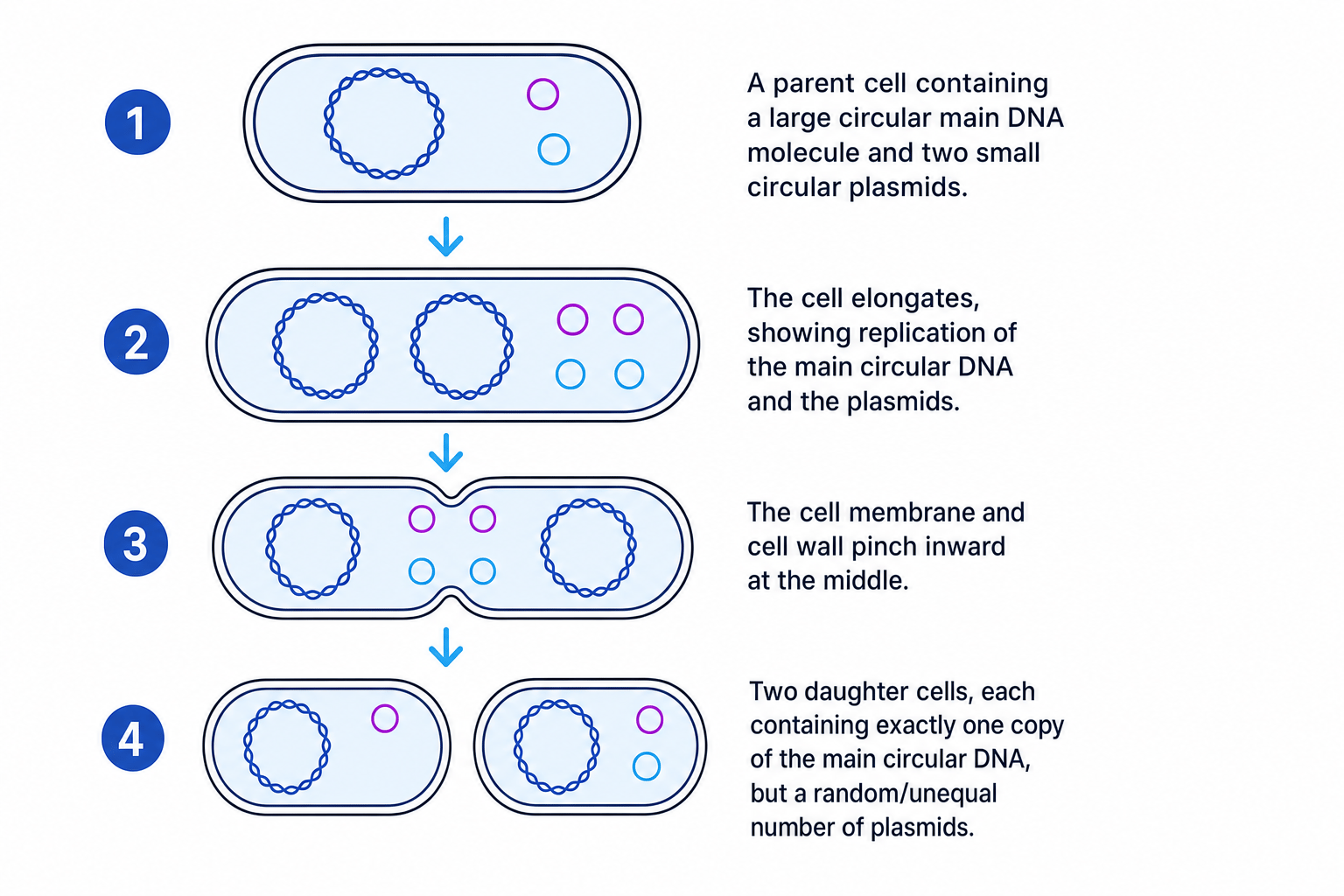
Prokaryotes (like bacteria) do not have a nucleus or linear chromosomes, so they do not undergo mitosis. Instead, they divide by a much simpler process called binary fission.
- Replication: The main circular DNA molecule replicates. Any smaller loops of DNA (plasmids) also replicate.
- Cell growth: The cell elongates, and the two copies of the main circular DNA move to opposite poles of the cell.
- Cytokinesis: The cell membrane pinches inward at the equator, and a new cell wall forms down the middle, dividing the cytoplasm.
- Daughter cells: Two new daughter cells are produced.
Plasmid distribution is random
Each daughter cell receives exactly one copy of the main circular DNA. However, the distribution of plasmids is random! One daughter cell might receive four copies of a plasmid, while the other might receive only one. They are not entirely identical in their plasmid content.
Viral Replication
Viruses are fundamentally different from both eukaryotes and prokaryotes.
Viruses are non-living
Because viruses are acellular (not made of cells) and non-living, they do not undergo cell division. They cannot reproduce on their own.
To multiply, a virus must find a host cell. The virus attaches to the surface of a host cell and injects its nucleic acid (DNA or RNA) into it. The host cell's own internal machinery (its ribosomes and enzymes) reads the viral genetic code and is essentially "hijacked" into synthesizing new viral proteins and replicating the viral nucleic acid. These components self-assemble into new virus particles, which are eventually released to infect other cells.
In the exam
- If asked why you push down hard on the cover slip during the root tip practical, state it is to squash the tissue into a single layer of cells so light can pass through. Do not say "to squash the cells" (squashing individual cells bursts them, which ruins the slide).
- Be precise when describing anaphase: mention the centromeres dividing and the spindle fibres contracting to pull chromatids to the poles.
- When using the magnification formula, always do your unit conversions (e.g., mm\text{mm}mm to μm\mu\text{m}μm) before putting the numbers into the division.
Check yourself
- During which main phase of the cell cycle does DNA replication occur?
- What is the difference between a chromosome and a chromatid?
- How does the distribution of genetic material in binary fission differ between the main circular DNA and the plasmids?
- Why do viruses require a host cell to replicate?
