What you'll learn
- How skeletal muscles work with bones in antagonistic pairs to produce movement.
- The structure of skeletal muscle from whole muscle down to the sarcomere.
- How actin, myosin, calcium ions, tropomyosin and ATP produce contraction.
- Why slow and fast skeletal muscle fibres are specialised for different activities.
Muscles as effectors
An effector is a cell, tissue or organ that carries out a response after receiving a signal from the nervous system or hormonal system. Skeletal muscles are effectors because they contract when stimulated by motor neurones, producing movement.
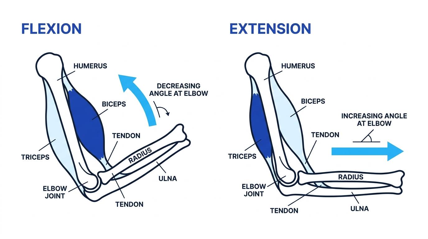
Skeletal muscle is attached to bones by tendons, which are strong, non-elastic connective tissues. When a skeletal muscle contracts, it pulls on a bone. It cannot push, so movement needs pairs of muscles working in opposite ways.
Antagonistic pair
An antagonistic pair is a pair of muscles that work against each other: when one contracts, the other relaxes.
The skeleton is described as incompressible because bones do not squash shorter when muscles pull on them. Instead, bones act as rigid levers, usually moving around joints.
For example, at the elbow:
- The biceps contracts to flex the arm, decreasing the angle at the elbow.
- The triceps contracts to extend the arm, increasing the angle at the elbow.
Working out the muscle action at a joint
A student lifts a cup from the table towards their mouth. Decide what happens to the biceps and triceps.
- Identify the movement at the elbow: the forearm moves upwards, so the elbow is flexing.
- Link the movement to the muscle that causes it: flexion at the elbow is caused by contraction of the biceps.
- Apply antagonism: because the biceps contracts, the triceps relaxes so it does not oppose the movement.
Muscles do not push bones
Skeletal muscles only produce force by contracting and pulling. If an exam answer says a muscle “pushes” a bone, it is usually biologically incorrect.
Gross structure of skeletal muscle
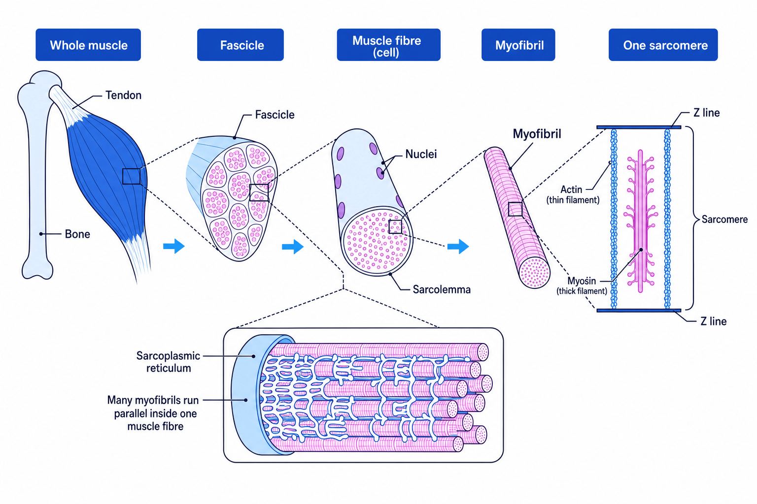
Gross structure means the larger-scale structure you could see by dissection or low-power microscopy. A whole skeletal muscle is made of bundles called fascicles. Each fascicle contains many muscle fibres.
Muscle fibre
A muscle fibre is a single skeletal muscle cell. It is long, cylindrical, multinucleate and packed with contractile threads called myofibrils.
Important terms:
- The sarcolemma is the cell-surface membrane of a muscle fibre.
- The sarcoplasm is the cytoplasm of a muscle fibre.
- The sarcoplasmic reticulum is a specialised endoplasmic reticulum that stores calcium ions.
- A myofibril is a long cylindrical organelle made of repeating contractile units.
This hierarchy is worth learning carefully because exam questions often move between levels: whole muscle, fascicle, fibre, myofibril and sarcomere.
In practical work, you may examine prepared slides of skeletal muscle using an optical microscope. Skeletal muscle has a striated appearance, meaning it shows alternating light and dark bands due to the regular arrangement of actin and myosin filaments.
The sarcomere: the contractile unit
A sarcomere is the section of a myofibril between two Z lines. It is the smallest unit that contracts in skeletal muscle.
Sarcomere
A sarcomere is one repeating contractile unit of a myofibril, bounded by two Z lines and containing overlapping actin and myosin filaments.
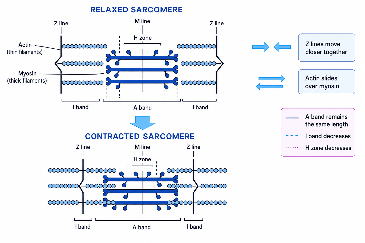
The main protein filaments are:
- Actin: thin filaments attached to the Z lines.
- Myosin: thick filaments found mainly in the centre of the sarcomere.
The main labelled regions are:
- Z line: boundary of each sarcomere.
- A band: dark band containing the full length of myosin filaments.
- I band: light band containing actin only.
- H zone: central region containing myosin only.
- M line: middle of the sarcomere.
When a sarcomere contracts, the actin and myosin filaments do not get shorter. Instead, actin slides over myosin, pulling the Z lines closer together. This is called the sliding filament theory.
What changes during contraction?
During contraction, the sarcomere shortens, the I band decreases, the H zone decreases, but the A band stays the same length because the myosin filaments do not shorten.
Interpreting band changes in a contracting sarcomere
A myofibril is stimulated and its sarcomeres become shorter. Predict what happens to the A band, I band and H zone.
- Use the sliding filament theory: actin slides further between myosin filaments, so overlap between actin and myosin increases.
- Decide what happens to regions containing actin only or myosin only: the I band and H zone both get smaller because there is less non-overlap.
- Decide what happens to the full length of myosin: myosin filaments do not shorten, so the A band remains the same length.
From nerve stimulation to calcium release
A skeletal muscle fibre is stimulated by a motor neurone, which carries an electrical impulse called an action potential. The junction between a motor neurone and muscle fibre is the neuromuscular junction.
When the muscle fibre is stimulated, the sarcolemma becomes depolarised. This leads to calcium ions being released from the sarcoplasmic reticulum into the sarcoplasm.
You do not need the full role of troponin for this AQA section. For this specification, focus on calcium ions causing tropomyosin to move so that myosin-binding sites on actin are exposed.
Tropomyosin
Tropomyosin is a protein associated with actin filaments. In a relaxed muscle, it blocks the myosin-binding sites on actin.
The actinomyosin bridge cycle
The contraction mechanism depends on repeated formation and breaking of actinomyosin bridges.
Actinomyosin bridge
An actinomyosin bridge is a temporary attachment between a myosin head and a binding site on an actin filament.
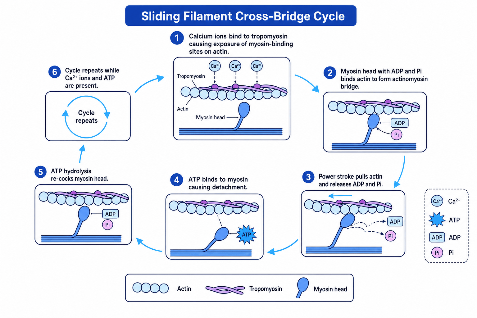
The cycle works like this:
- Calcium ions are released from the sarcoplasmic reticulum.
- Calcium ions cause tropomyosin to move away from the myosin-binding sites on actin.
- Myosin heads bind to exposed binding sites on actin, forming actinomyosin bridges.
- Myosin heads bend, pulling actin along. This is the power stroke.
- ATP binds to myosin, causing myosin to detach from actin.
- ATP is hydrolysed to ADP and inorganic phosphate, releasing energy to return the myosin head to its original position.
- The cycle repeats as long as calcium ions and ATP are available.
ATP has two key roles
ATP is needed to detach myosin from actin and to re-cock the myosin head so another power stroke can occur.
ATP is not just for the power stroke
Many students write that ATP directly causes the power stroke. More precisely, ATP binding causes detachment, and ATP hydrolysis provides energy to reset the myosin head. The power stroke occurs as the myosin head changes angle while attached to actin.
Predicting the effect of no calcium ions
A muscle fibre has plenty of ATP, but calcium ions remain inside the sarcoplasmic reticulum. Predict whether the fibre contracts.
- Check whether myosin-binding sites are exposed: without calcium ions in the sarcoplasm, tropomyosin remains blocking the binding sites on actin.
- Decide whether actinomyosin bridges can form: myosin heads cannot bind effectively to actin because the binding sites are not exposed.
- Link bridge formation to contraction: without repeated actinomyosin bridge formation and power strokes, the sarcomere does not shorten.
ATP and phosphocreatine
Muscle contraction uses ATP very rapidly. However, muscle fibres only store a small amount of ATP, so ATP must be regenerated quickly.
One short-term ATP reserve is phosphocreatine.
Phosphocreatine
Phosphocreatine is a high-energy compound stored in muscle fibres. It can rapidly transfer phosphate to ADP to regenerate ATP during intense contraction.
The reaction can be summarised in words:
phosphocreatine plus ADP forms creatine plus ATP
This is especially useful at the start of intense exercise, such as sprinting, because it regenerates ATP quickly without needing oxygen. However, phosphocreatine stores are limited and are depleted rapidly.
Remember the order of energy supply
For a sudden sprint, muscle fibres use stored ATP first, then phosphocreatine, then anaerobic respiration, with aerobic respiration becoming increasingly important if exercise continues.
Slow and fast skeletal muscle fibres
Skeletal muscles contain a mixture of fibre types. The two key types for this topic are slow fibres and fast fibres.
Slow skeletal muscle fibres
Slow fibres are adapted for sustained, endurance-based activity.
They have:
- Many mitochondria.
- A rich blood supply with many capillaries.
- Lots of myoglobin, an oxygen-binding pigment.
- Mainly aerobic respiration.
- A slower contraction speed.
- High resistance to fatigue.
They are common in muscles used for posture and endurance, such as muscles in the back and legs.
Fast skeletal muscle fibres
Fast fibres are adapted for rapid, powerful contractions.
They have:
- Fewer mitochondria than slow fibres.
- Less myoglobin, so they appear paler.
- A greater store of phosphocreatine and glycogen.
- Faster contraction speed.
- Greater force production.
- Fatigue more quickly.
They are useful in sprinting, jumping and other short bursts of high-intensity movement.
| Feature | Slow fibres | Fast fibres |
|---|---|---|
| Contraction speed | Slow | Fast |
| Fatigue resistance | High | Low |
| Main respiration type | Aerobic | More anaerobic |
| Mitochondria | Many | Fewer |
| Myoglobin | Lots | Less |
| Capillary supply | Dense | Less dense |
| Typical role | Endurance and posture | Rapid, powerful movement |
Identifying a muscle fibre type from evidence
A muscle biopsy shows fibres with many mitochondria, lots of myoglobin and a dense capillary network. Identify the likely fibre type.
- Use the mitochondria evidence: many mitochondria suggest a high rate of aerobic respiration.
- Use the myoglobin and capillary evidence: both improve oxygen supply, again supporting aerobic respiration.
- Match the evidence to fibre type: fibres specialised for sustained aerobic respiration are slow skeletal muscle fibres.
Muscles are mixtures
Do not describe a whole muscle as purely slow or purely fast. Most skeletal muscles contain a mixture of fibre types, but the proportions vary depending on function.
Muscle fatigue
Muscle fatigue is the reduced ability of a muscle to contract after repeated stimulation. It can be investigated in human volunteers, for example by repeatedly squeezing a hand grip and recording how force or number of contractions changes over time.
Fatigue may involve several factors, including depletion of ATP or phosphocreatine, accumulation of metabolites, and reduced ability to release or respond to calcium ions.
Fatigue is not simply running out of oxygen
Low oxygen availability can contribute to fatigue, but exam answers should link fatigue to the muscle’s ability to keep producing ATP and maintaining the contraction cycle.
In the exam
- For sliding filament questions, name the molecules and sequence: calcium ions, tropomyosin movement, actinomyosin bridge formation, power stroke, ATP binding, detachment, ATP hydrolysis.
- For sarcomere diagrams, state that actin and myosin filaments do not shorten; the sarcomere shortens because the filaments slide past each other.
- For fibre-type questions, justify your answer using evidence such as mitochondria, myoglobin, capillary supply, contraction speed and fatigue resistance.
Check yourself
- Why must skeletal muscles work in antagonistic pairs?
- During contraction, why does the A band stay the same length while the I band decreases?
- What would happen to the actinomyosin bridge cycle if ATP were unavailable?