Control of blood water potential (A-level only)
What you'll learn:
- What osmoregulation is and why maintaining blood water potential is crucial.
- The structure of the kidney nephron and how it filters your blood.
- How the loop of Henle acts as a counter-current multiplier to create a sodium ion gradient.
- How the hypothalamus, pituitary gland, and the hormone ADH control the volume of water lost in your urine.
Introduction to Osmoregulation
Your cells need to be bathed in tissue fluid that has the exact same water potential as their cytoplasm. If the water potential of your blood and tissue fluid drops too low, water leaves your cells by osmosis, causing them to shrink and shrivel. If the water potential rises too high, water enters the cells, causing them to swell and potentially burst (lysis).
Osmoregulation
Osmoregulation is the homeostatic control of the water potential of the blood. It ensures that the water content of the body is kept constant, regardless of how much water you drink or how much you lose through sweating.
The main organs responsible for osmoregulation are the kidneys. They achieve this by filtering the blood and then selectively reabsorbing the substances the body needs, allowing the rest to be excreted as urine.
The Structure of the Nephron
Each kidney contains about a million tiny filtering units called nephrons. A nephron is a long, highly coiled tube surrounded by a complex network of blood capillaries.
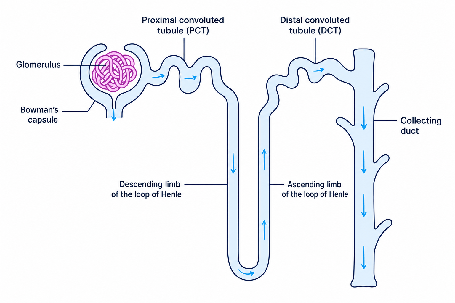
Blood enters the kidney through the renal artery, which branches into tiny afferent arterioles. Each afferent arteriole supplies a knot of capillaries called the glomerulus, which sits inside a cup-shaped structure called the Bowman's capsule.
From the Bowman's capsule, the fluid travels through the nephron tube in this order:
- Proximal convoluted tubule (PCT)
- Loop of Henle (consisting of a descending limb and an ascending limb)
- Distal convoluted tubule (DCT)
- Collecting duct (which empties the final urine into the center of the kidney to be carried to the bladder)
Arteriole names
To remember which arteriole is which: Afferent arrives at the glomerulus. Efferent exits the glomerulus.
Stage 1: The Formation of Glomerular Filtrate
The process of making urine begins in the Bowman's capsule with a process called ultrafiltration.
The afferent arteriole (bringing blood in) has a wider diameter than the efferent arteriole (taking blood away). This bottleneck creates a high hydrostatic pressure inside the glomerulus.
This high pressure forces small molecules out of the capillary, through a selectively permeable basement membrane, and into the Bowman's capsule. The fluid that ends up in the capsule is called the glomerular filtrate. It contains:
- Water
- Glucose
- Mineral ions (like sodium and chloride)
- Urea
Large molecules, such as plasma proteins and red blood cells, are too large to pass through the basement membrane and remain in the blood.
Stage 2: Selective Reabsorption by the PCT
The body cannot afford to lose all the water, glucose, and useful ions present in the glomerular filtrate. Therefore, they must be reabsorbed back into the blood. Around 85% of the filtrate is reabsorbed in the proximal convoluted tubule (PCT).
Crucially, all of the glucose is reabsorbed here by co-transport with sodium ions:
- Sodium ions (Na+\text{Na}^+Na+) are actively transported out of the PCT epithelial cells and into the blood capillaries.
- This dramatically lowers the concentration of Na+\text{Na}^+Na+ inside the PCT epithelial cells.
- Na+\text{Na}^+Na+ from the lumen of the PCT diffuses down its concentration gradient into the epithelial cells through specific co-transporter proteins, bringing a molecule of glucose with it each time.
- The glucose then diffuses from the epithelial cell into the blood via facilitated diffusion.
Because all these dissolved solutes are leaving the filtrate and entering the cells, the water potential of the filtrate rises, while the water potential of the blood drops. Consequently, water moves from the PCT into the blood by osmosis.
Stage 3: Maintaining a Sodium Gradient in the Medulla
The next structure is the loop of Henle. Its main job is not to reabsorb water directly, but to create a very low water potential in the tissue fluid of the kidney medulla (the inner region of the kidney). This allows water to be reabsorbed later.
The loop of Henle acts as a counter-current multiplier:
- "Counter-current" because the fluid flows in opposite directions in the two limbs.
- "Multiplier" because the gradient becomes steeper and steeper as you go deeper into the medulla.
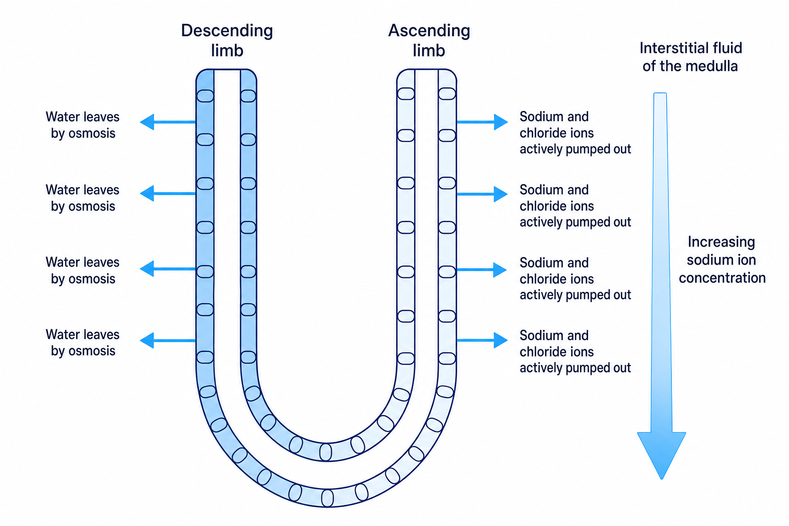
Here is how the mechanism works:
- The Ascending Limb: This limb is impermeable to water. Its walls actively transport sodium (Na+\text{Na}^+Na+) and chloride (Cl−\text{Cl}^-Cl−) ions out of the filtrate and into the surrounding interstitial fluid of the medulla.
- Lowering the water potential: Because ions are accumulating in the medulla, the water potential of the medulla tissue fluid becomes highly negative.
- The Descending Limb: This limb is highly permeable to water. As the filtrate flows down this limb into the salty medulla, water leaves the tubule by osmosis into the tissue fluid (and is then carried away by blood capillaries).
- Concentrating the filtrate: Because water is leaving but ions cannot enter, the filtrate reaches its lowest water potential (highest concentration of ions) at the very bottom of the loop.
Water is never actively transported
In exam answers, never write that water is "pumped" or "actively transported". Water only ever moves across biological membranes passively via osmosis down a water potential gradient. The kidney moves ions actively to set up the gradient, and water follows by osmosis.
Stage 4: Reabsorption of Water by the DCT and Collecting Duct
By the time the filtrate reaches the distal convoluted tubule (DCT) and the collecting duct, it is very dilute again (because ions were actively pumped out of the ascending limb). Now, the body can finely tune exactly how much water is reabsorbed.
This is controlled by hormones, specifically depending on the water potential of the blood.
The Role of the Hypothalamus and ADH
If you are dehydrated (e.g. from sweating), your blood water potential drops.
- Detection: Special cells in the brain called osmoreceptors (located in the hypothalamus) detect this drop. Because the blood has a lower water potential than the osmoreceptors, water leaves the osmoreceptor cells by osmosis, causing them to shrink.
- Secretion: This shrinking stimulates the hypothalamus to send nerve impulses to the posterior pituitary gland.
- Release: The posterior pituitary gland releases a hormone called antidiuretic hormone (ADH) directly into the blood.
The action of ADH
ADH travels in the blood to the kidneys and binds to receptors on the cell surface membranes of the DCT and the collecting ducts. It causes vesicles containing water channels called aquaporins to fuse with the cell membrane. This dramatically increases the permeability of these tubules to water.
Because the collecting duct passes straight through the salty medulla (which has a very low water potential thanks to the loop of Henle), water rapidly leaves the collecting duct by osmosis and is reabsorbed into the blood.
The result? The body saves water, and produces a small volume of highly concentrated urine.
Conversely, if you drink plenty of water, blood water potential rises. Osmoreceptors swell, less ADH is released, fewer aquaporins are inserted, the collecting duct remains largely impermeable to water, and you produce a large volume of dilute urine.
Evaluating the effect of Loop of Henle length on urine concentration
Many exam questions ask you to compare the kidneys of animals from different habitats. For instance:
The kangaroo rat is a desert mammal. It has a much longer loop of Henle than a human. Explain how this structural adaptation helps the kangaroo rat survive in dry conditions.
Here is the step-by-step reasoning you should apply:
- Identify the active mechanism: The ascending limb actively pumps out Na+\text{Na}^+Na+ and Cl−\text{Cl}^-Cl− ions into the medulla.
- Relate the structural change to the mechanism: Because the loop of Henle is longer, the ascending limb is longer. This means more ions can be actively pumped out into the surrounding interstitial fluid of the medulla.
- State the consequence for the gradient: This builds up a much higher concentration of ions deep in the medulla, resulting in an extremely low (highly negative) water potential in the tissue fluid.
- Connect to the collecting duct: A steeper water potential gradient is maintained between the filtrate in the collecting duct and the medulla.
- State the final outcome: More water is reabsorbed from the collecting duct into the blood by osmosis, resulting in highly concentrated urine and minimizing water loss.
In the exam
When writing long-answer responses on osmoregulation, keep your terminology tight:
- Always state that water moves by osmosis.
- Always refer to water potential (rather than "water concentration"). Say "the water potential becomes more negative" or "the water potential decreases".
- When describing the effect of ADH, clearly state that it increases the permeability of the DCT and collecting duct cell membranes (mentioning aquaporins often gains you a specific mark).
- Be clear on the direction of movement: "from the collecting duct, into the interstitial fluid, and into the blood capillaries".
Check yourself
- What difference in arteriole diameter generates the high hydrostatic pressure needed for ultrafiltration?
- By what mechanism is all glucose reabsorbed from the proximal convoluted tubule?
- Which limb of the loop of Henle is impermeable to water?
- Where in the brain are the osmoreceptors located, and which gland releases ADH?
- How does ADH increase the water permeability of the collecting duct?