What you'll learn
- How amino acids join to form dipeptides, polypeptides and proteins.
- Why the R group, peptide bonds and different protein bonds matter.
- How primary, secondary, tertiary and quaternary structure link to function.
- How to test for proteins using the biuret test, and identify amino acids by chromatography.
Proteins are polymers made from amino acids
Proteins are large biological molecules found in all living organisms. They are involved in almost everything cells do: catalysing reactions, transporting substances, forming structures, signalling and defending the body.
Monomer and polymer
A monomer is a small repeating unit. A polymer is a large molecule made from many monomers joined together. In proteins, the monomers are amino acids.
A polypeptide is a chain of many amino acids. A functional protein may contain one polypeptide or several polypeptides folded and arranged together.
Amino acids: the building blocks
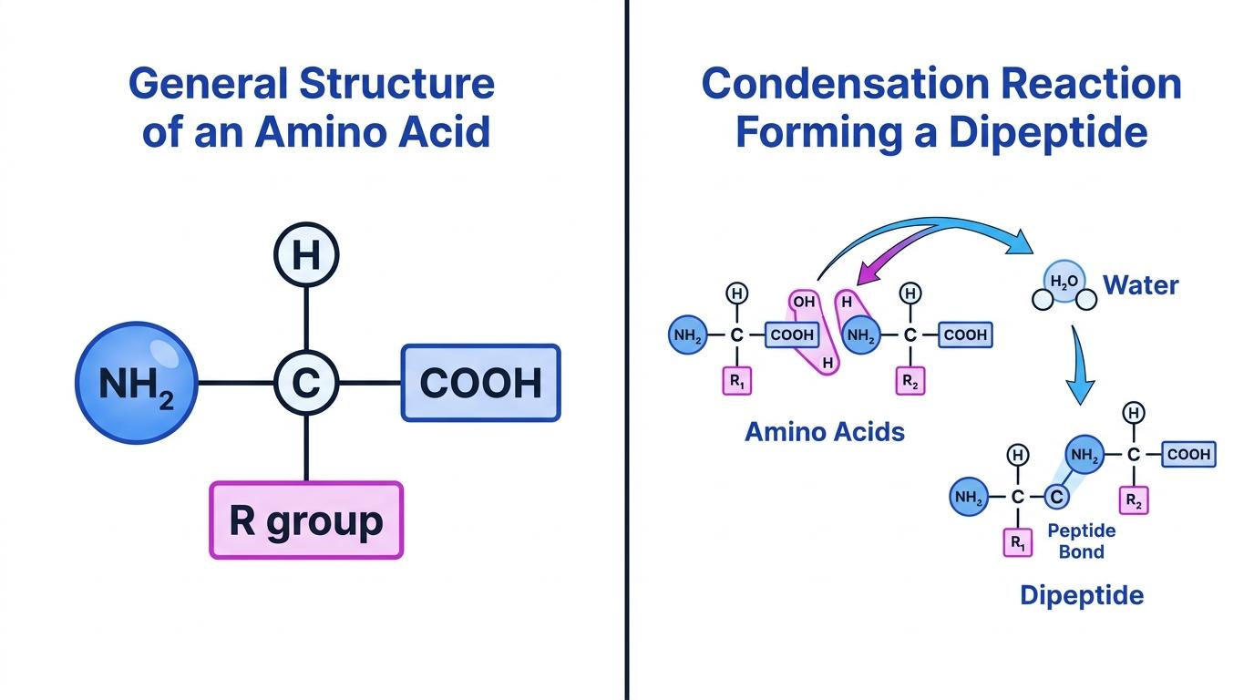
Each amino acid has the same general structure: a central carbon atom bonded to an amine group, a carboxyl group, a hydrogen atom and a variable R group.
Amino acid
An amino acid is an organic molecule containing an amine group (NH2), a carboxyl group (COOH), a hydrogen atom and an R group attached to a central carbon atom.
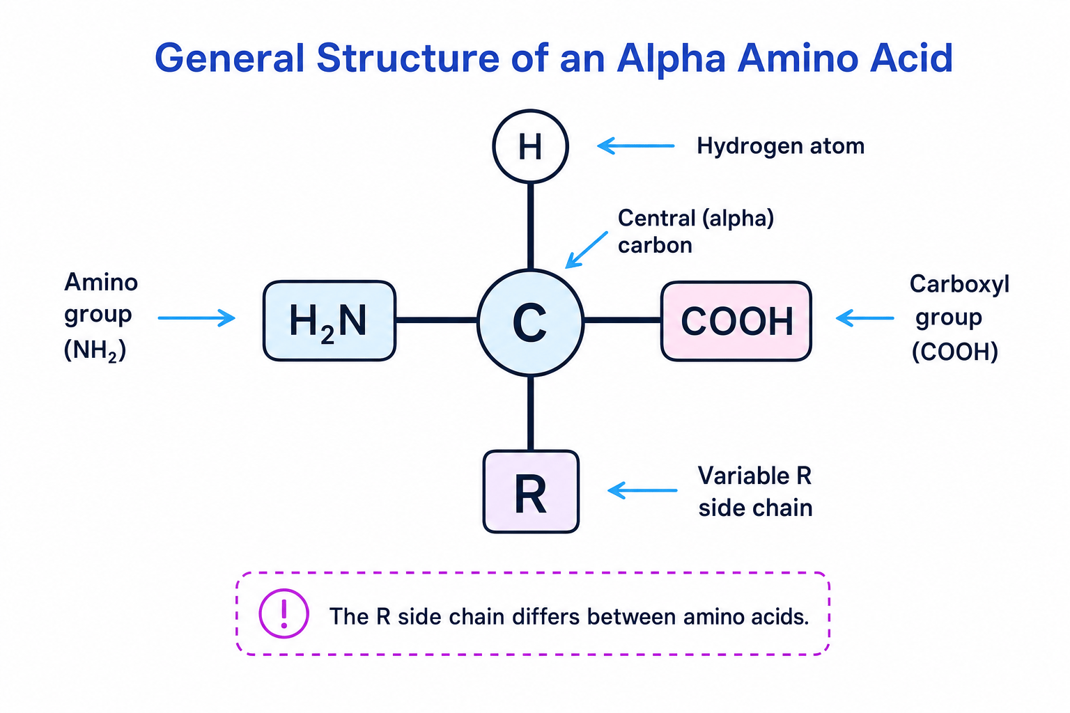
The 20 amino acids common in all organisms differ only in their R group, also called the side chain. This side chain can be non-polar, polar, charged, or contain sulfur.
Side chains matter
The R group determines how each amino acid behaves, so the order of R groups in a polypeptide helps determine how the protein folds and what it can do.
Joining amino acids: condensation and peptide bonds
Amino acids join together by a condensation reaction. In a condensation reaction, two molecules join and a molecule of water is released.
When two amino acids join:
- an OH is removed from the carboxyl group of one amino acid
- an H is removed from the amine group of another amino acid
- these form water, H2O
- a peptide bond forms between the two amino acids
Peptide bond
A peptide bond is the covalent bond formed between the carboxyl group of one amino acid and the amine group of another amino acid.
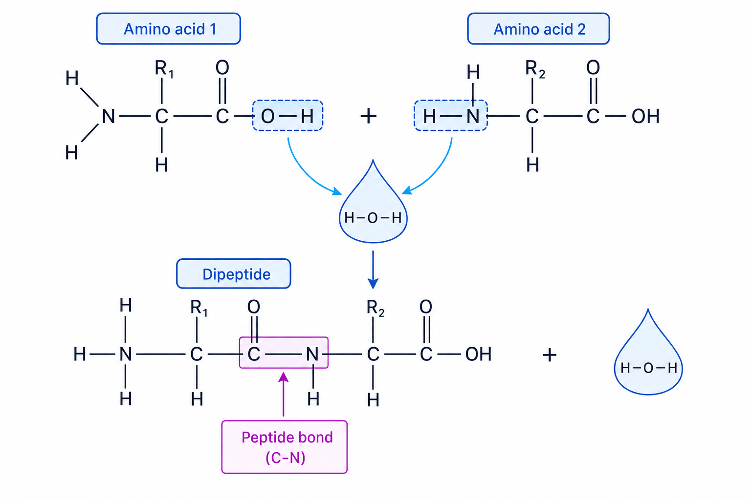
A dipeptide contains two amino acids joined by one peptide bond. A polypeptide contains many amino acids joined by many peptide bonds.
Counting peptide bonds
A linear polypeptide contains 120 amino acids. How many peptide bonds were formed, and how many water molecules were released?
- In a linear chain, each peptide bond joins two neighbouring amino acids, so the number of joins is one fewer than the number of amino acids: n−1n - 1n−1.
- Substitute n=120n = 120n=120: 120−1=119120 - 1 = 119120−1=119 peptide bonds.
- Each condensation reaction releases one water molecule, so 119 water molecules are released.
The reverse reaction is hydrolysis, where water is used to break a peptide bond.
Levels of protein structure
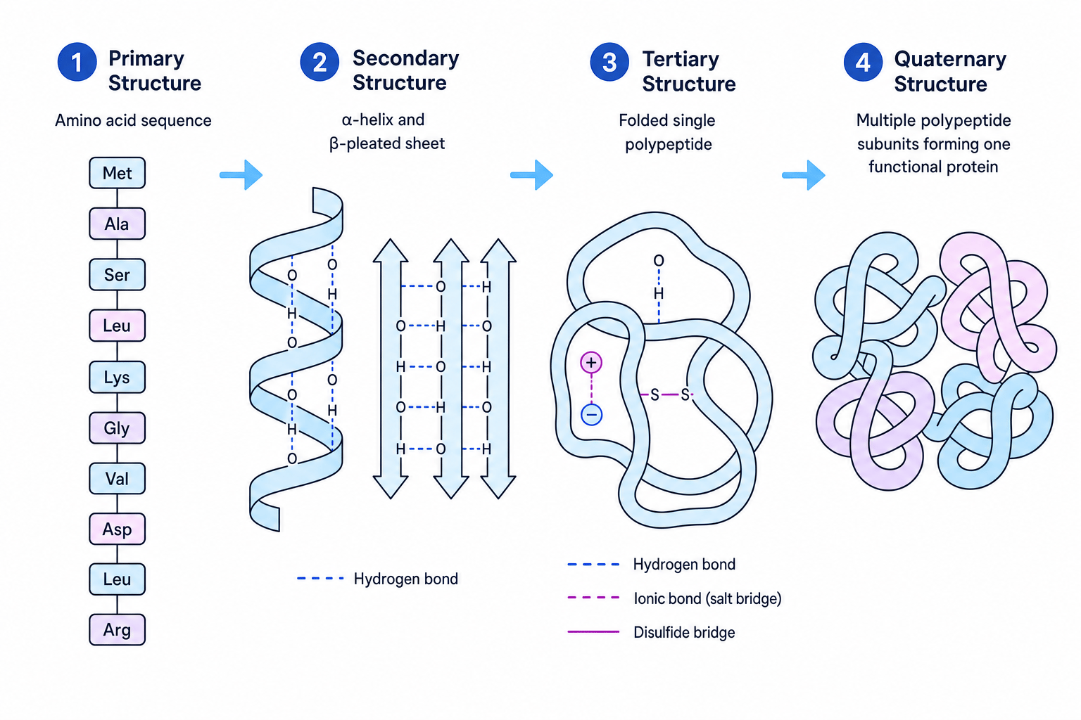
Protein structure is described in four levels. These levels build on each other: the amino acid sequence affects folding, and folding affects function.
Primary structure
The primary structure is the sequence of amino acids in a polypeptide chain. It is held together by peptide bonds.
Even a small change in primary structure can change the final shape of a protein, because different R groups form different interactions.
Secondary structure
The secondary structure is the local coiling or folding of the polypeptide chain. Common examples are:
- an alpha helix
- a beta pleated sheet
These shapes are held by hydrogen bonds between parts of the peptide backbone, not usually between R groups.
Tertiary structure
The tertiary structure is the overall three-dimensional shape of one polypeptide chain. It is held in place by interactions between R groups.
Bonds in tertiary structure
- A hydrogen bond is a weak attraction between slightly positive and slightly negative parts of molecules.
- An ionic bond is an attraction between oppositely charged R groups.
- A disulfide bridge is a strong covalent bond between sulfur atoms in two cysteine R groups.
Quaternary structure
The quaternary structure is the arrangement of two or more polypeptide chains in a functional protein. Haemoglobin is a good example because it contains four polypeptide subunits.
Polypeptide vs protein
Do not assume every polypeptide is already a functional protein. Many proteins only work after the polypeptide has folded correctly, or after several polypeptides have joined together.
Protein shape determines protein function
A protein’s function depends on its shape. That shape depends on its primary structure and the bonds that stabilise its secondary, tertiary and quaternary structure.
For example:
- enzymes have an active site, a region with a specific shape that binds the substrate
- transport proteins have shapes that let them bind or move specific molecules
- structural proteins, such as collagen, have shapes that give strength
- haemoglobin’s quaternary structure helps it bind and transport oxygen
Denaturation
Denaturation is a change in a protein’s three-dimensional shape so that it can no longer function properly. It usually happens because bonds holding the secondary, tertiary or quaternary structure are disrupted.
High temperature increases vibration in the molecule, which can disrupt hydrogen bonds. Changes in pH can alter charges on R groups, disrupting ionic bonds and changing the protein’s shape.
Explaining loss of enzyme activity after a pH change
An enzyme works best at pH 7. Its activity falls sharply at pH 3. Explain why.
- A large pH change alters the charges on some R groups, so ionic bonds and hydrogen bonds in the tertiary structure may break or form in different places.
- The tertiary structure changes, so the shape of the active site changes.
- The substrate is no longer complementary to the active site, so fewer enzyme-substrate complexes form.
- The enzyme’s activity decreases because fewer reactions are catalysed per second.
Denaturation does not usually break peptide bonds
In typical enzyme denaturation, the primary structure usually remains intact. The peptide bonds are not normally broken; the problem is the change in folding and three-dimensional shape.
The biuret test for proteins
The biuret test is used to test for proteins. It detects peptide bonds in an alkaline solution.
Biuret test
A positive biuret test changes from blue to lilac or purple, showing that protein is present.
Method:
- Add the sample to a test tube.
- Add sodium hydroxide solution to make the solution alkaline.
- Add a few drops of dilute copper(II) sulfate solution, or use prepared biuret reagent.
- Mix gently. No heating is needed.
A negative result stays blue. Free amino acids do not give a positive protein result because they are not joined by peptide bonds.
Interpreting biuret test results
A student tests three solutions using the biuret test. Tube A stays blue, tube B turns lilac, and tube C turns deep purple.
- Tube A staying blue means no protein is detected.
- Tube B turning lilac means peptide bonds are present, so protein is present.
- Tube C turning a deeper purple suggests more protein than tube B, assuming the same sample volume and reagent volumes were used.
Biuret test wording
For full marks, name the reagent conditions and the colour change: alkaline sodium hydroxide plus copper(II) sulfate, blue to lilac or purple.
Separating amino acids by chromatography
Chromatography can separate a mixture of amino acids. The stationary phase is the paper or chromatography plate. The mobile phase is the solvent that moves through it.
Amino acids are often colourless, so a locating agent such as ninhydrin may be used to make the spots visible. Unknown amino acids can be identified by comparing their positions with known standard solutions run on the same chromatogram.
The Rf value can also help identify substances:
Rf=distance moved by spotdistance moved by solvent frontR_f = \frac{\text{distance moved by spot}}{\text{distance moved by solvent front}}Rf=distance moved by solvent frontdistance moved by spotIdentifying amino acids from Rf values
On a chromatogram, the solvent front moves 80 mm. An unknown amino acid spot moves 48 mm. A known alanine standard has an Rf value of 0.60.
- Substitute the distances into the formula: Rf=48 mm80 mmR_f = \frac{48\ \text{mm}}{80\ \text{mm}}Rf=80 mm48 mm.
- Calculate the value: Rf=0.60R_f = 0.60Rf=0.60 because the distance units cancel.
- Compare with the standard: the unknown spot matches alanine, so the unknown amino acid is likely to be alanine.
In the exam
- Link structure to function in order: primary sequence affects bonds, bonds affect three-dimensional shape, and shape affects function.
- Use precise bond names: peptide bonds for primary structure; hydrogen bonds, ionic bonds and disulfide bridges for folding.
- For practical questions, give both method and interpretation: biuret is blue to lilac/purple for protein; chromatography uses standards or Rf values for identification.
Check yourself
- Which part of the amino acid differs between the 20 common amino acids?
- How can a change in primary structure lead to an enzyme no longer working?
- What colour change shows a positive biuret test, and what is being detected?