

What you'll learn
- How diffusion makes gas exchange possible, and why exchange surfaces need specific adaptations.
- How gas exchange happens in single-celled organisms, insects, fish and dicotyledonous leaves.
- How human lungs and alveoli are adapted for gas exchange, including the mechanism of breathing.
- How to use pulmonary ventilation rate and interpret lung disease data.
The starting point: diffusion
Gas exchange is mainly about moving oxygen into an organism and carbon dioxide out. These gases move by diffusion, which is the net movement of particles from a region of higher concentration to a region of lower concentration.
A concentration gradient is the difference in concentration between two areas. The steeper the gradient, the faster diffusion happens.
Gas exchange surface
A gas exchange surface is a boundary across which gases such as oxygen and carbon dioxide diffuse between an organism and its environment.
Efficient gas exchange surfaces usually have:
- a large surface area for more diffusion at once
- a thin diffusion pathway so gases travel a short distance
- a moist, permeable surface so gases can dissolve and pass through
- ventilation, meaning movement of air or water over the surface to maintain the concentration gradient
- sometimes a mass transport system, such as blood, to carry gases away and keep the gradient steep
The core pattern
Most gas exchange questions are really asking: “How does this feature increase surface area, decrease diffusion distance, or maintain a concentration gradient?”
Gas exchange across the body surface of a single-celled organism
A single-celled organism, such as an amoeba, has a very large surface area compared with its volume. This is called a high surface area-to-volume ratio.
Because every part of the cell is close to the external environment, oxygen can diffuse directly through the cell surface membrane into the cytoplasm, and carbon dioxide can diffuse out.
Larger multicellular organisms cannot rely on diffusion across the body surface alone because many cells are too far from the surface. They need specialised gas exchange surfaces and transport systems.
Gas exchange in insects
Insects use a tracheal system, which is a network of air-filled tubes that carries oxygen directly to tissues.
Air enters through pores called spiracles on the insect’s body surface. The spiracles lead into larger tubes called tracheae, which branch into smaller tubes called tracheoles. Tracheoles extend between cells, giving a short diffusion pathway to respiring tissues.
Oxygen diffuses from the tracheoles into cells. Carbon dioxide diffuses in the opposite direction. In active insects, body movements can ventilate the tracheal system by moving air in and out.
Insects do not use blood for oxygen transport
In insects, oxygen is delivered mainly through the tracheal system, not by haemoglobin in the blood. Their blood-like fluid, haemolymph, is not the main oxygen carrier.
The water-loss compromise in insects
Insects live on land, so exposing moist gas exchange surfaces to air risks water loss by evaporation. They reduce this by having:
- spiracles that can close
- a waterproof cuticle
- small spiracle openings
- hairs around spiracles in some species, which trap humid air
The compromise is that closing spiracles reduces water loss but also reduces gas exchange.
Gas exchange in fish gills
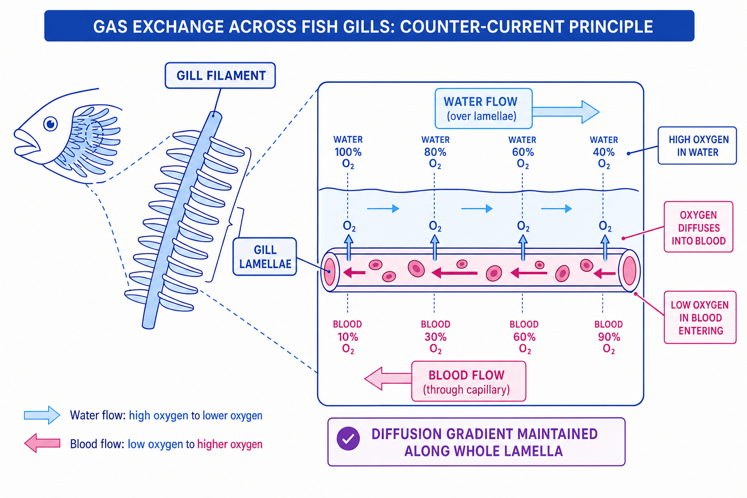
Fish exchange gases with water using gills. Each gill has many gill filaments, and each filament has many thin gill lamellae. The lamellae provide a very large surface area and contain capillaries, giving a short diffusion pathway between water and blood.
Water flows over the lamellae, while blood flows through capillaries inside them. Fish use the counter-current principle, where water and blood flow in opposite directions. This maintains a diffusion gradient for oxygen along the whole length of the lamella.
Explaining counter-current exchange
- Compare the oxygen concentrations at each point along the lamella: in counter-current flow, the water always has a higher oxygen concentration than the blood next to it.
- Because oxygen concentration is higher in the water than in the blood at every point, oxygen continues to diffuse into the blood along the full length of the lamella.
- This means blood leaving the gill can become highly oxygenated, whereas in parallel flow the gradient would fall quickly and diffusion would be less efficient.
Gas exchange in dicotyledonous leaves
A dicotyledonous leaf is the typical broad leaf of many flowering plants. Plants need carbon dioxide for photosynthesis and release oxygen as a product.
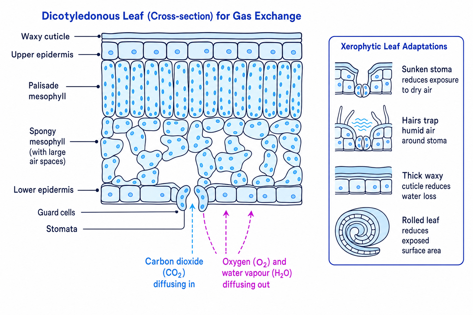
Gases enter and leave mainly through stomata. A single pore is a stoma. Each stoma is surrounded by guard cells, which can open or close the pore.
Inside the leaf, mesophyll tissue contains many air spaces. The spongy mesophyll has large air spaces, allowing carbon dioxide to diffuse through the leaf and reach photosynthesising cells. The moist surfaces of mesophyll cells allow gases to dissolve before diffusing into cells.
Xerophytes and water loss
A xerophyte is a plant adapted to dry conditions. Leaves need stomata for gas exchange, but open stomata allow water vapour to leave by transpiration.
Xerophytes reduce water loss using features such as:
- a thick waxy cuticle
- sunken stomata
- hairs that trap humid air
- rolled leaves
- reduced leaf surface area, such as spines
The unavoidable trade-off
Efficient gas exchange often needs a large, moist surface exposed to air, but that also increases water loss. Terrestrial insects and xerophytic plants show adaptations that balance these opposing needs.
The human gas exchange system
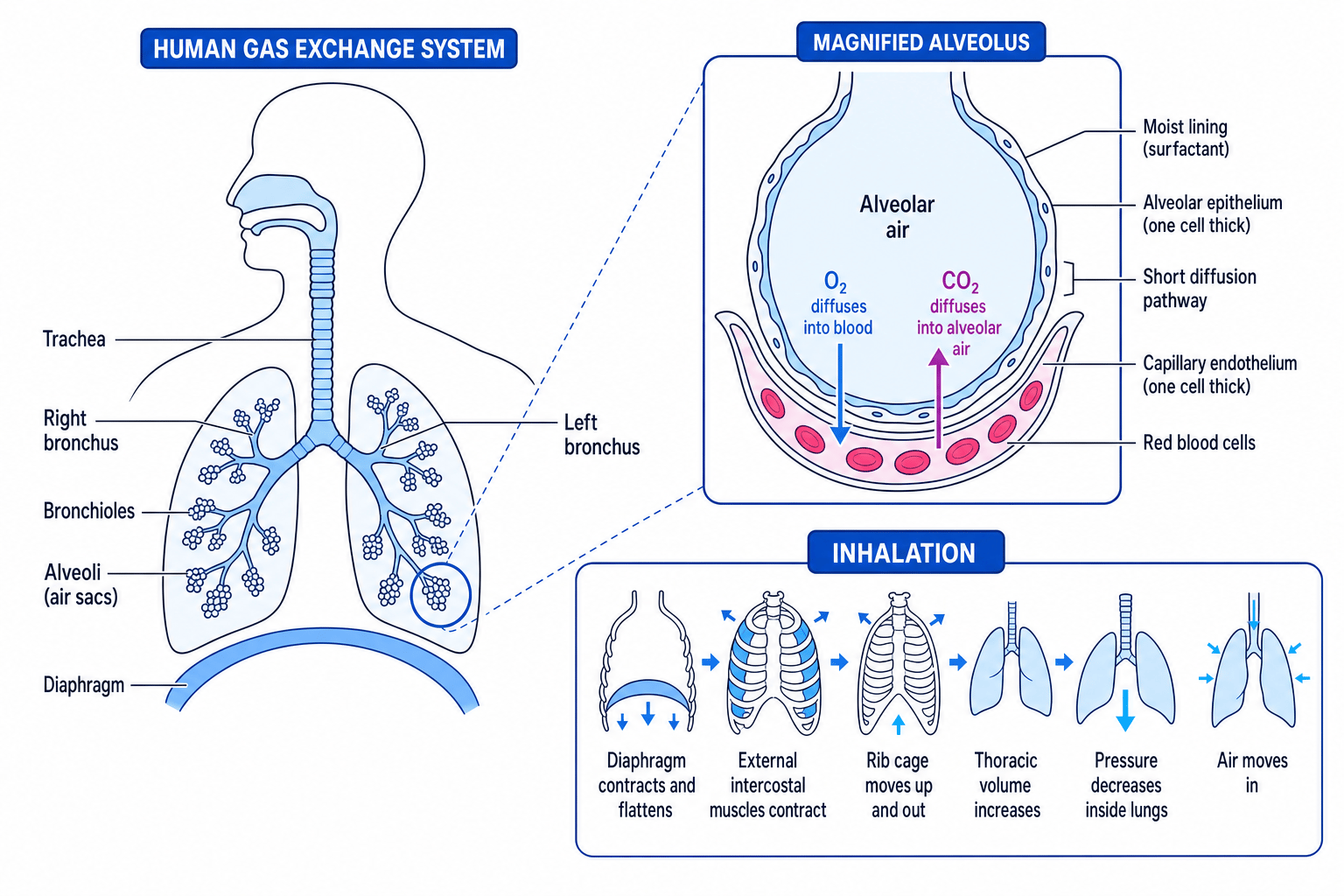
In humans, air passes down the trachea, into two bronchi, then into smaller bronchioles, and finally into tiny air sacs called alveoli inside the lungs.
The alveolar epithelium is the thin layer of cells lining each alveolus. Alveoli are adapted for gas exchange because they have:
- a very large total surface area
- a one-cell-thick epithelium
- a moist lining, so gases dissolve
- a rich blood supply from capillaries
- elastic tissue to help recoil during exhalation
- ventilation to maintain steep concentration gradients
Ventilation: how breathing moves air
Ventilation is the movement of air into and out of the lungs. It maintains oxygen and carbon dioxide concentration gradients at the alveoli.
The thoracic cavity is the chest cavity containing the lungs. Breathing works by changing its volume, which changes pressure.
Inhalation
During inhalation:
- The diaphragm contracts and flattens.
- The external intercostal muscles contract.
- The rib cage moves up and out.
- The volume of the thoracic cavity increases.
- Pressure inside the lungs decreases below atmospheric pressure.
- Air moves into the lungs.
Exhalation
During normal exhalation:
- The diaphragm relaxes and becomes dome-shaped.
- The external intercostal muscles relax.
- Elastic recoil helps the lungs and rib cage move back.
- Thoracic volume decreases.
- Pressure inside the lungs increases.
- Air moves out.
During forced exhalation, the internal intercostal muscles contract and the external intercostal muscles relax, pulling the rib cage down and in. The external and internal intercostal muscles are antagonistic muscles, meaning they have opposite effects.
Pressure wording
Air moves from higher pressure to lower pressure. Inhalation happens because pressure inside the lungs becomes lower than atmospheric pressure; exhalation happens because it becomes higher.
Pulmonary ventilation rate
Tidal volume is the volume of air moved into or out of the lungs in one breath. Breathing rate is the number of breaths per minute.
Pulmonary ventilation rate is the total volume of air moved into or out of the lungs per minute.
PVR=tidal volume×breathing rate\text{PVR} = \text{tidal volume} \times \text{breathing rate}PVR=tidal volume×breathing rateCommon units are cubic decimetres per minute, written as dm³ min⁻¹.
Calculating pulmonary ventilation rate
A student has a tidal volume of 0.50 dm³ breath⁻¹ and a breathing rate of 14 breaths min⁻¹.
- Substitute the values into the equation:
- Cancel “breath” because one value is per breath and the other is breaths per minute:
- State the answer with units: the pulmonary ventilation rate is 7.0 dm³ min⁻¹.
Lung disease and gas exchange
Lung disease can affect gas exchange, ventilation, or both.
- Emphysema destroys alveolar walls, reducing surface area and elastic recoil.
- Fibrosis thickens and stiffens alveolar walls, increasing diffusion distance and reducing ventilation.
- Asthma narrows bronchioles, making ventilation harder.
- Chronic bronchitis increases mucus production and can damage cilia, reducing airflow and increasing infection risk.
- Lung cancer can obstruct airways and destroy healthy lung tissue.
When interpreting data, distinguish correlation from causation. A correlation means two variables are associated. Causation means one variable directly brings about the change in the other.
Interpreting smoking and disease data
A study shows lung disease incidence of 160 cases per 10 000 people per year in smokers and 20 cases per 10 000 people per year in non-smokers.
- Compare incidence using a simple ratio:
So the incidence is 8 times higher in smokers.
- Decide what the data alone shows: this is a correlation between smoking and lung disease.
- Judge whether causation is supported by extra evidence, such as a dose-response pattern, large sample size, control of age and occupation, repeated studies, and a biological mechanism involving tar, carcinogens and damaged cilia.
Strong evidence from many studies has led to statutory restrictions such as bans on smoking in enclosed public places, age restrictions on tobacco sales, and limits on sources of air pollution.
Practical links
You may be shown microscope images or dissection material. Look for the same principles each time: large surface area, short diffusion pathway, and a maintained gradient.
For example, fish gills should show many lamellae, mammalian lung tissue should show many alveoli close to capillaries, and a leaf section should show stomata plus air spaces in the spongy mesophyll.
In the exam
- Link every adaptation to its effect: larger surface area, shorter diffusion distance, steeper concentration gradient, or reduced water loss.
- For breathing questions, write the chain in order: muscle action → thoracic volume change → pressure change → air movement.
- For disease and risk-factor data, separate correlation from causation and comment on sample size, controls, confounding variables and biological mechanism.
Check yourself
- Why does counter-current flow in fish gills maintain a diffusion gradient along the whole lamella?
- How do spiracles help insects balance gas exchange with water conservation?
- What would happen to pulmonary ventilation rate if tidal volume stayed the same but breathing rate doubled?
