What you'll learn:
- How the heart generates its own electrical impulses (myogenic stimulation).
- The specific pathway of electrical activity through the heart's nodes and tissues.
- How the autonomic nervous system uses chemoreceptors and pressure receptors to speed up or slow down heart rate.
- How to calculate cardiac output using heart rate and stroke volume.
The Heart's Internal Pacemaker
Skeletal muscle only contracts when it receives an impulse from a motor neurone. Cardiac muscle is different; it beats entirely on its own.
Myogenic
Cardiac muscle is myogenic, meaning its contraction is initiated from within the muscle itself, rather than relying on external nerve impulses.
Because the heart cells are myogenic, if left to their own devices they would all contract at slightly different rates. To produce an effective, coordinated heartbeat, the heart has an internal electrical conduction system to act as a pacemaker.
The wave of electrical activity
The heartbeats you feel are the result of a highly coordinated wave of electrical excitation sweeping through the heart tissue. This happens in a specific sequence:
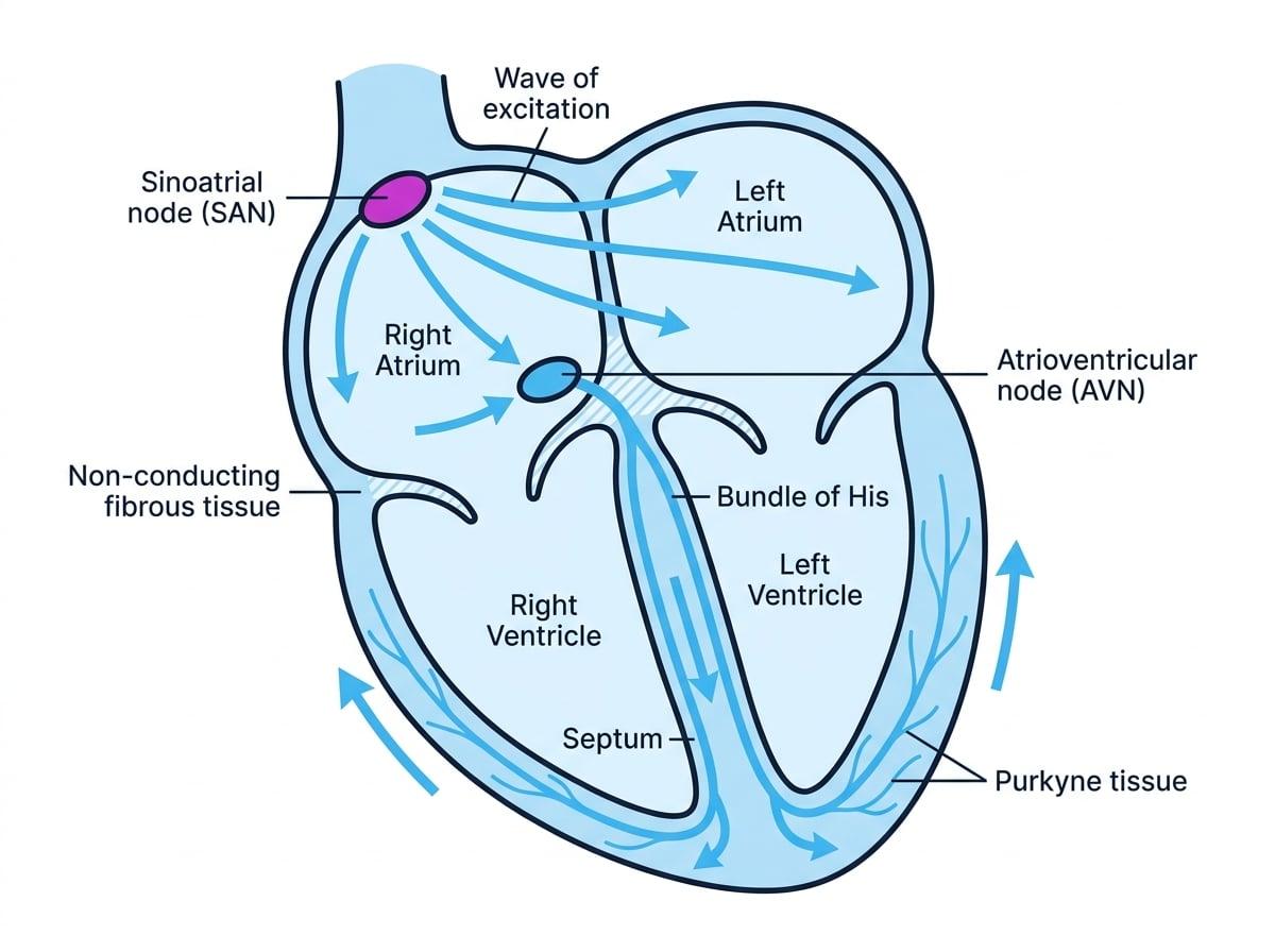
- The Sinoatrial Node (SAN): Located in the wall of the right atrium, the SAN acts as the heart's primary pacemaker. It initiates a wave of electrical excitation that spreads rapidly across the walls of both atria.
- Atrial contraction: This electrical wave causes both atria to contract simultaneously, pushing blood down into the ventricles.
- The non-conducting layer: A layer of non-conducting fibrous tissue separates the atria from the ventricles. This prevents the electrical wave from crossing directly into the ventricular walls, ensuring the ventricles don't contract at the exact same time as the atria.
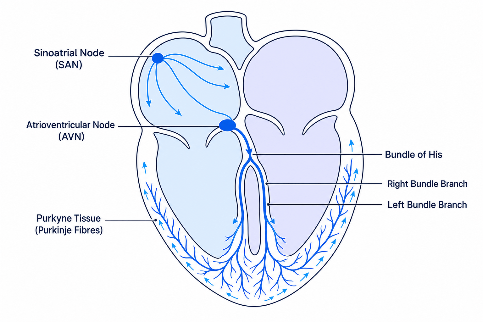
- The Atrioventricular Node (AVN): The only route for the electrical wave to pass down to the ventricles is through the AVN, located between the atria. The AVN deliberately delays the electrical impulse. This short delay is crucial: it allows the atria to fully empty and the ventricles to fully fill with blood before the ventricles contract.
- The bundle of His and Purkyne tissue: After the delay, the AVN passes the wave of excitation down a bundle of specialised conductive muscle fibres called the bundle of His, which runs right down the central septum of the heart to the apex (the bottom).
- Ventricular contraction: At the apex, the bundle of His splits into finer muscle fibres called Purkyne tissue (or Purkinje fibres), which carry the wave of excitation upwards through the muscular walls of both ventricles. This causes the ventricles to contract simultaneously from the bottom upwards, squeezing blood up and out of the heart through the arteries.
Why the delay?
Examiners frequently ask about the AVN. The key points to remember are the delay (to allow ventricles to fill) and the routing of the impulse to the apex (so contraction happens from the bottom up, pushing blood out efficiently).
Modifying the Heart Rate
Although the heart is myogenic, your body still needs to alter your heart rate to meet changing demands (like running for a bus or sleeping). This fine-tuning is controlled by the brain, specifically a region called the medulla oblongata.
Autonomic nervous system
The part of the nervous system responsible for controlling involuntary body functions. It is split into two antagonistic (opposing) pathways: the sympathetic nervous system (which stimulates and speeds up activity) and the parasympathetic nervous system (which inhibits and slows down activity).
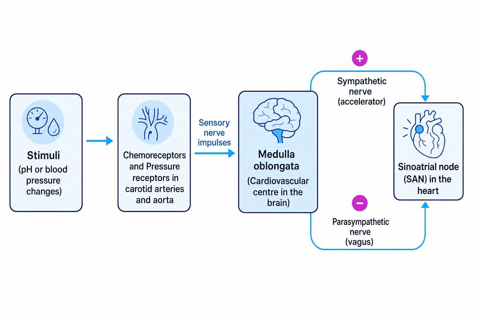
The medulla oblongata has two "centres" linked to the SAN:
- One centre increases heart rate by sending impulses down the sympathetic nervous system.
- One centre decreases heart rate by sending impulses down the parasympathetic nervous system.
To know whether to speed the heart up or slow it down, the medulla relies on sensory information from two types of receptors located in the walls of the carotid arteries (the arteries in the neck supplying the brain) and the aorta.
Chemoreceptors
Chemoreceptors detect changes in the chemical composition of the blood—specifically changes in pH\text{pH}pH caused by carbon dioxide (CO2\text{CO}_{2}CO2).
When you exercise heavily, your rate of respiration increases, releasing more CO2\text{CO}_{2}CO2 into the blood. CO2\text{CO}_{2}CO2 dissolves in the blood to form a weak acid, which lowers blood pH\text{pH}pH.
- Chemoreceptors in the carotid arteries and aorta detect this drop in pH\text{pH}pH and increase the frequency of nerve impulses sent to the medulla oblongata.
- The medulla increases the frequency of impulses sent via the sympathetic nervous system to the SAN.
- The SAN increases the heart rate.
- This increased blood flow rushes the excess CO2\text{CO}_{2}CO2 to the lungs to be exhaled. As CO2\text{CO}_{2}CO2 levels drop, blood pH\text{pH}pH returns to normal, and the chemoreceptors reduce their signaling.
Pressure receptors (Baroreceptors)
Pressure receptors detect changes in blood pressure.
- If blood pressure is too high: Pressure receptors transmit more nerve impulses to the medulla oblongata. The medulla sends impulses via the parasympathetic nervous system to the SAN, slowing the heart rate and bringing pressure back down.
- If blood pressure is too low: Pressure receptors transmit more impulses to the medulla. The medulla sends impulses via the sympathetic nervous system to the SAN, increasing the heart rate to raise blood pressure back to normal.
Nerves vs Hormones
Do not confuse this nervous pathway with adrenaline. While adrenaline is a hormone that also increases heart rate by acting on the SAN, the control loop involving the medulla, chemoreceptors, and pressure receptors is entirely nervous (electrical impulses). If an exam question asks about the nervous control of heart rate, do not mention adrenaline.
Calculating Cardiac Output
The amount of blood the heart pumps out in a given minute is called the cardiac output (COCOCO). It is determined by two factors:
- Heart rate (RRR): The number of beats per minute.
- Stroke volume (VVV): The volume of blood pumped out of the left ventricle during one cardiac cycle (one beat).
The specification outlines a specific mathematical skill (MS 2.2) requiring you to use these values to calculate cardiac output using the formula:
CO=R×V CO = R \times V CO=R×VTypically, cardiac output is measured in dm3 min−1\text{dm}^{3} \text{ min}^{-1}dm3 min−1, heart rate in beats per minute, and stroke volume in dm3\text{dm}^{3}dm3.
Calculating cardiac output during exercise
A student takes their pulse during an investigation into heart rate and records 125 beats per minute. Their estimated stroke volume is 82 cm382 \text{ cm}^{3}82 cm3. Calculate their cardiac output in dm3 min−1\text{dm}^{3} \text{ min}^{-1}dm3 min−1.
- Convert the stroke volume from cm3\text{cm}^{3}cm3 to dm3\text{dm}^{3}dm3 by dividing by 1000.
- Substitute the values for heart rate (RRR) and stroke volume (VVV) into the cardiac output formula.
- Calculate the final value, ensuring the units match the required dm3 min−1\text{dm}^{3} \text{ min}^{-1}dm3 min−1.
Checking your units
In exam questions, stroke volume is almost always given in cm3\text{cm}^{3}cm3 but cardiac output is almost always requested in dm3 min−1\text{dm}^{3} \text{ min}^{-1}dm3 min−1. Remember to divide your stroke volume by 1000 before multiplying, or divide your final answer by 1000 at the end.
In the exam
- Be precise with anatomy: State that the SAN is in the right atrium. State that receptors are in the carotid arteries and aorta. Examiners look for these specific locations.
- Track the pathway clearly: When explaining how heart rate increases due to exercise, map the sequence logically: Exercise →\rightarrow→ more CO2\text{CO}_{2}CO2 →\rightarrow→ lower pH\text{pH}pH →\rightarrow→ chemoreceptors detect change →\rightarrow→ sensory impulses to medulla →\rightarrow→ sympathetic impulses to SAN →\rightarrow→ SAN increases rate.
- Use the phrase "frequency of impulses": Neurones do not send "bigger" impulses when stimulus intensity increases; they send more frequent impulses. Always write that the medulla sends an "increased frequency of impulses" down the sympathetic nerve, never a "stronger impulse".
Check yourself
- Why does the AVN delay the electrical impulse before passing it to the bundle of His?
- What specific change in the blood is detected by chemoreceptors during intense exercise, and why does this change occur?
- Which branch of the autonomic nervous system is responsible for slowing down the heart rate?
- If a patient has a cardiac output of 4.8 dm3 min−14.8 \text{ dm}^{3} \text{ min}^{-1}4.8 dm3 min−1 and a resting heart rate of 60 beats per minute, what is their stroke volume in cm3\text{cm}^{3}cm3?