Receptors (A-level only)
What you'll learn
- What receptors are, and why each receptor responds only to a specific type of stimulus.
- How a Pacinian corpuscle converts pressure into a generator potential.
- How rods and cones in the retina explain sensitivity to light, colour vision and visual acuity.
- How to approach practical questions about human skin receptors.
Starting point: stimuli, receptors and responses
A stimulus is a detectable change in the internal or external environment, such as light intensity, pressure, temperature or blood glucose concentration.
A receptor is a cell, or part of a cell, that detects a stimulus and converts it into a nervous impulse. This conversion of one form of energy into electrical energy is called transduction.
Receptor
A receptor detects a specific stimulus and converts the energy of that stimulus into a nervous signal.
Different receptors are specialised for different stimuli. For example:
- Photoreceptors in the retina detect light.
- Mechanoreceptors detect mechanical pressure, stretch or vibration.
- Chemoreceptors detect chemicals, such as carbon dioxide or glucose.
The type of stimulus a receptor is best adapted to detect is sometimes called its adequate stimulus.
Specificity of receptors
A receptor responds only to a specific type of stimulus because its structure and membrane proteins are specialised for that stimulus.
Generator potentials
When a receptor is stimulated, its cell surface membrane may become depolarised. This means the inside of the cell becomes less negative compared with the outside, usually because positive ions enter the cell.
A generator potential is a small, local change in potential difference across the membrane of a receptor.
Generator potential
A generator potential is a depolarisation produced in a receptor when it detects a stimulus. If it is large enough to reach threshold, it can trigger action potentials in a sensory neurone.
Generator potentials are graded, meaning their size depends on the strength of the stimulus. A stronger stimulus usually causes a larger generator potential.
This is different from an action potential, which is an all-or-nothing nerve impulse. Once threshold is reached, action potentials all have the same size, but stronger stimuli usually produce a higher frequency of action potentials.
Generator potential vs action potential
Do not write that a receptor immediately “makes an action potential” in every case. First, stimulation produces a generator potential. Action potentials occur only if the generator potential reaches threshold.
Predicting whether action potentials are produced
A pressure receptor is stimulated twice. Stimulus A causes a generator potential of -55 mV. Stimulus B causes a generator potential of -45 mV. The threshold is -50 mV.
- Compare stimulus A with the threshold: -55 mV is more negative than -50 mV, so threshold has not been reached.
- Compare stimulus B with the threshold: -45 mV is less negative than -50 mV, so the membrane has depolarised past threshold.
- Therefore, stimulus A will not produce action potentials, but stimulus B will produce action potentials in the sensory neurone.
The Pacinian corpuscle
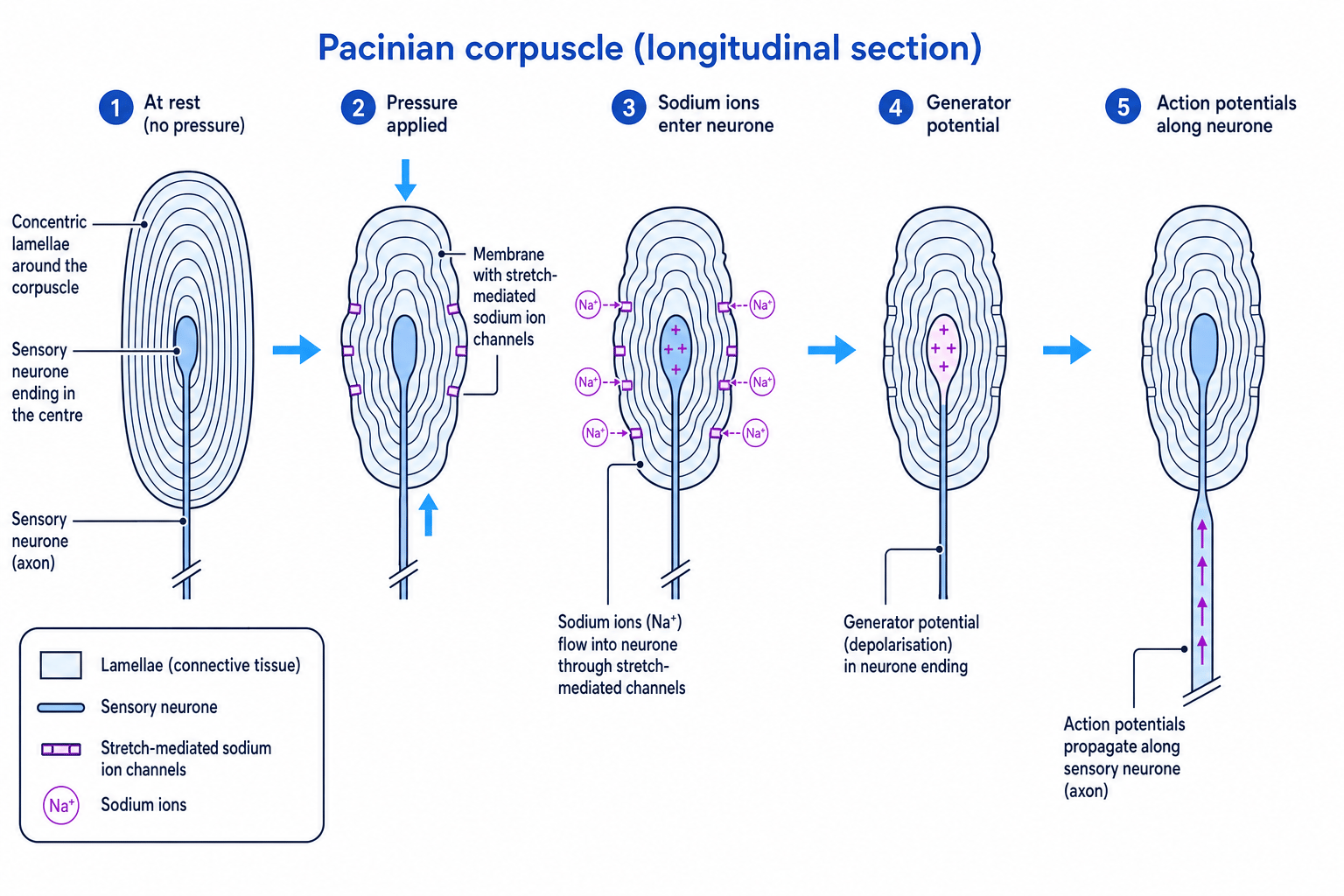
The Pacinian corpuscle is a mechanoreceptor found deep in the skin, as well as in joints, tendons and some internal organs. It detects mechanical pressure and vibration.
It is a useful A-Level example because it shows two important principles:
- receptors respond only to specific stimuli
- stimulation of a receptor leads to a generator potential
The Pacinian corpuscle contains:
- a sensory neurone ending in the centre
- many concentric layers of connective tissue called lamellae
- stretch-mediated sodium ion channels in the membrane of the sensory neurone ending
Stretch-mediated sodium ion channel
A stretch-mediated sodium ion channel is a membrane channel that opens when the membrane is mechanically deformed, allowing sodium ions to diffuse into the neurone.
The lamellae give the Pacinian corpuscle its onion-like structure. They help transmit pressure to the neurone ending and make the receptor especially sensitive to changes in pressure.
How pressure produces a generator potential
At rest, the stretch-mediated sodium ion channels in the sensory neurone membrane are closed. Sodium ions cannot diffuse into the neurone through these channels.
When pressure is applied:
- The Pacinian corpuscle is deformed.
- The membrane of the sensory neurone ending is stretched.
- Stretch-mediated sodium ion channels open.
- Sodium ions diffuse into the neurone down their electrochemical gradient.
- The inside of the neurone becomes less negative, so the membrane is depolarised.
- This depolarisation is the generator potential.
- If threshold is reached, action potentials travel along the sensory neurone.
Pacinian corpuscle mechanism
Pressure deforms the Pacinian corpuscle, opening stretch-mediated sodium ion channels. Sodium ions enter, causing depolarisation and establishing a generator potential.
Explaining why a Pacinian corpuscle responds to pressure
A Pacinian corpuscle is exposed to pressure and then to light. Only pressure produces action potentials in its sensory neurone.
- Pressure mechanically deforms the lamellae and the sensory neurone ending.
- This deformation opens stretch-mediated sodium ion channels, so sodium ions enter and produce a generator potential.
- Light does not deform these channels, so it does not produce the same depolarisation in the Pacinian corpuscle.
- Therefore, the Pacinian corpuscle responds specifically to pressure, not light.
The retina: receptors for light
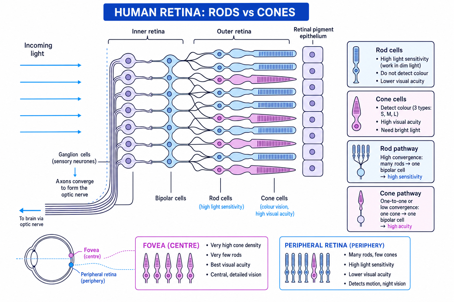
The retina is the light-sensitive layer at the back of the eye. It contains photoreceptors, which convert light energy into nervous impulses.
There are two main types of photoreceptor:
- rod cells
- cone cells
Both rods and cones contain optical pigments. An optical pigment is a light-sensitive molecule that changes when it absorbs light, leading to a generator potential in the photoreceptor.
Optical pigment
An optical pigment is a light-sensitive molecule in a photoreceptor that absorbs particular wavelengths of light and helps trigger a nervous signal.
Light passes through the inner layers of the retina before reaching rods and cones. The signals then pass to bipolar cells, then to sensory neurones. The axons of these sensory neurones form the optic nerve, which carries impulses to the brain.
Rod cells: high sensitivity, low acuity
Rod cells are very sensitive to light, so they allow you to see in dim light. They contain the optical pigment rhodopsin, which is broken down even at low light intensities.
However, rods do not detect colour. They mainly give black-and-white vision.
Rods also have low visual acuity because many rod cells often connect to one bipolar neurone. This is called convergence.
Visual acuity
Visual acuity is the ability to distinguish between two points that are close together, giving sharp, detailed vision.
If many rods share the same neurone pathway, the brain receives a signal but cannot tell exactly which rod was stimulated. This improves sensitivity but reduces detail.
Rod cells
Rod cells are highly sensitive to light because many rods converge onto the same neurone pathway, but this convergence gives low visual acuity.
Cone cells: colour and high acuity
Cone cells are less sensitive to light, so they need brighter light to work effectively. However, they provide colour vision and high visual acuity.
Humans have three types of cone, each containing a different optical pigment:
- cones most sensitive to red light
- cones most sensitive to green light
- cones most sensitive to blue light
The brain compares stimulation from the three cone types to produce colour vision.
Cones give high visual acuity because cone pathways show little or no convergence. In the fovea, many cone cells connect to their own bipolar cells and sensory neurones. This means the brain can identify the exact area of the retina that was stimulated.
The fovea is the central region of the retina with a very high density of cones. It gives the sharpest vision, especially in bright light.
Cone cells
Cone cells give colour vision and high visual acuity because they contain different optical pigments and have little convergence in their neurone pathways.
Explaining dim-light and detailed colour vision
A student can detect movement at the edge of their vision in dim light, but they need to look directly at small coloured text to read it clearly.
- In dim light, rod cells are most useful because they are very sensitive to light.
- The peripheral retina contains many rods, so movement at the edge of vision can be detected even when light intensity is low.
- Small coloured text needs cone cells because cones detect colour and provide high visual acuity.
- The fovea has the highest density of cones, so looking directly at the text places its image on the fovea and gives the clearest vision.
Comparing rods and cones
| Feature | Rod cells | Cone cells |
|---|---|---|
| Light sensitivity | High | Lower |
| Work best in | Dim light | Bright light |
| Colour vision | No | Yes |
| Optical pigment | Rhodopsin | Three different cone pigments |
| Visual acuity | Low | High |
| Convergence | Many rods to one bipolar cell | Little or no convergence |
| Main location | More common in peripheral retina | Densest in fovea |
Remember rods and cones
Rods are for low light: think “rods for the dark”. Cones are for colour and clarity.
Practical angle: investigating skin receptors
You may meet questions about designing investigations into human skin receptors, such as:
- sensitivity of temperature receptors
- habituation of touch receptors
- resolution of touch receptors
Habituation is a reduced response to a repeated, harmless stimulus. For example, you may stop noticing the feeling of clothing on your skin.
Resolution of touch receptors can be investigated using two-point discrimination. This tests the minimum distance at which two separate points touching the skin can be felt as two points rather than one.
Good experimental design includes:
- controlling the force applied
- using the same skin area for comparisons, or clearly comparing different named areas
- preventing the participant from seeing the stimulus
- repeating measurements and calculating a mean
- considering safety and consent, especially when working with human participants
Interpreting two-point discrimination
A student compares touch resolution on the fingertip and forearm. The fingertip detects two points as separate when they are 3 mm apart. The forearm needs 35 mm.
- A smaller two-point threshold means the skin can distinguish closer points, so it has better touch resolution.
- The fingertip threshold, 3 mm, is much smaller than the forearm threshold, 35 mm.
- Therefore, the fingertip has higher touch receptor density or more precise sensory neurone connections than the forearm.
Sensitivity is not the same as acuity
High sensitivity means detecting a weak stimulus. High acuity means distinguishing fine detail or close points. Rods have high sensitivity but low visual acuity.
In the exam
- For Pacinian corpuscles, always link pressure → deformation → sodium ion channels open → sodium ions enter → depolarisation → generator potential.
- For retina questions, explain differences using both optical pigments and neurone connections. Pigments explain colour and light sensitivity; convergence explains acuity and sensitivity.
- Use precise terms: write generator potential for the initial receptor depolarisation, and action potential only when threshold has been reached.
Check yourself
- Why does a Pacinian corpuscle respond to pressure but not light?
- How does convergence make rods more sensitive but reduce visual acuity?
- Why is the fovea the best part of the retina for reading small coloured text?