What you'll learn
- Why blood glucose concentration must be controlled by negative feedback.
- How the liver, insulin, glucagon and adrenaline regulate glucose.
- How the second messenger model explains glucagon and adrenaline action.
- How to use a dilution series, colorimeter and calibration curve to estimate glucose concentration in an unknown “urine” sample.
Why blood glucose must be controlled
Your cells need glucose for respiration, which releases energy for processes such as active transport, protein synthesis and muscle contraction. But blood glucose concentration cannot be allowed to swing too far.
Blood glucose concentration
Blood glucose concentration is the amount of glucose dissolved in a given volume of blood, often measured in millimoles per cubic decimetre, mmol dm⁻³.
If blood glucose is too low, cells may not receive enough glucose for respiration. If it is too high, it can lower the water potential of the blood and cause water to move out of cells by osmosis.
Homeostasis and negative feedback
Homeostasis is the maintenance of a stable internal environment. Negative feedback is a control mechanism in which a change from the normal level triggers responses that reverse the change.
Blood glucose is affected by several factors: carbohydrate intake, glucose absorption from the small intestine, respiration during exercise, storage or release of glycogen, and hormones such as insulin, glucagon and adrenaline.
The overall pattern
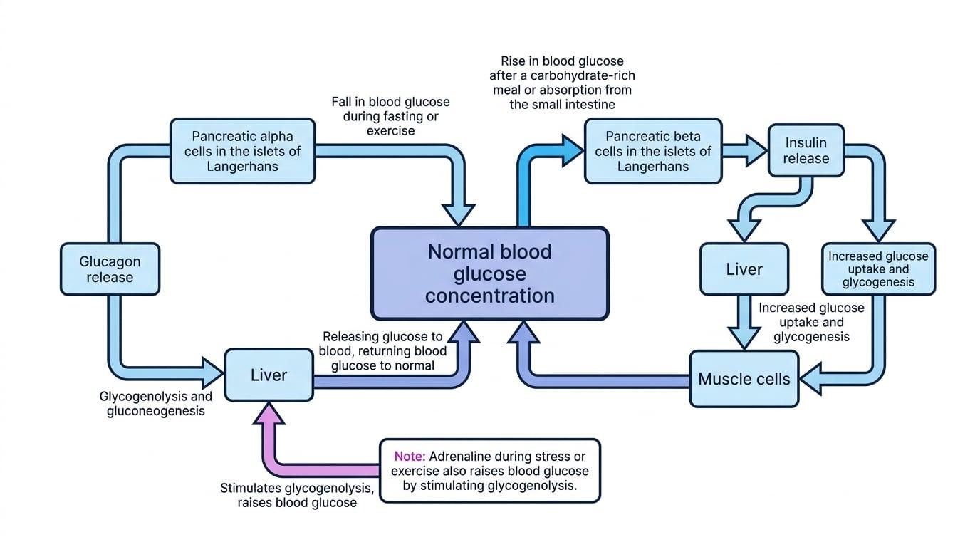
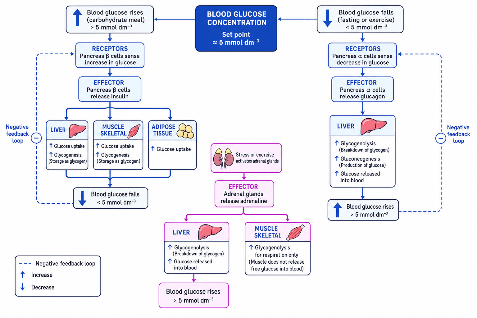
When blood glucose rises, insulin lowers it. When blood glucose falls, glucagon raises it. Adrenaline also raises blood glucose during stress or exercise.
Predicting the response after a carbohydrate meal
- A carbohydrate meal is digested to glucose, which is absorbed into the blood, so blood glucose concentration rises above the normal level.
- Pancreatic beta cells detect this rise and secrete insulin into the blood.
- Insulin causes target cells, especially liver and muscle cells, to take up more glucose and convert some of it to glycogen, so blood glucose falls back towards the normal level.
The pancreas and liver
The pancreas contains groups of hormone-secreting cells called the islets of Langerhans. Beta cells secrete insulin. Alpha cells secrete glucagon.
Hormone and target cell
A hormone is a chemical messenger released into the blood. A target cell is a cell with specific receptors that the hormone can bind to.
The liver is central because it can store glucose as glycogen and release glucose back into the blood.
Three key liver processes
Glycogenesis
Glycogenesis is the conversion of glucose into glycogen. This lowers blood glucose concentration.
Glycogenolysis
Glycogenolysis is the breakdown of glycogen into glucose. This raises blood glucose concentration.
Gluconeogenesis
Gluconeogenesis is the production of glucose from non-carbohydrate molecules such as glycerol and amino acids. This raises blood glucose concentration.
Glucagon is not glycogen
Glucagon is a hormone. Glycogen is a storage polysaccharide made from glucose. Their names are similar, but they are completely different things.
Insulin lowers blood glucose
Insulin is released when blood glucose concentration is too high, such as after a meal.
Insulin acts by attaching to receptors on the surfaces of target cells. This changes the activity of the cell without insulin needing to enter the cell.
Its main effects are:
- It increases the uptake of glucose into target cells.
- It regulates the inclusion of glucose channel or transporter proteins in the surface membranes of target cells.
- It activates enzymes that convert glucose into glycogen by glycogenesis.
In muscle and liver cells, this means glucose is removed from the blood and stored as glycogen.
Insulin does not become glycogen
Do not write that insulin is converted to glycogen. Insulin is a hormone that activates processes causing glucose to be converted into glycogen.
Glucagon raises blood glucose
Glucagon is released when blood glucose concentration is too low, such as during fasting.
Glucagon attaches to receptors on the surfaces of target cells, mainly liver cells. It activates enzymes involved in:
- converting glycogen into glucose by glycogenolysis
- converting glycerol and amino acids into glucose by gluconeogenesis
The liver then releases glucose into the blood.
Adrenaline raises blood glucose quickly
Adrenaline is a hormone released from the adrenal glands during stress, fear or exercise. It prepares the body for rapid action.
Adrenaline attaches to receptors on target cells and activates enzymes involved in converting glycogen to glucose. In the liver, glucose can be released into the blood. In muscle, glucose from glycogen is mainly used locally for respiration.
The second messenger model
Glucagon and adrenaline are examples of hormones that use a second messenger inside the cell.
Second messenger
A second messenger is a molecule inside a cell that transmits the signal from a hormone bound to a receptor on the cell surface.
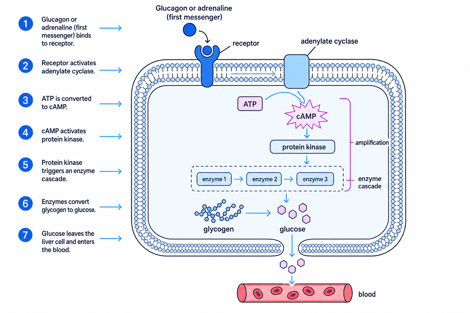
The hormone is the first messenger because it carries the signal through the blood. It binds to a receptor on the surface membrane. This activates an enzyme called adenylate cyclase, which converts ATP into cyclic AMP, usually shortened to cAMP.
cAMP activates protein kinase, an enzyme that phosphorylates other proteins. This triggers an enzyme cascade, leading to glycogen being broken down into glucose.
Amplification
One hormone molecule can lead to many cAMP molecules being made, which can activate many enzyme molecules. This amplifies the original signal.
Diabetes mellitus
Diabetes mellitus is a condition in which the body cannot control blood glucose concentration effectively. It often causes prolonged high blood glucose, called hyperglycaemia.
Type I diabetes
Type I diabetes is usually caused by an autoimmune response that destroys pancreatic beta cells. This means little or no insulin is produced.
It is controlled by:
- insulin injections or an insulin pump
- monitoring blood glucose concentration
- matching insulin dose to carbohydrate intake and activity level
- managing diet to avoid large glucose spikes
Type II diabetes
Type II diabetes is usually caused by target cells becoming less responsive to insulin. This is called insulin resistance. The pancreas may still produce insulin, especially early in the condition, but the target cells do not respond properly.
Risk factors include obesity, physical inactivity, age, genetic factors and diet. It may be controlled by:
- reducing energy intake if overweight
- eating a balanced diet with controlled carbohydrate intake
- increasing physical activity
- medication that improves insulin sensitivity or insulin release
- insulin treatment in some cases
Type II is not simply eating sugar once
Type II diabetes develops over time and involves insulin resistance. High sugar intake can contribute indirectly by increasing energy intake and obesity risk, but it is not the only factor.
Health advisers and the food industry
You may be asked to evaluate views about the increased incidence of type II diabetes.
Health advisers often argue for reduced intake of free sugars, clearer food labelling, smaller portion sizes, more fibre-rich foods and more physical activity. The food industry may argue for consumer choice, reformulation of products and personal responsibility, but may also be criticised for marketing energy-dense foods and resisting regulation.
A balanced answer should recognise that type II diabetes is multifactorial: diet matters, but so do activity level, genetics, age, income, education and access to healthy food.
Required practical 11: glucose concentration by colorimetry
In this practical, you produce known glucose concentrations, measure their colour intensity using a colorimeter, then use a calibration curve to estimate the concentration of glucose in an unknown “urine” sample.
Colorimeter
A colorimeter is an instrument that measures how much light is absorbed by a coloured solution. Absorbance is often recorded in arbitrary units.
Calibration curve
A calibration curve is a graph made using known concentrations. You use it to estimate the concentration of an unknown sample from its absorbance.
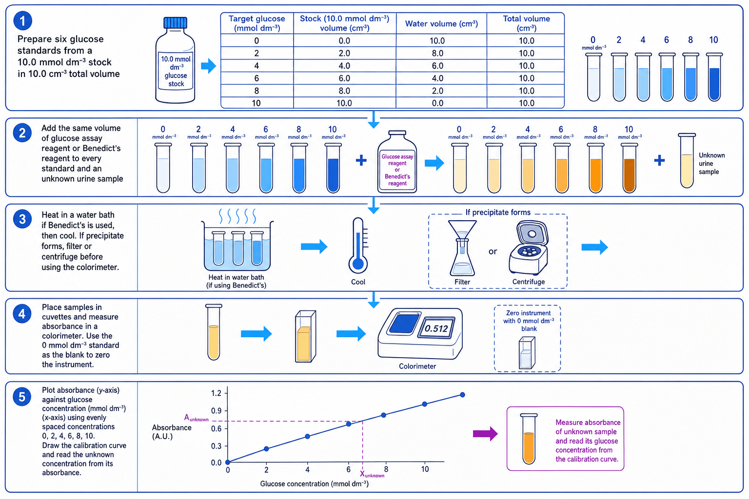
The basic method is:
- Prepare a dilution series of glucose standards with known concentrations.
- Add the same volume of reagent to each standard and to the unknown sample.
- Heat in a water bath if using Benedict’s reagent, then cool.
- If a precipitate forms, filter or centrifuge before measuring absorbance.
- Zero the colorimeter using a blank.
- Measure absorbance for each standard and the unknown.
- Plot absorbance against glucose concentration.
- Read the unknown concentration from the calibration curve.
Preparing a glucose standard
You have a 10.0 mmol dm⁻³ glucose stock solution. You need 10.0 cm³ of a 6.0 mmol dm⁻³ standard.
- Use the dilution equation, making sure both volumes use the same unit:
- Substitute the known values:
- Rearrange and calculate the volume of stock solution:
- Add water to make the final volume up to 10.0 cm³, so add 4.0 cm³ of distilled water.
Good practical control
Keep reagent volume, heating time, temperature, cooling time, cuvette orientation and colorimeter wavelength the same for every sample.
Do not extrapolate
If the unknown absorbance is higher than your highest standard, dilute the unknown and repeat. Reading beyond the calibration curve is unreliable.
In the exam
- For hormone control questions, always name the stimulus, receptor cells, hormone, target organ and effect on blood glucose.
- For insulin, mention cell-surface receptors, increased glucose uptake and activation of glycogenesis enzymes.
- For glucagon or adrenaline, include the second messenger sequence: receptor, adenylate cyclase, cAMP, protein kinase and enzyme activation.
- For the practical, describe known standards, a blank, absorbance readings, a calibration curve and how the unknown concentration is read.
Check yourself
- What happens in glycogenesis, glycogenolysis and gluconeogenesis?
- How does insulin increase glucose uptake into target cells?
- Why does the second messenger model allow a small amount of hormone to produce a large response?