What you'll learn
- How light microscopes, slides, cover slips and stains are used to view cells.
- How to calculate magnification and handle small units like micrometres.
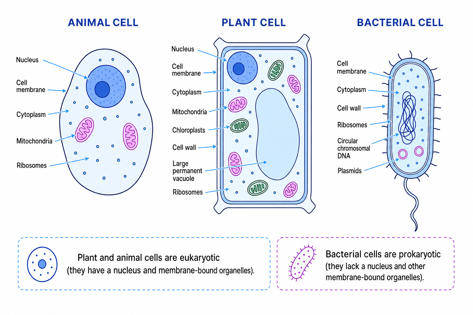
- The main sub-cellular structures in plant, animal and bacterial cells.
- Why electron microscopes increased our understanding of cells.
Cells: the basic building blocks of life
A living organism is any living thing, such as a bacterium, plant or animal. All living organisms are made of cells.
Cell
A cell is the smallest basic unit of a living organism. Cells carry out life processes such as respiration, growth and making proteins.
Inside cells are smaller parts called sub-cellular structures. “Sub-cellular” just means “smaller than a cell”. These structures are not just decoration — each one has a job that helps the whole cell function.
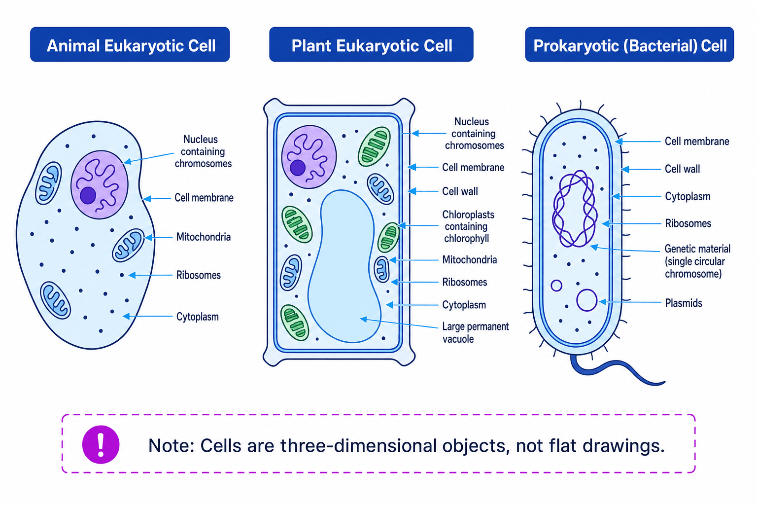
Cells are also three-dimensional objects. Diagrams often look flat because they show a simplified slice or view of the cell, but real cells have depth.
Cells are not flat
Do not imagine a cell as a flat circle on a page. A real animal cell is more like a tiny blob with structures inside it, and a plant cell is more like a box-shaped 3D compartment.
Viewing cells with a light microscope
A microscope is an instrument used to view objects that are too small to see clearly with the naked eye.
Light microscope
A light microscope uses visible light and lenses to form a magnified image of a specimen.
A specimen is the object or material being viewed, such as onion epidermis cells or cheek cells. In GCSE practical work, specimens are often placed on a glass slide and covered with a thin glass cover slip.
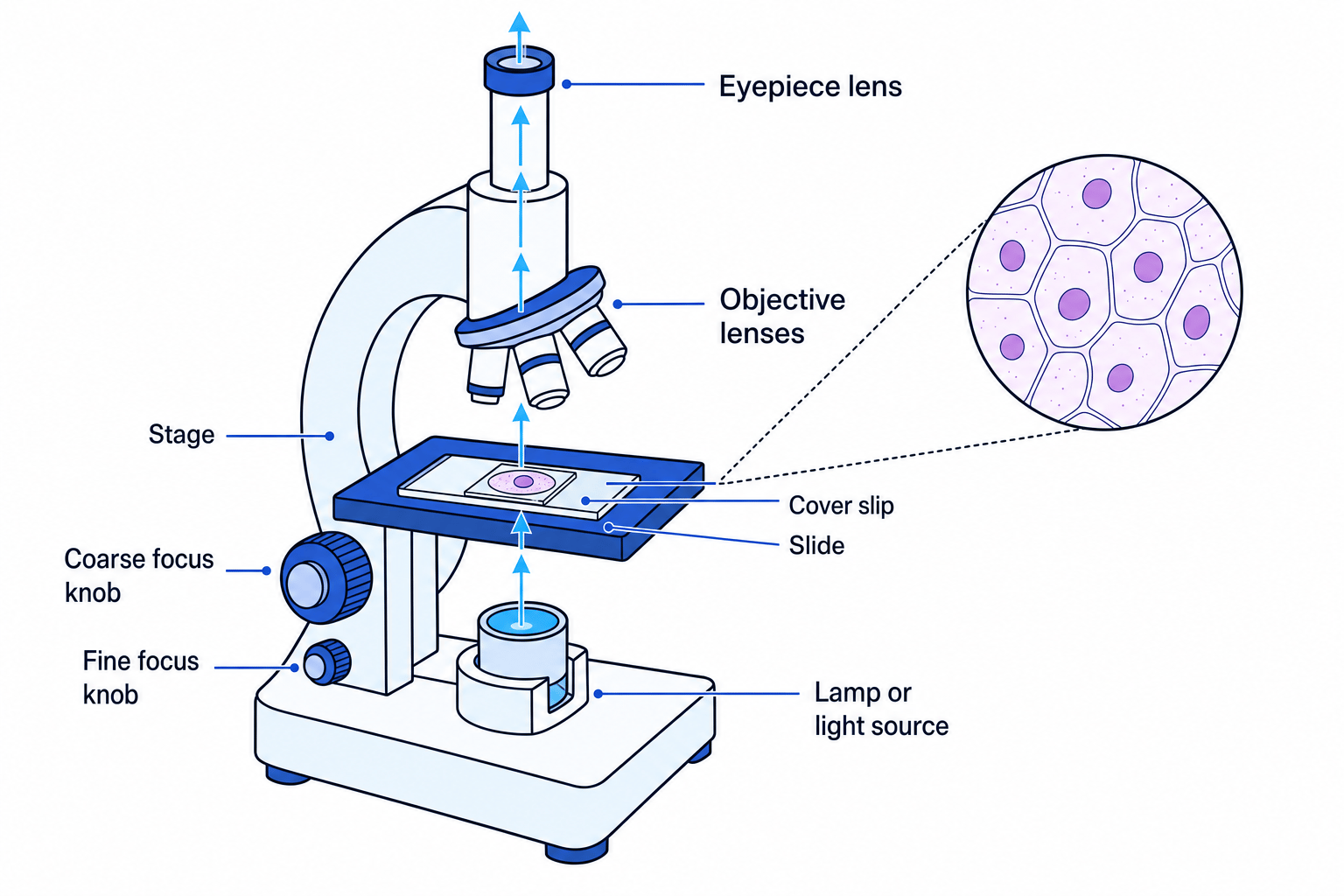
This labelled microscope shows the parts you need to recognise and use.
Main microscope parts
- Lenses bend light to make the image appear larger. A typical light microscope has an eyepiece lens and objective lenses.
- The stage supports the slide.
- The lamp or light source shines light through the specimen.
- The coarse focus knob moves the stage or lenses a larger distance to bring the image roughly into focus.
- The fine focus knob makes smaller adjustments to sharpen the image.
Preparing and viewing a slide
To view cells using a light microscope:
- Place a thin specimen on a clean slide, often in a drop of water.
- Add a stain if needed. A stain is a coloured chemical that makes colourless structures easier to see, or highlights different tissues or structures.
- Gently lower a cover slip over the specimen to protect it and reduce air bubbles.
- Put the slide on the stage and start with the lowest-power objective lens.
- Use the coarse focus knob first, then the fine focus knob.
- Move to a higher-power objective lens if you need to see more detail.
Stains do not magnify
A stain makes structures easier to see by improving contrast. It does not make the specimen larger — magnification comes from the lenses.
Size, scale and magnification
Cells are tiny, so biologists often use very small units:
- A micrometre is one thousandth of a millimetre. Its symbol is µm.
- A nanometre is one thousandth of a micrometre. Its symbol is nm.
A typical animal cell might be around 10–30 µm across. Some sub-cellular structures are smaller than this, so using the right units matters.
Magnification
Magnification tells you how many times larger the image is than the actual object.
The key equation is:
M=IAM = \frac{I}{A}M=AIwhere:
- MMM is magnification
- III is image size
- AAA is actual size
Image size and actual size must be in the same unit before you divide.
For a microscope, total magnification is found by multiplying the eyepiece magnification by the objective lens magnification:
Mtotal=Meyepiece×MobjectiveM_{\text{total}} = M_{\text{eyepiece}} \times M_{\text{objective}}Mtotal=Meyepiece×MobjectiveEstimating cell size
If an exact measurement is difficult, estimate sensibly. For example, if about five similar cells fit across a field of view that is 500 µm wide, each cell is about 100 µm wide.
Calculating magnification
A cell appears 40 mm long in a drawing. Its actual length is 80 µm. Calculate the magnification.
- Convert the image size into micrometres so both sizes use the same unit: 40 mm is 40,000 µm.
- Substitute into the magnification equation: M=40,000 μm80 μmM = \frac{40,000\ \mu\text{m}}{80\ \mu\text{m}}M=80 μm40,000 μm.
- Divide the numbers and cancel the units: M=500M = 500M=500, so the cell has been magnified 500 times.
If you are taking Higher Tier, very small measurements may also be written in standard form. For example, a length of 2 µm can be written as 2×10⁻⁶ m2 \times 10^{-6}\ \text{m}2×10⁻⁶ m.
Eukaryotic and prokaryotic cells
Cells can be grouped into two main types.
Eukaryotic and prokaryotic cells
A eukaryotic cell has a nucleus containing genetic material. Plant and animal cells are eukaryotic. A prokaryotic cell is smaller and simpler, with no nucleus; bacterial cells are prokaryotic.
Genetic material is the DNA that carries instructions for the cell. In eukaryotic cells, this DNA is arranged into chromosomes inside the nucleus. A chromosome is a long DNA molecule carrying many genes.
Here is a comparison of the key structures in animal, plant and bacterial cells.
Structure links to function
In cell biology, always connect the name of a structure to what it does. The function explains why that structure matters to the cell.
Main sub-cellular structures and their functions
| Structure | Found in | Main function |
|---|---|---|
| Nucleus | Plant and animal cells | Contains genetic material arranged as chromosomes; helps control cell activities. |
| Chromosomes | Plant and animal cell nuclei | Carry genetic instructions in DNA. |
| Cell membrane | Plant, animal and bacterial cells | Acts as a selective barrier, controlling movement of substances in and out; contains receptor molecules. |
| Receptor molecules | Cell membranes | Detect chemical signals outside the cell. |
| Mitochondria | Plant and animal cells | Contain enzymes for cellular respiration, which transfers energy for cell processes. |
| Chloroplasts | Plant cells | Contain chlorophyll, which absorbs light for photosynthesis. |
| Ribosomes | Plant, animal and bacterial cells | Site of protein synthesis, meaning where proteins are made. |
| Plasmids | Bacterial cells | Small loops of DNA, separate from the main bacterial chromosome. |
A few extra structures are useful to know too. Plant cells have a cell wall, which strengthens and supports the cell, and often a large permanent vacuole, which contains cell sap and helps keep the cell firm. Bacterial cells also usually have a cell wall, but they do not have a nucleus, mitochondria or chloroplasts.
Using structures to identify a cell
A micrograph shows cells with a cell wall, chloroplasts and a nucleus. Decide what type of cell is shown.
- The nucleus shows the cells are eukaryotic, because prokaryotic cells do not have a nucleus.
- The chloroplasts show the cells are plant cells, because animal cells do not contain chloroplasts.
- The cell wall supports this conclusion, because plant cells have a cell wall whereas animal cells do not.
Bacteria are not tiny animal cells
Bacterial cells are prokaryotic. They do have a cell membrane, cytoplasm, ribosomes and genetic material, but they do not have a nucleus.
Electron microscopy and resolution
Light microscopes are useful, but they cannot show every detail inside a cell. This is because they have limited resolution.
Resolution
Resolution is the ability to distinguish two points that are very close together as two separate points.
Magnification and resolution are different. Magnification makes an image larger. Resolution decides whether the detail is clear enough to separate.
A transmission electron microscope, often shortened to TEM, uses a beam of electrons transmitted through a very thin specimen. Electrons can give much higher resolution than visible light, so TEM images reveal smaller sub-cellular structures in much greater detail.
This increased resolution helped scientists understand cells better because structures that looked blurred or invisible with light microscopes could be seen more clearly, including tiny structures such as ribosomes and details inside organelles.
Comparing microscope resolution
Two tiny structures in a cell are 100 nm apart. A light microscope has a resolution of about 200 nm, while a TEM can resolve much smaller distances. Decide which microscope can show the two structures separately.
- Compare the separation with the light microscope resolution: 100 nm is smaller than 200 nm.
- Because the structures are closer together than the light microscope can resolve, they would appear blurred together.
- A TEM has much higher resolution, so it can distinguish structures much closer than this; it could show them separately.
Electron microscope limitations
Electron microscopes are powerful, but specimens usually have to be specially prepared and are not viewed alive. Electron micrographs are also often black and white unless colour is added afterwards.
In the exam
- If asked for a function, link the structure to the job: for example, mitochondria contain enzymes for cellular respiration.
- In magnification questions, convert image size and actual size into the same unit before dividing.
- Do not confuse magnification with resolution: a bigger image is not automatically a clearer image.
Check yourself
- What structures would you expect to find in a plant cell but not an animal cell?
- Why is a stain useful when viewing cheek cells or onion cells with a light microscope?
- How is genetic material arranged differently in eukaryotic and prokaryotic cells?