Defence cells in the blood
White blood cell
A white blood cell is a blood cell with a nucleus that defends the body against pathogens.
Platelet
A platelet is a fragment of a cell in the blood that helps blood to clot at a wound.
Pathogen
A microorganism that causes a communicable disease, such as a virus, a bacterium, a fungus or a protist.
- Blood is made of a liquid called plasma carrying red blood cells, white blood cells and platelets.
- Red blood cells carry oxygen, while white blood cells and platelets are the parts of the blood that defend the body against pathogens.
- White blood cells are much less common than red blood cells, with roughly one white blood cell for every 600600600 red blood cells in a healthy person.
- Unlike red blood cells, every white blood cell has a nucleus, and the shape and size of that nucleus is one way of telling the different types apart.
- The number of white blood cells rises during an infection, because the body makes more of them to fight the pathogen.
- There are two main types of white blood cell, the phagocytes and the lymphocytes, and each has a structure suited to a different job.
- Platelets are not whole cells but tiny fragments of cells, and their job is to start the formation of a blood clot when a blood vessel is damaged.
- A cell is adapted to its function when its structure makes it able to carry out that function well, so every feature described below should be linked to what it lets the cell do.
Phagocytes
Phagocyte
A white blood cell that engulfs and digests pathogens by phagocytosis.
Phagocytosis
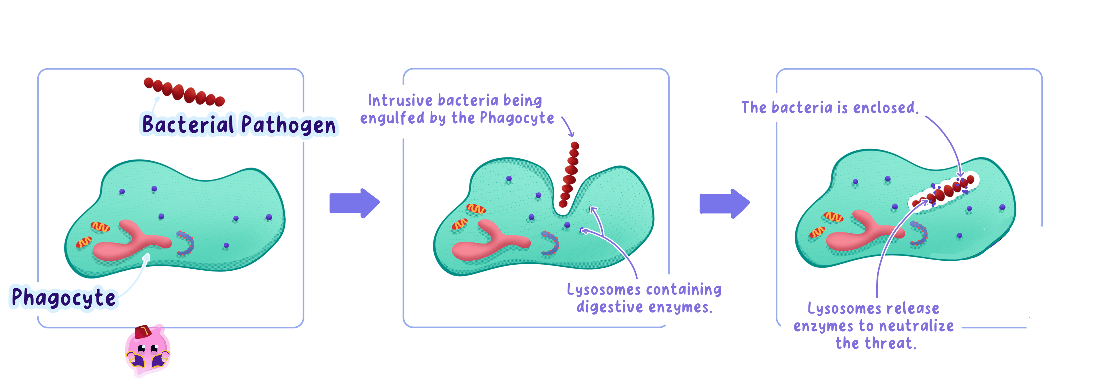
The process in which a phagocyte surrounds a pathogen, takes it into the cell and digests it with enzymes.
- A phagocyte destroys pathogens by phagocytosis, which means it takes a pathogen inside itself and digests it.
- Phagocytes are non-specific, so the same phagocyte will engulf a bacterium, a virus, a fungal spore or any other cell it recognises as foreign.
- Phagocytosis happens in a fixed sequence of steps.
- The phagocyte is attracted towards the pathogen by chemicals released by the pathogen and by damaged tissue.
- The phagocyte's cell membrane flows around the pathogen until the pathogen is completely engulfed inside a small bag of membrane called a vacuole.
- Small sacs called lysosomes fuse with the vacuole and release digestive enzymes onto the pathogen.
- The enzymes break the pathogen down into small harmless molecules, which the phagocyte absorbs or releases.
- Each feature of a phagocyte is linked to one of these steps.
- A flexible cell membrane and fluid cytoplasm let the phagocyte change shape so it can wrap around and engulf a pathogen.
- Receptors on the membrane detect chemicals from the pathogen, so the phagocyte can move towards the site of infection.
- Large numbers of lysosomes in the cytoplasm provide the enzymes needed to digest the engulfed pathogen.
- A lobed nucleus makes the cell flexible enough to squeeze through the gaps between the cells of capillary walls, so it can leave the blood and reach pathogens in infected tissue.
- Many phagocytes die after engulfing large numbers of bacteria, and the pus in an infected cut is largely made of dead phagocytes and the remains of pathogens.
- Do not write that a phagocyte "eats" a pathogen, because the marks are given for the words engulfs and digests.
- Do not write that phagocytes make antibodies, because antibodies are made only by lymphocytes.
- Do not describe phagocytosis as specific, because a phagocyte engulfs any pathogen it meets.
Lymphocytes
Lymphocyte
A white blood cell that produces antibodies and antitoxins in response to a specific antigen.
Antibody
A protein produced by lymphocytes that binds to a specific antigen with a complementary shape.
Antitoxin
A protein produced by lymphocytes that binds to a specific toxin and neutralises it.
- A lymphocyte is a white blood cell with a large, round nucleus that fills most of the cell and leaves only a thin layer of cytoplasm around it.
- Lymphocytes defend the body by producing antibodies and antitoxins, which are both proteins.
- Antibodies bind to antigens, the molecules on the surface of a pathogen, and each antibody has a shape that fits only one type of antigen.
- Antitoxins bind to the toxins released by some bacteria and stop the toxins damaging body cells.
- Each feature of a lymphocyte is linked to making these proteins in large amounts.
- The large nucleus contains the genes that code for the antibody proteins.
- The cytoplasm contains many ribosomes, which are the sites of protein synthesis, so the cell can make antibody molecules quickly.
- A well-developed network of membranes inside the cytoplasm packages the antibodies so they can be released from the cell in large numbers.
- A lymphocyte that meets its matching antigen can divide rapidly by mitosis, producing many identical lymphocytes that all make the same antibody.
- Because each lymphocyte makes only one kind of antibody, lymphocytes give a specific defence that targets one pathogen at a time.
- Lymphocytes work more slowly than phagocytes the first time a pathogen is met, because the right lymphocytes must first multiply before enough antibody is made.
Platelets and blood clotting
Fibrin
An insoluble protein that forms a mesh of fibres at a wound and traps blood cells to form a clot.
Blood clot
A solid mass of fibrin, platelets and trapped red blood cells that seals a damaged blood vessel.
- Platelets are small, disc-shaped fragments of large cells in the bone marrow, and they have no nucleus.
- Their small size lets huge numbers of them circulate in every blood vessel, so there are always platelets close to any wound.
- When a blood vessel is cut, platelets stick to the damaged vessel wall and to one another, forming a loose platelet plug over the gap.
- The platelets and the damaged tissue then release chemicals that set off a chain of reactions in the plasma.
- These reactions convert the soluble plasma protein fibrinogen into insoluble threads of fibrin.
- The fibrin threads form a mesh across the wound that traps red blood cells and more platelets, producing a blood clot.
- At the surface of the skin the clot dries and hardens into a scab, which stays in place while new skin grows underneath.
- The clot has two defence functions.
- It stops blood loss, which prevents the loss of blood volume and the fall in blood pressure that would follow.
- It forms a barrier that stops pathogens entering the body through the break in the skin.
- A person with too few platelets bruises easily and bleeds for longer from small cuts, which shows how important platelets are to sealing wounds quickly.
Clotting after a grazed knee
- A student falls on a playground and grazes her knee, breaking the skin and many small capillaries.
- Within seconds, platelets stick to the damaged capillary walls and clump together to plug the smallest gaps.
- Chemicals released at the wound convert fibrinogen into fibrin, and a mesh of fibrin threads traps red blood cells to form a clot.
- Over the next hour the clot dries into a scab, which seals the graze so bacteria from the playground surface cannot enter the blood.
- Any bacteria that entered before the scab formed are engulfed by phagocytes that squeeze out of nearby capillaries into the tissue.
Explaining how a cell is adapted
- Name the feature and then state what it allows the cell to do, such as "a lobed nucleus lets the phagocyte squeeze out of capillaries to reach an infection".
- Use the precise verbs examiners look for, which are engulf, ingest and digest for phagocytes, and produce or release for antibodies and antitoxins.
- When asked about platelets, go beyond "they clot the blood" and say that the clot prevents pathogens entering the wound, because that is the defence mark.
- Keep the two white blood cells separate in your answer, since mixing up which cell engulfs and which makes antibodies is the most common way to lose marks.
- State two ways in which white blood cells differ from red blood cells.
- Describe the steps of phagocytosis in the correct order.
- Explain how a lobed nucleus helps a phagocyte to defend the body.
- Explain why a lymphocyte needs a large number of ribosomes.
- Describe how platelets and fibrin form a clot, and give two reasons why the clot helps defend the body.