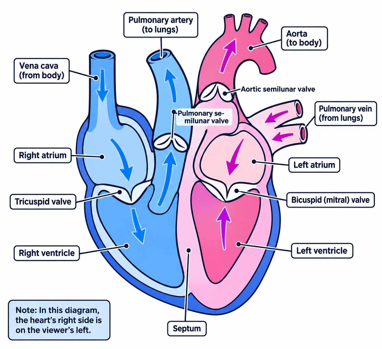
Structure of the heart
Cardiac muscle
Cardiac muscle is the muscle tissue of the heart wall, which contracts and relaxes rhythmically throughout life without becoming fatigued.
Atrium
An atrium is one of the two upper chambers of the heart, which receives blood returning to the heart from a vein.
Ventricle
A ventricle is one of the two lower chambers of the heart, which pumps blood out of the heart into an artery.
Septum
The septum is the wall of muscle that separates the right and left sides of the heart.
- The heart is a double pump built almost entirely from cardiac muscle, a tissue that contracts and relaxes rhythmically for a lifetime without becoming fatigued.
- It has four chambers: a right atrium and a right ventricle on one side, and a left atrium and a left ventricle on the other.
- The septum is a solid wall of muscle down the middle that keeps oxygenated and deoxygenated blood completely separate.
- The atria have thin walls because they only push blood a few centimetres down into the ventricles below them.
- The ventricles have much thicker walls because they have to generate enough pressure to drive blood out of the heart altogether.
- The right ventricle wall is moderately thick because it pumps only as far as the lungs, which sit alongside the heart.
- Its pressure is deliberately kept low, because the thin capillary walls in the lungs would burst under a high pressure.
- The left ventricle wall is the thickest of all, roughly three times the thickness of the right, because it pumps blood all the way round the body.
- More muscle produces a more forceful contraction, which produces a higher blood pressure and so a faster flow to the tissues.
- Four vessels are attached at the top: the vena cava into the right atrium, the pulmonary artery out of the right ventricle, the pulmonary vein into the left atrium and the aorta out of the left ventricle.
- The cardiac muscle itself is supplied with oxygen and glucose by the coronary arteries that run across the outside of the heart.
The valves of the heart
Atrioventricular valve
An atrioventricular valve is a valve between an atrium and a ventricle that stops blood flowing back into the atrium when the ventricle contracts.
Semilunar valve
A semilunar valve is a valve at the base of the aorta or the pulmonary artery that stops blood flowing back into the ventricle when it relaxes.
- A valve is a flap of tissue that opens in one direction only, so blood can pass forwards but cannot slip back.
- Each valve opens when the pressure behind it is higher than the pressure in front of it, and snaps shut when that difference reverses.
- The atrioventricular valves sit between each atrium and the ventricle below it.
- The one on the right has three flaps and is called the tricuspid valve.
- The one on the left has two flaps and is called the bicuspid valve, or the mitral valve.
- Both are anchored to the ventricle wall by tough tendons that stop the flaps turning inside out when the ventricle contracts hard.
- The semilunar valves sit at the base of the aorta and the base of the pulmonary artery, each made of three half-moon pockets.
- They stop blood that has just been forced into an artery falling back into the ventricle as it relaxes.
- Without valves the heart would push blood back and forth in the same place and no net flow would leave it.
How one heartbeat works
- Blood flows into the two atria from the vena cava and the pulmonary vein while the heart is relaxed.
- Both atria then contract together, raising the pressure inside them so the atrioventricular valves are pushed open.
- Blood passes down into the ventricles, which fill and stretch.
- Both ventricles then contract, and the pressure inside them rises sharply.
- That pressure forces the atrioventricular valves shut and forces the semilunar valves open.
- Blood is driven out of the right ventricle into the pulmonary artery and out of the left ventricle into the aorta.
- The ventricles then relax, the pressure inside them falls below the pressure in the arteries and the semilunar valves close.
- The two sets of valves closing are what produce the two sounds of a heartbeat.
Dissecting a mammalian heart
- Aim: to examine the outside and inside of a mammalian heart and relate the thickness of each chamber wall and the position of each valve to the job that chamber does.
- Apparatus: a fresh or fully defrosted sheep or pig heart, a dissecting board or tray, a scalpel, blunt-nosed scissors, forceps, a blunt seeker, a ruler, paper towel, disposable gloves, eye protection, disinfectant and a sealed bag for waste.
- Variables: this is an observational dissection rather than a fair test, so there is no independent variable, and comparability comes from following the same sequence of cuts and measuring each wall at the same height.
- Method, examining the outside:
- Put on gloves and eye protection and work on a board covered with paper towel.
- Rinse the heart and blot it dry, because surface blood hides the vessels.
- Find the front by the diagonal groove running across the surface, and follow the coronary arteries lying in that groove.
- Squeeze each side gently between finger and thumb. The left side feels distinctly firmer because its wall is thicker.
- Identify the vessels at the top: the aorta is the thickest and springs back when squeezed, the pulmonary artery lies in front of it, and the vena cava and pulmonary vein have thin, floppy walls.
- Slide a blunt seeker into each vessel to find which chamber it opens into, rather than naming it from its position alone.
- Method, opening the chambers:
- Cut down the wall of the right ventricle from the pulmonary artery towards the point of the heart, using scissors so the blade does not slice the structures inside.
- Fold the flap back and find the three-flapped tricuspid valve and the tough white tendons anchoring it to the muscle.
- Make the matching cut on the left side, from the aorta down towards the point, and find the two-flapped bicuspid valve.
- Measure the thickness of each ventricle wall with a ruler at the same height and record both values in millimetres.
- Look into the base of the aorta for the three half-moon pockets of the semilunar valve, and pour a little water down the aorta to watch them fill and close.
- Trace the septum between the two ventricles and confirm that no opening passes through it.
- Results: the left ventricle wall is the thickest, the right ventricle wall is thinner and the atria walls are thinnest of all, which matches how far each chamber has to pump blood.
- Maths: express the two ventricle measurements as a ratio, so a left wall of 12 mm12\ \text{mm}12 mm against a right wall of 4 mm4\ \text{mm}4 mm gives 124=3\dfrac{12}{4}=3412=3, written 3:13:13:1.
- Watch out: cutting with a scalpel inside the heart destroys the valve flaps, a partly frozen heart cannot be opened cleanly, and the heart's own right side lies on your left when the front of it faces you.
- Safety: wear gloves and eye protection, cut away from your fingers on a board, treat the heart as a source of bacteria, seal the waste in the bag provided, and disinfect the instruments, the board and your hands afterwards.
Arteries
Artery
An artery is a blood vessel that carries blood away from the heart.
Lumen
The lumen is the central space inside a blood vessel through which the blood flows.
- An artery carries blood away from the heart, and that blood leaves at high pressure in surges.
- The wall is thick so that it can withstand that pressure without bursting.
- The wall contains a great deal of muscle and elastic fibres, so it stretches as each surge arrives and recoils afterwards.
- That stretching and recoil smooths the flow between beats and keeps the blood moving forwards, and it is what you feel as a pulse.
- The lumen is narrow compared with the thickness of the wall, which helps to keep the pressure high.
- An artery has no valves along its length, because the pressure behind the blood is high enough to keep it flowing one way.
Veins
Vein
A vein is a blood vessel that carries blood towards the heart.
- A vein carries blood back towards the heart, and by this stage the blood is at low pressure and flowing steadily rather than in surges.
- The wall is thin with little muscle or elastic tissue, because there is no high pressure to resist.
- The lumen is wide, which reduces the resistance to flow so that low-pressure blood still returns quickly.
- Veins contain valves along their length, which is the clearest structural difference from an artery.
- Those valves stop blood flowing backwards, which matters most in the legs where blood has to travel upwards against gravity.
- Many veins run between skeletal muscles, and each time those muscles contract they squeeze the vein and push blood on towards the heart.
Capillaries
Capillary
A capillary is a blood vessel with a wall one cell thick across which substances are exchanged between the blood and the surrounding tissue.
- A capillary links the smallest arteries to the smallest veins and is the only vessel where substances are actually exchanged with the tissues.
- Its wall is one cell thick, giving the shortest possible diffusion distance between the blood and the surrounding cells.
- Its lumen is so narrow that red blood cells pass through in single file, which presses them against the wall and shortens the distance further.
- Capillaries are extremely numerous and branch through every tissue, so no cell is far from a supply.
- That branching also gives a very large total surface area for exchange.
- Blood flows slowly through a capillary bed, which allows time for diffusion to take place.
- The wall is also slightly leaky, so plasma can seep out to bathe the tissues and white blood cells can squeeze through it.
Identifying a vessel from a cross-section
- A cross-section shows a vessel whose outside diameter is 4.0 mm4.0\ \text{mm}4.0 mm and whose lumen diameter is 1.0 mm1.0\ \text{mm}1.0 mm.
- The total wall thickness is 4.0−1.0=3.0 mm4.0-1.0=3.0\ \text{mm}4.0−1.0=3.0 mm, so each side of the wall is 1.5 mm1.5\ \text{mm}1.5 mm thick.
- The wall on each side is therefore 1.51.51.5 times the width of the lumen.
- A thick wall around a narrow lumen identifies the vessel as an artery.
- A vein of the same outside diameter would show a wall well under 0.5 mm0.5\ \text{mm}0.5 mm thick and a lumen filling most of the vessel.
Explaining an adaptation of a vessel
- Name the feature, then say what it does, because a feature on its own scores nothing on an explain question.
- Use comparative words when two vessels are compared, so write thicker wall and narrower lumen rather than thick wall and narrow lumen.
- Link the thickness of a ventricle wall to the distance blood is pumped, not to the volume of blood, since distance is the reason the left wall is thicker.
- For any valve question, say which way blood is prevented from moving, because backflow is the word the answer turns on.
- Do not say the left ventricle pumps more blood than the right, because both pump the same volume and the left simply pumps it at a higher pressure.
- Do not write that valves pump blood, because they are flaps moved by pressure and it is the muscle that does the pumping.
- Do not describe capillaries as having thick walls or valves, because a one-cell-thick wall is the whole point of them.
- Do not label the tricuspid valve on the left of the heart, because three flaps go with the right side and two flaps with the left.
- Name the four chambers of the heart and the vessel attached to each.
- Explain why the wall of the left ventricle is thicker than the wall of the right ventricle.
- State where the atrioventricular valves and the semilunar valves are found and what each prevents.
- Compare an artery and a vein in terms of wall thickness, lumen diameter and the presence of valves.
- Explain two ways in which a capillary is adapted for the exchange of substances.