What blood is made of
- Blood is a tissue, because it is a group of cells working together, but its cells are carried in a liquid rather than held in a solid block.
- Just over half of blood by volume is plasma, and the rest is made up of cells and cell fragments.
- The four components are plasma, red blood cells, white blood cells and platelets.
- Red blood cells and plasma are the two components that carry out the transport work of the blood.
- Red blood cells vastly outnumber the others, which is why whole blood looks red rather than straw-coloured.
- All of them are made in the bone marrow, the soft tissue inside the larger bones.
How a red blood cell is built
Definition
Red blood cell
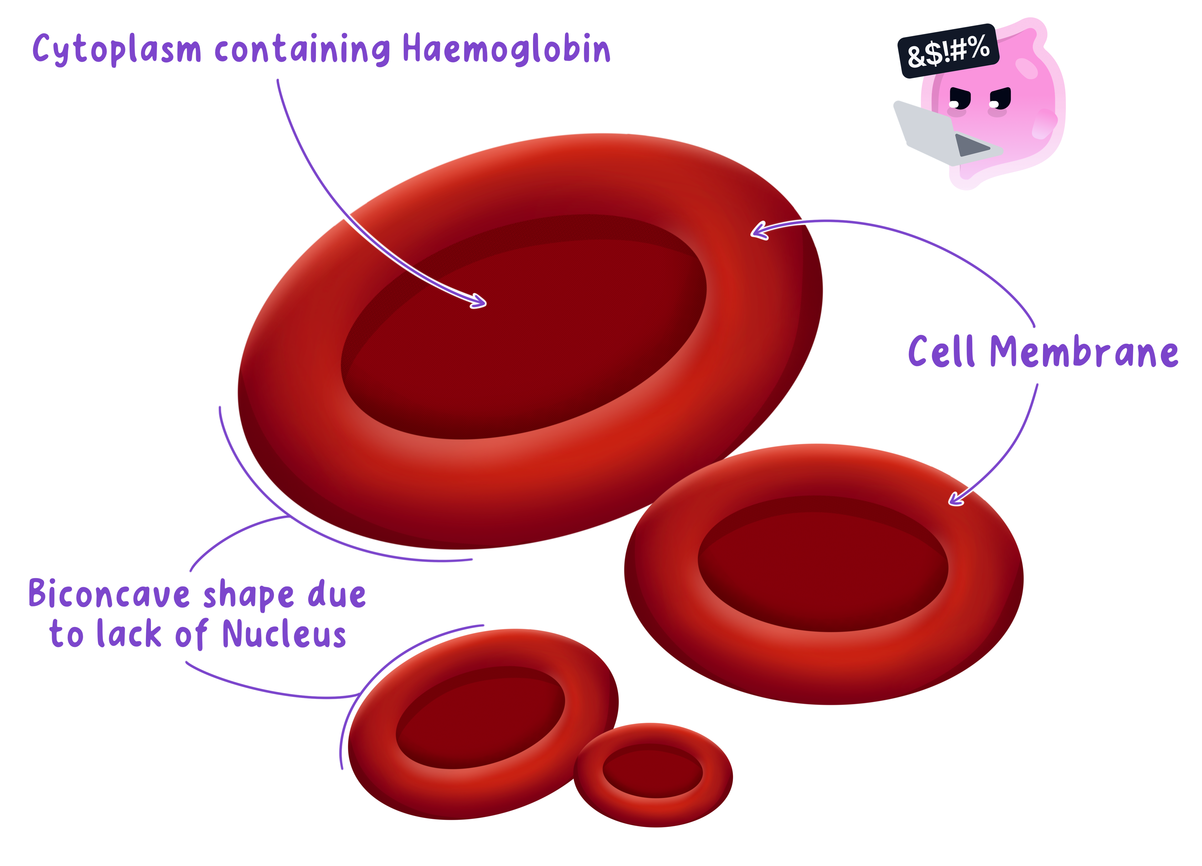
A red blood cell is a biconcave blood cell with no nucleus that is packed with haemoglobin and carries oxygen from the lungs to respiring tissues.
Definition
Haemoglobin
Haemoglobin is the red, iron-containing protein inside red blood cells that binds oxygen reversibly.
- A red blood cell has no nucleus, which frees the whole of the inside of the cell for carrying oxygen.
- That space is filled with haemoglobin, so a single cell holds hundreds of millions of these oxygen-binding molecules.
- The cell is a biconcave disc, which means it is dented inwards on both flat faces.
- That shape gives a larger surface area than a sphere of the same volume, so oxygen can diffuse in and out over a bigger area.
- It also makes the cell thin through the middle, so no haemoglobin molecule sits far from the surface and the diffusion distance stays short.
- The cell is small, roughly 7 μm7\ \mu\text{m}7 μm across, and flexible, so it can bend and squeeze single file along a capillary narrower than itself.
- Being pressed against the capillary wall as it passes shortens the distance oxygen has to diffuse to reach the tissue.
- Having no nucleus means the cell cannot repair itself or divide, so it survives only about three months and is replaced continuously.
Carrying oxygen
Definition
Oxyhaemoglobin
Oxyhaemoglobin is the bright red compound formed when haemoglobin binds oxygen in the lungs.
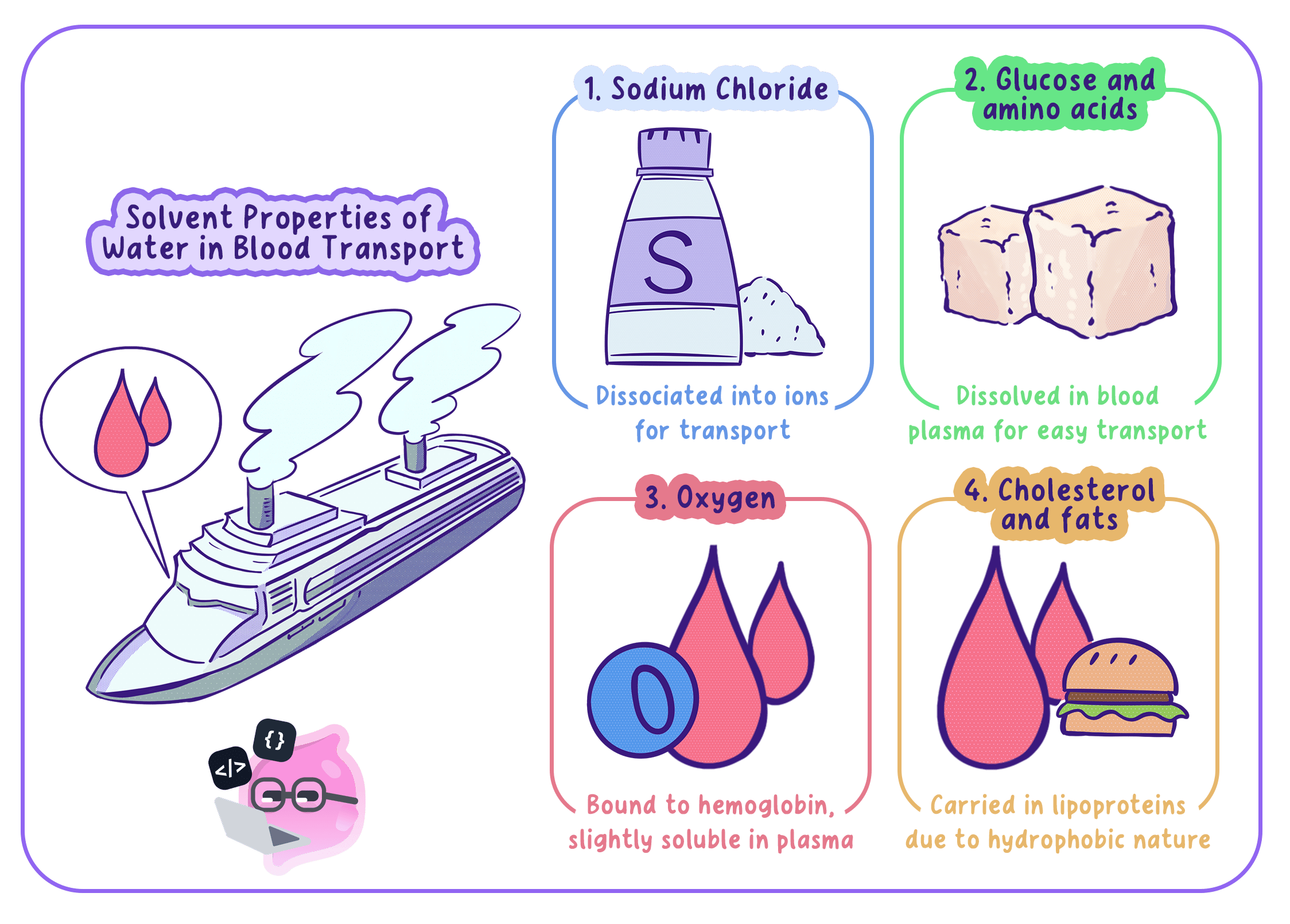
- Oxygen dissolves only slightly in water, so plasma on its own could not carry enough of it to keep a mammal alive.
- Haemoglobin solves that problem by binding oxygen instead of dissolving it, which raises the amount blood can carry many times over.
- In the lungs the oxygen concentration is high, so haemoglobin binds oxygen and becomes bright red oxyhaemoglobin.
- The reaction is written as haemoglobin+oxygen⇌oxyhaemoglobin\text{haemoglobin}+\text{oxygen}\rightleftharpoons\text{oxyhaemoglobin}haemoglobin+oxygen⇌oxyhaemoglobin
- The double arrow matters, because the binding is reversible and the oxygen has to be released again.
- In a respiring tissue the oxygen concentration is low, so oxyhaemoglobin breaks apart and releases oxygen into the cells.
- The blood turns the darker red of deoxygenated blood as it does so.
- Each haemoglobin molecule contains iron, which is why a shortage of iron in the diet lowers how much oxygen the blood can carry and leaves a person tired and short of breath.
Example
Comparing oxygen carried in plasma and in red blood cells
- Each 100 cm3100\ \text{cm}^{3}100 cm3 of blood carries about 0.3 cm30.3\ \text{cm}^{3}0.3 cm3 of oxygen dissolved in the plasma and about 20.0 cm320.0\ \text{cm}^{3}20.0 cm3 bound to haemoglobin.
- The total is 0.3+20.0=20.3 cm30.3+20.0=20.3\ \text{cm}^{3}0.3+20.0=20.3 cm3.
- The percentage carried by haemoglobin is 20.020.3×100=98.5%\dfrac{20.0}{20.3}\times100=98.5\%20.320.0×100=98.5%.
- Haemoglobin therefore raises the oxygen-carrying capacity of blood by a factor of about 20.30.3=68\dfrac{20.3}{0.3}=680.320.3=68.
- Almost all the oxygen in the blood is therefore travelling inside red blood cells rather than dissolved in the plasma.
What plasma carries
Definition
Plasma
Plasma is the straw-coloured liquid part of the blood in which the blood cells, platelets and dissolved substances are carried.
- Plasma is about nine-tenths water, and water is an excellent solvent, so a great many substances dissolve in it and travel together.
- Being a liquid, plasma can be pumped, so everything dissolved in it is carried by mass flow rather than relying on diffusion.
- Plasma carries carbon dioxide from the respiring tissues to the lungs, mostly as hydrogencarbonate ions.
- It carries urea from the liver to the kidneys.
- It carries the soluble products of digestion, such as glucose and amino acids, from the small intestine to the liver and then to every tissue.
- It carries mineral ions, hormones, antibodies and the proteins that make blood clot.
- It also carries the blood cells and the platelets themselves, which are suspended in it rather than dissolved.
- Plasma distributes heat as well as substances, moving warmth from busy organs such as the liver and the muscles to cooler parts of the body.
- Because water needs a great deal of energy to change temperature, plasma can absorb heat without the body temperature swinging about.
The other cells in the blood
Definition
White blood cell
A white blood cell is a blood cell with a nucleus that defends the body against pathogens.
Definition
Platelet
A platelet is a fragment of a cell in the blood that helps blood to clot at a wound.
- White blood cells are larger than red blood cells and, unlike them, each one keeps its nucleus.
- They defend the body against pathogens rather than transporting substances, so they are not adapted for carrying oxygen.
- Some engulf and digest pathogens, while lymphocytes produce antibodies that stick pathogens together.
- Because a white blood cell can change shape, it can squeeze out through a capillary wall and reach an infection in the surrounding tissue.
- Platelets are small fragments of larger cells and have no nucleus of their own.
- They gather at a wound and trigger the reactions that turn a soluble plasma protein into a mesh of insoluble fibres, and that mesh traps red blood cells to form a clot.
- A clot stops blood loss and seals the skin against pathogens while the wound heals.
Practical
Examining a prepared blood smear
- Aim: to view a prepared, stained blood smear under a light microscope and compare the shape, size and number of red and white blood cells.
- Apparatus: a light microscope with ×4\times4×4, ×10\times10×10 and ×40\times40×40 objectives, a commercially prepared and stained blood smear slide, lens tissue, a sharp pencil, a ruler and plain paper.
- Variables: this is an observational study, so there is no independent variable, and the counts are made comparable by using the same slide, the same magnification and fields of the same thickness.
- Method, focusing the slide:
- Clip the prepared slide to the stage with the smear centred over the hole and the ×4\times4×4 objective in place.
- Watch from the side while the coarse focus brings the objective close to the slide, then look down the eyepiece and focus upwards until the smear appears as a pink haze.
- Move up to ×10\times10×10 and then ×40\times40×40, using the fine focus only so the objective cannot be driven into the glass.
- Close the iris diaphragm slightly until the cell outlines look crisp rather than washed out.
- Track across the slide to the thin part of the smear where the cells lie in a single layer, because piled-up cells cannot be counted.
- Method, observing and recording:
- Find the red blood cells, which appear as small pink discs with a paler centre because the biconcave dip lets more light through the middle.
- Find the white blood cells, which are much larger and are easy to spot because the stain colours their nucleus dark purple.
- Count the red cells and the white cells in one whole field of view, then move to a fresh field and repeat for at least three fields.
- Draw a small group of cells with single unbroken pencil lines and no shading, and label a red blood cell, its pale centre and the nucleus of a white blood cell.
- Write the eyepiece and objective magnifications beside the drawing.
- Results: red blood cells outnumber white blood cells by several hundred to one, they carry no nucleus, and each is roughly a third of the diameter of a white blood cell.
- Maths: calculate the mean count of each cell type per field and give the answer as a ratio of red to white cells; total magnification is the eyepiece value multiplied by the objective value, so ×10\times10×10 with ×40\times40×40 gives ×400\times400×400.
- Watch out: counting in a thick region double counts stacked cells, focusing downwards at ×40\times40×40 cracks the slide, and only commercially prepared slides may be used because nobody should make a smear from their own blood.
- Safety: handle slides by their edges because the glass edges are sharp, carry the microscope with one hand under the base, and report a broken slide straight away rather than clearing it yourself.
Exam technique
Explaining a red blood cell adaptation
- Pair every feature with what it achieves, so no nucleus is followed by more room for haemoglobin and so more oxygen carried.
- Use the word biconcave rather than describing the shape loosely, because it is the term the mark scheme looks for.
- Give both consequences of the biconcave shape, the larger surface area and the shorter diffusion distance, since they are separate marking points.
- When a question asks about plasma, name the actual substances carried rather than writing nutrients and waste, because a general answer earns nothing.
Common Mistake
- Do not write that red blood cells have no organelles at all, because the point being tested is that they have no nucleus.
- Do not say haemoglobin dissolves oxygen, because it binds oxygen and forms oxyhaemoglobin.
- Do not confuse plasma with platelets, because plasma is the liquid and platelets are cell fragments floating in it.
- Do not state that plasma carries oxygen around the body, because almost all of it travels bound to haemoglobin.
Self review
- Give three ways a red blood cell is adapted to carry oxygen and state what each adaptation achieves.
- Write the word equation for the reversible reaction between haemoglobin and oxygen.
- Explain why oxyhaemoglobin releases its oxygen in a respiring muscle but not in the lungs.
- Name four substances carried by plasma and state where each is going.
- Explain why plasma can transport substances that would take far too long to reach a tissue by diffusion.