The cell cycle
Cell cycle
The repeating sequence of events in which a cell grows, replicates its DNA, grows again and then divides to give two genetically identical daughter cells.
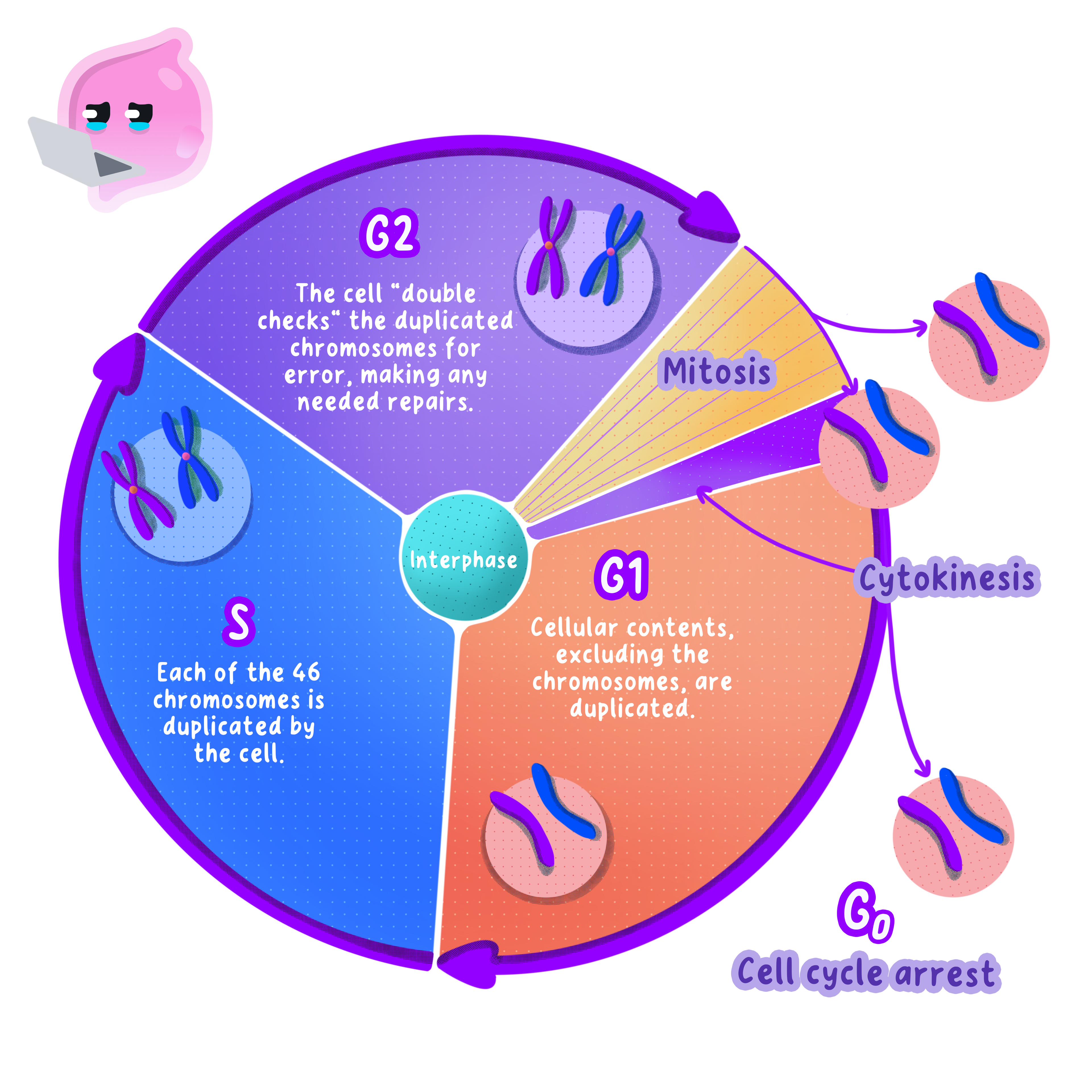
- Body cells divide by repeating a fixed sequence of events called the cell cycle.
- The cycle runs in four steps: cell growth, DNA replication, more cell growth and then movement of the chromosomes as the cell divides.
- In the first step the cell grows and builds more sub-cellular structures, including extra mitochondria and ribosomes.
- In the second step every chromosome is copied, so the cell holds two identical copies of each one.
- In the third step the cell grows again and checks the copied DNA before committing to division.
- In the final step the copied chromosomes are moved to opposite ends of the cell during mitosis, and the cell then splits in two.
- One turn of the cycle produces two genetically identical daughter cells from one parent cell.
- How long a cycle takes depends on the cell: a cell lining the small intestine completes one in roughly 242424 hours, while a mature nerve cell may never divide again.
Interphase
Interphase
The stage of the cell cycle before division, in which the cell grows, replicates its DNA and makes extra sub-cellular structures.
- The first three steps of the cycle together are called interphase, and interphase takes up most of the cycle.
- The cell increases in size, so each daughter cell will be large enough to work normally.
- Extra mitochondria are made so that both daughter cells can release enough energy by respiration.
- Extra ribosomes are made so that both daughter cells can build the proteins they need.
- DNA replication copies every chromosome exactly, which is what allows both daughter cells to end up with identical genetic information.
- Under a microscope an interphase nucleus looks like an evenly stained blob, because the DNA is still unwound and no separate chromosomes can be picked out.
Chromosomes and chromatids
Chromosome
A chromosome is a structure made of a long DNA molecule associated with proteins that carries many genes.
Sister chromatids
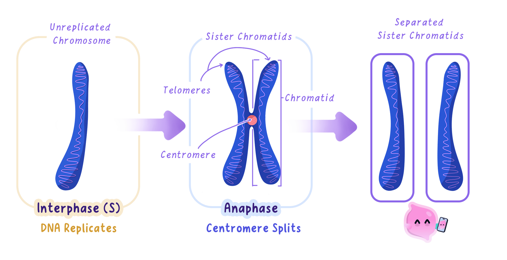
The two identical copies of a chromosome made during DNA replication, joined to each other at the centromere.
- A chromosome is a long molecule of DNA coiled around proteins, and it carries many genes.
- A human body cell contains 464646 chromosomes, arranged as 232323 pairs.
- During DNA replication each chromosome is copied to give two identical copies.
- The two copies stay joined at a narrow point called the centromere, and together they are called sister chromatids.
- A chromosome becomes visible only when it coils up tightly at the start of mitosis, which is why the familiar X shape is seen then and not during interphase.
Mitosis
Mitosis
Mitosis is the stage of the cell cycle in which the nucleus divides to produce two nuclei with identical sets of chromosomes.
- Mitosis is the stage in which the nucleus divides, and it runs through four phases: prophase, metaphase, anaphase and telophase.
- In prophase the chromosomes coil up until they are short and thick enough to be seen, and the membrane around the nucleus breaks down.
- Protein threads called spindle fibres form and stretch across the cell from one end to the other.
- In metaphase the chromosomes line up in a single row across the middle of the cell, and a spindle fibre attaches to the centromere of each one.
- In anaphase the spindle fibres shorten and pull the two sister chromatids of each chromosome apart, to opposite ends of the cell.
- Each separated chromatid now counts as a chromosome in its own right, so each end of the cell holds one complete set.
- In telophase a new nuclear membrane forms around each group of chromosomes, giving two nuclei, and the chromosomes uncoil again.
Cytokinesis
Cytokinesis
The final stage of the cell cycle, in which the cytoplasm and cell membrane divide to form two separate daughter cells.
- Once the nucleus has divided, the cytoplasm and cell membrane divide as well.
- In an animal cell the membrane pinches inwards around the middle until the cell is cut in two.
- In a plant cell a new cell wall and membrane are built across the middle instead, because the rigid wall cannot pinch inwards.
- The mitochondria, ribosomes and other structures built during interphase are shared between the two new cells.
- The result is two daughter cells, each holding a full set of chromosomes identical to those of the parent cell.
Why mitosis matters
- Mitosis increases the number of cells, which is how a multicellular organism grows.
- Mitosis repairs damaged tissue, for example when skin cells divide to close a cut.
- Mitosis replaces cells that wear out, such as the lining of the small intestine, which is renewed every few days.
- In plants, mitosis in the root and shoot tips is what makes roots and stems longer.
- Because every daughter cell is genetically identical, every body cell of an organism carries the same genetic information.
- For the same reason, mitosis on its own cannot produce genetic variation.
Observing mitosis in root tip cells
- Aim: to prepare a stained squash of a growing root tip and identify cells in the phases of mitosis.
- Apparatus: garlic cloves or onion bulbs grown over water, a watch glass, forceps, a scalpel, a white tile, 1 mol dm−31\ \text{mol dm}^{-3}1 mol dm−3 hydrochloric acid, a water bath at 60 ∘C60\ ^{\circ}\text{C}60 ∘C, distilled water, toluidine blue or acetic orcein stain, a microscope slide and cover slip, a mounted needle, filter paper, a pencil with a rubber end and a light microscope.
- Method, growing the roots:
- Suspend a garlic clove or onion bulb so that its flat base just touches the surface of the water in a beaker.
- Leave it at room temperature for three to five days, until the new roots are 222 to 3 cm3\ \text{cm}3 cm long.
- Method, preparing the slide:
- Cut off the final 5 mm5\ \text{mm}5 mm of a root with a scalpel on a white tile, because the dividing cells sit in the meristem just behind the root cap.
- Place the tip in a watch glass with a few drops of 1 mol dm−31\ \text{mol dm}^{-3}1 mol dm−3 hydrochloric acid and warm it at 60 ∘C60\ ^{\circ}\text{C}60 ∘C for about five minutes, which breaks down the pectin holding the cells together so they can later be spread out.
- Rinse the tip in distilled water and blot it, so the acid stops acting and does not spoil the stain.
- Transfer the tip to a slide, add a few drops of toluidine blue or acetic orcein and leave it for about two minutes, because the stain binds to DNA and makes the chromosomes show up dark against the pale cytoplasm.
- Lower a cover slip onto the tip with a mounted needle, one edge first, so that no air bubbles are trapped.
- Cover the slide with filter paper and press straight down with your thumb or the rubber end of a pencil, without twisting, to squash the tip into a single layer of cells that light can pass through.
- Method, viewing:
- Start on the lowest power objective and find the region where the cells are small, square and densely stained.
- Switch to a higher power objective and refocus with the fine adjustment only.
- What you see: most cells show one plain dark nucleus and are in interphase, while a smaller number show dark thread-like chromosomes, some lined up across the middle and some being drawn towards the ends.
- Maths: count at least 100100100 cells and calculate the mitotic index using number of cells in mitosistotal number of cells×100\dfrac{\text{number of cells in mitosis}}{\text{total number of cells}}\times100total number of cellsnumber of cells in mitosis×100.
- Watch out: press vertically and never twist the cover slip, because twisting tears the cells apart and breaks up the chromosomes you are trying to count.
- Safety: hydrochloric acid is an irritant, so wear eye protection and wash any splashes off at once, and cut away from your fingers on a tile.
Calculating a mitotic index
- A student counts 120120120 cells in one field of view of a stained root tip squash.
- Of those, 181818 cells show visible chromosomes and are therefore in mitosis.
- Mitotic index =number of cells in mitosistotal number of cells×100=\dfrac{\text{number of cells in mitosis}}{\text{total number of cells}}\times100=total number of cellsnumber of cells in mitosis×100.
- Substituting gives 18120×100=15%\dfrac{18}{120}\times100=15\%12018×100=15%.
- The other 85%85\%85% of the cells are in interphase, which confirms that interphase takes up most of the cell cycle.
- If one full cycle lasts 202020 hours, mitosis lasts about 0.15×20=3.00.15\times20=3.00.15×20=3.0 hours.
Describing the cell cycle
- Give the steps of the cycle in order and say what happens in each, because a list of names on its own rarely scores.
- Write genetically identical rather than the same, since the mark is for the genetic point.
- Say that the DNA is replicated before mitosis begins, not during it.
- Describe anaphase as chromatids being pulled apart by spindle fibres, and name the centromere as the point they attach to.
- Give a mitotic index as a percentage unless the question asks for a decimal, and show the division before you multiply by 100100100.
- Do not say the chromosome number halves in mitosis, because each daughter cell receives a full set.
- Do not confuse mitosis with meiosis; mitosis gives two identical cells with a full set of chromosomes, while meiosis gives four non-identical sex cells with half a set.
- Do not describe chromosomes as being split down the middle, because the two chromatids are separated whole.
- Do not call the whole cell cycle mitosis, because mitosis is only the stage in which the nucleus divides.
- Do not expect to see chromosomes in an interphase cell, because the DNA is still unwound at that point.
- Name the steps of the cell cycle in order and say what happens in each.
- Explain why the DNA must be replicated before the cell divides.
- Describe what happens to the chromosomes in metaphase and in anaphase.
- Explain how the two daughter cells compare with the parent cell.
- Calculate the mitotic index of a root tip in which 242424 of 150150150 counted cells are in mitosis.