Resolution and the limit of light
Definition
Resolution
The smallest distance between two points that can still be seen as two separate points.
- Resolution is the smallest distance between two points at which they can still be seen as two separate points instead of merging into one blur.
- Resolution is measured as a distance, so a smaller number means a better resolution and finer detail.
- Magnification and resolution do different jobs: magnification sets how large the image is, and resolution sets how much detail that image can hold.
- Once two structures lie closer together than the resolution of the instrument, no extra magnification will separate them and the image simply becomes a larger blur.
- The resolution of a light microscope is limited by the wavelength of visible light, because two points closer together than about half a wavelength cannot be told apart.
- Visible light has wavelengths of roughly 400 nm400\ \text{nm}400 nm to 700 nm700\ \text{nm}700 nm, which fixes the best resolution of a light microscope at about 200 nm200\ \text{nm}200 nm.
- That limit also caps the useful magnification, which is why a light microscope is not taken far beyond about ×1500\times1500×1500.
- A structure smaller than 200 nm200\ \text{nm}200 nm, such as a ribosome at roughly 20 nm20\ \text{nm}20 nm across, is therefore invisible to light no matter how the microscope is set up.
Common Mistake
- Do not treat a larger magnification as more detail, because the detail available is fixed by the resolution.
- Do not describe a good resolution as a large number, because resolution is a distance and the smaller that distance is the finer the detail.
How an electron microscope works
Definition
Electron microscope
An instrument that uses a beam of electrons instead of light to produce a magnified image of a specimen.
Definition
Transmission electron microscope
An electron microscope in which electrons pass through a very thin specimen to reveal the detail inside it.
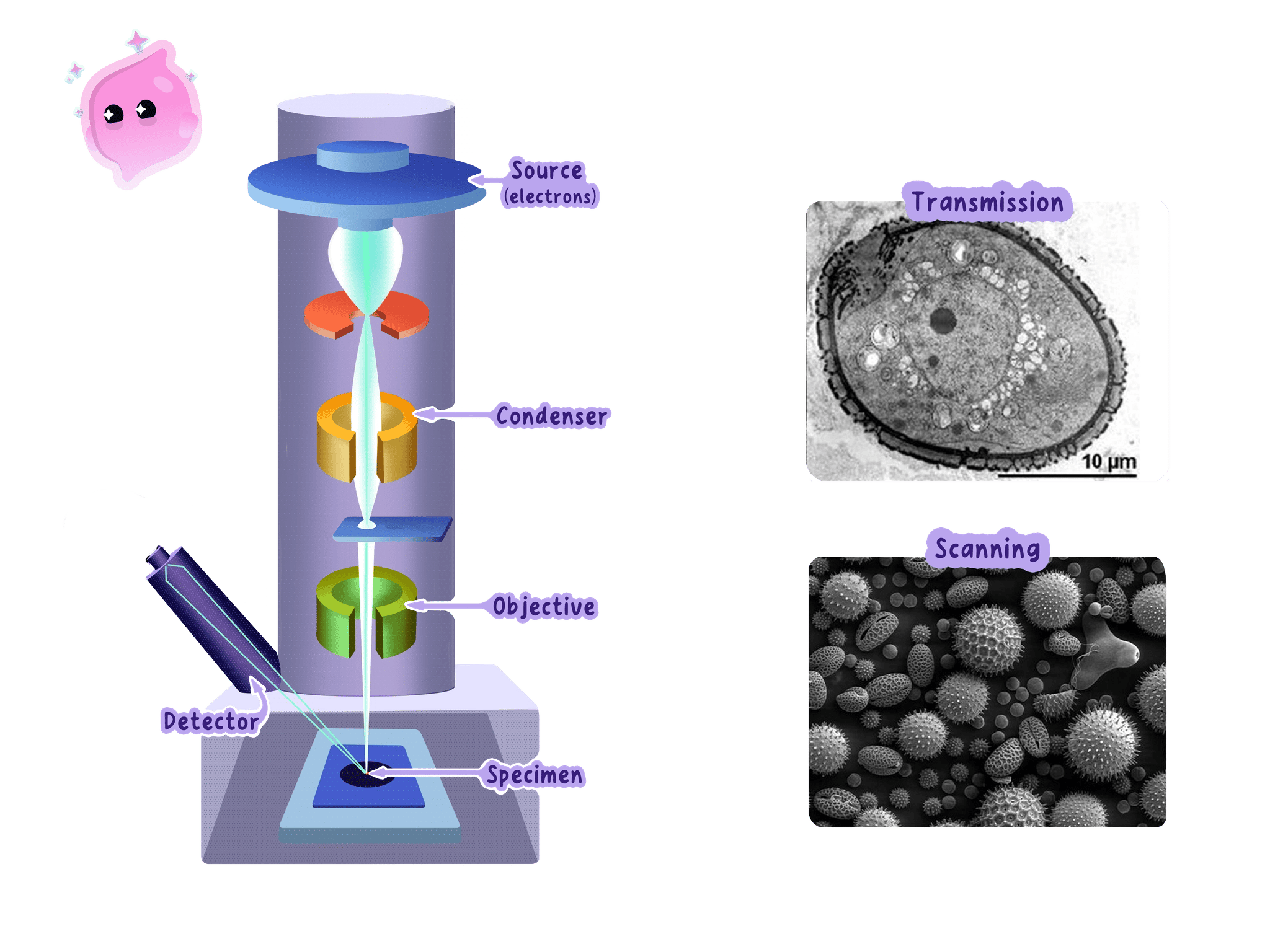
- An electron microscope replaces the beam of light with a beam of electrons.
- The electrons are released from a heated filament called the electron gun at the top of the column and are accelerated downwards by a high voltage.
- Electrons carry a negative charge, so the beam is focused by electromagnets rather than glass lenses, and changing the current through a magnet changes how strongly the beam is bent.
- The whole column is held under a vacuum, because electrons collide with air particles and would be scattered off course long before reaching the specimen.
- In a transmission electron microscope, the electrons pass straight through a specimen that has been cut into an extremely thin slice.
- The specimen is treated with heavy metal salts, which gather on particular structures and scatter electrons strongly, in the same way that a stain builds contrast on a light microscope slide.
- Dense parts of the specimen scatter more electrons and appear dark, while thinner or less dense parts let more electrons through and appear pale, and this difference is what forms the image.
- The electrons that get through strike a fluorescent screen or an electronic detector, producing an image in shades of grey because electrons carry no colour information.
- A beam of electrons behaves as though it has a far shorter wavelength than visible light, and that shorter wavelength is the reason the resolution is so much better.
- A transmission electron microscope resolves detail down to about 0.2 nm0.2\ \text{nm}0.2 nm and magnifies usefully to well over ×1 000 000\times1\,000\,000×1000000.
- A scanning electron microscope instead sweeps the beam back and forth across the surface of a whole specimen and builds a three-dimensional picture of that surface, at a lower resolution than a transmission instrument reaches.
Example
Comparing what each microscope can separate
- A light microscope resolves down to about 200 nm200\ \text{nm}200 nm and a transmission electron microscope down to about 0.2 nm0.2\ \text{nm}0.2 nm.
- Dividing one limit by the other gives 2000.2=1000\dfrac{200}{0.2}=10000.2200=1000.
- The electron microscope therefore separates points that are a thousand times closer together.
- A ribosome is about 20 nm20\ \text{nm}20 nm across, which is ten times smaller than the light microscope's limit but a hundred times larger than the electron microscope's, so only the electron microscope can show it.
What electron microscopy revealed
Definition
Ultrastructure
The fine internal detail of a cell that can only be seen using an electron microscope.
- Under a light microscope a cell showed a nucleus, cytoplasm, a membrane, chloroplasts and the outlines of mitochondria, and nothing finer than that.
- Mitochondria appeared as featureless rods, so their internal arrangement could only be guessed at.
- The far higher resolution of the electron microscope revealed the ultrastructure of the cell, the fine internal detail that light could never separate.
- It showed that a mitochondrion has two membranes and that the inner one is folded into shelves, which explained how the organelle packs a large surface area of respiration enzymes into a tiny volume.
- It revealed ribosomes for the first time, as tiny dense granules lying free in the cytoplasm and attached to membranes, which showed where proteins are assembled.
- It showed that a chloroplast contains stacks of flattened membrane sacs carrying the chlorophyll, which explained how the pigment is spread out so that as much light as possible is absorbed.
- It showed the cell membrane as two distinct dark lines rather than one, which supported the idea of a membrane built from a double layer of lipid.
- It revealed folded internal membrane systems that light had never shown at all, so the cytoplasm turned out to be organised rather than a featureless jelly.
- Each of these discoveries let biologists match a structure to the job it does, and it is that link between structure and function that transformed how cells were understood.
Note
- A specimen for an electron microscope has to be dead, dried out and held in a vacuum, so the instrument can never be used to watch a living process.
- The preparation can create artefacts, which are structures that appear in the image but were not present in the living cell.
- A newly seen structure is therefore only accepted once it shows up again after a different preparation method.
- Electron microscopes are also large, expensive and need trained operators, so a light microscope is still the practical choice for routine work.
Exam technique
Writing about what the electron microscope changed
- Answer in terms of higher resolution, because credit goes to the ability to separate close points and not to magnification on its own.
- Name a specific structure it revealed, such as ribosomes or the folded inner membrane of a mitochondrion, since a vague reference to seeing more detail rarely scores.
- Follow the structure with the job it does when the command word is explain, for example that folds give a large surface area for respiration enzymes.
- State a limitation as a consequence rather than a bare fact, for example that the specimen is dead so no living process can be observed.
Self review
- State what is meant by the resolution of a microscope.
- Explain why raising the magnification of a light microscope beyond a certain point reveals no extra detail.
- Describe how a transmission electron microscope produces an image of a specimen.
- Explain why the column of an electron microscope is kept under a vacuum.
- Name two structures the electron microscope revealed and state what each one told biologists about how a cell works.