The Heart Is a Double Pump
Definition
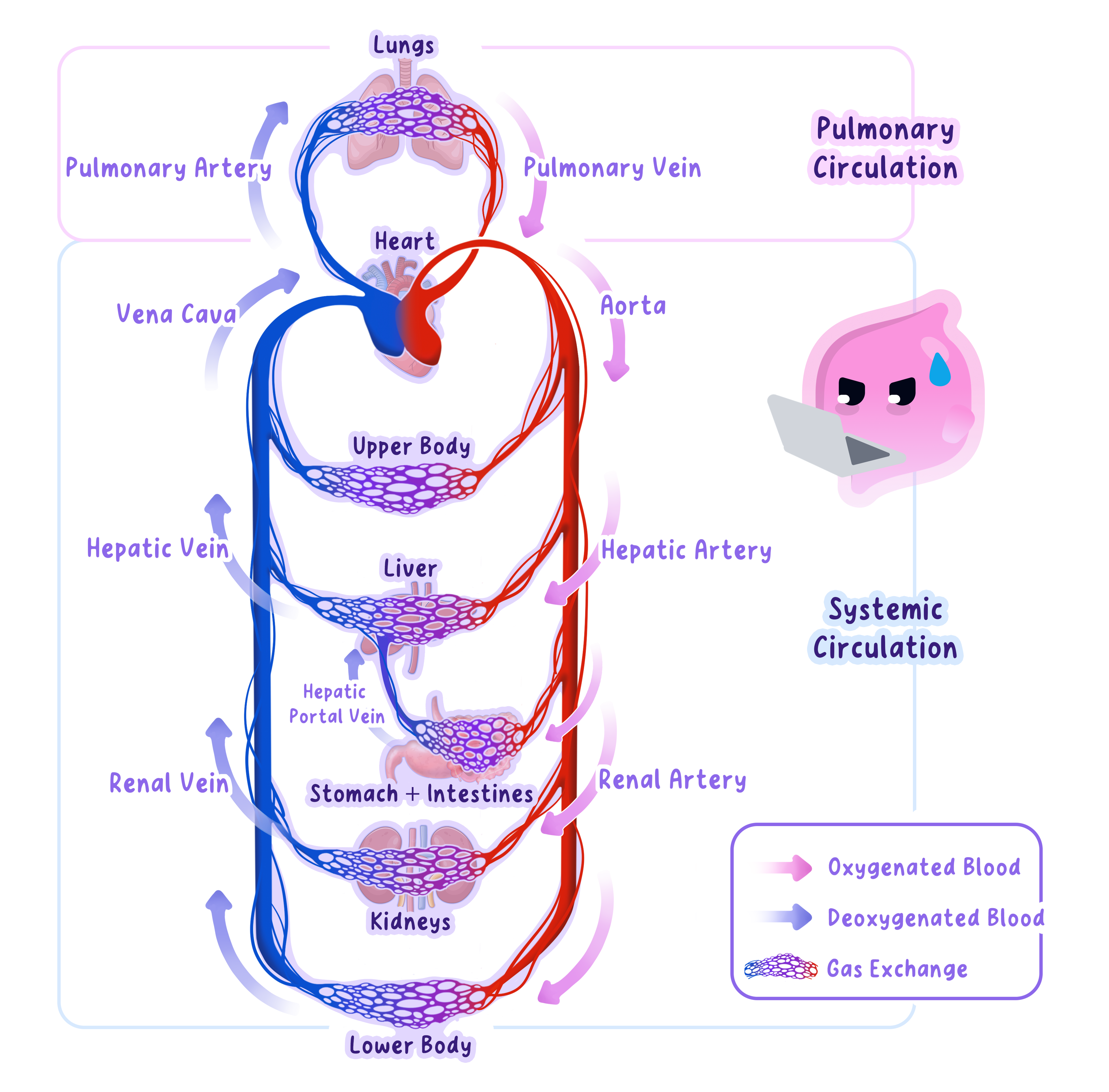
Double circulatory system
A circulatory system in which blood passes through the heart twice during one complete circuit of the body.
- The right side pumps deoxygenated blood to the lungs in the pulmonary circulation.
- The left side pumps oxygenated blood to the rest of the body in the systemic circulation.
- Blood returns to the heart between the two circuits, so it passes through the heart twice in one complete journey.
- Separate pumps allow lower pressure through delicate lung capillaries and higher pressure through the longer systemic circuit.
Chambers Control Blood Flow
Definition
Atrium
An upper chamber of the heart that receives blood returning to the heart.
Definition
Ventricle
A lower chamber of the heart that contracts to pump blood out of the heart.
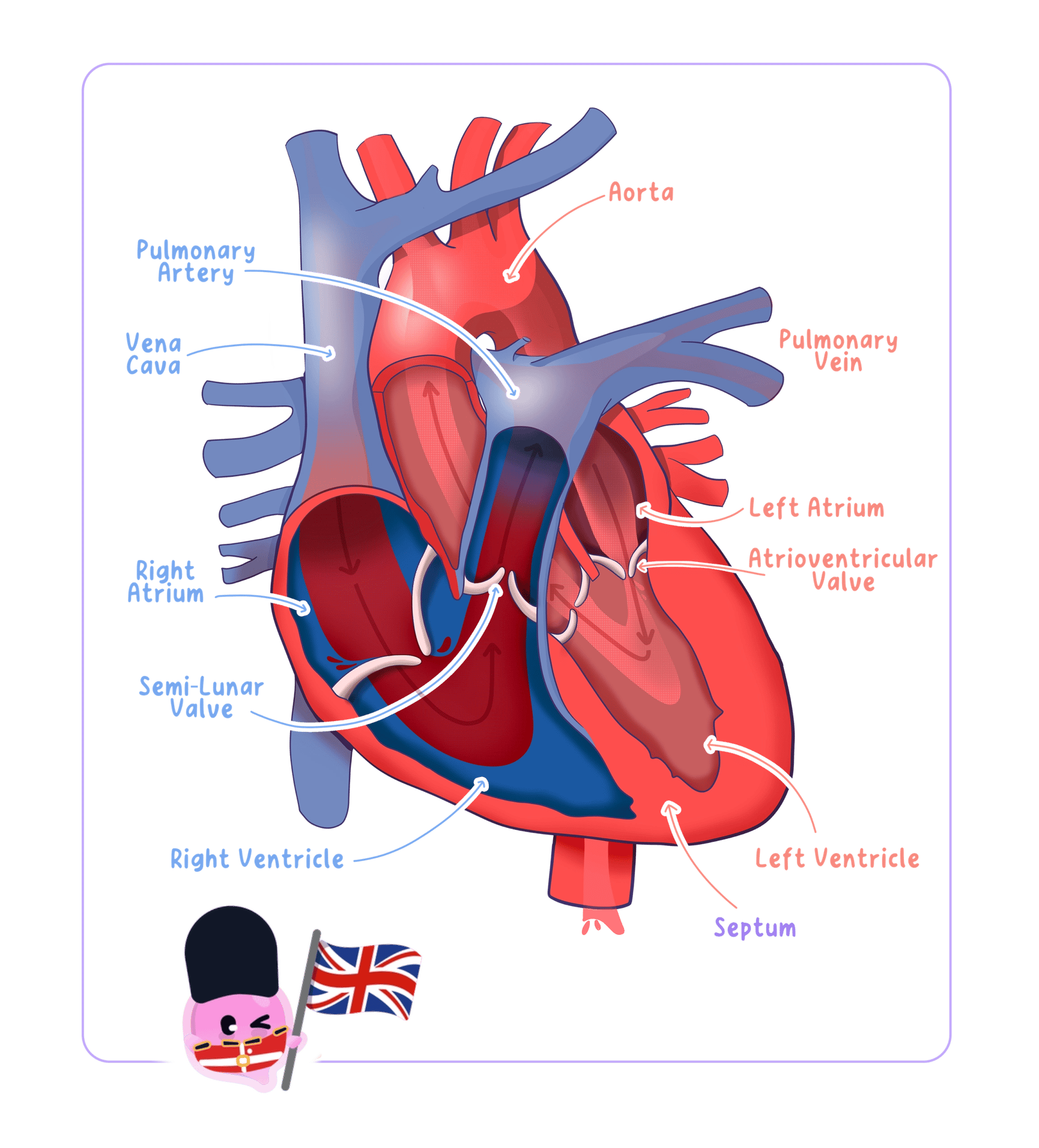
- The right atrium receives deoxygenated blood from the body through the vena cava.
- The right atrium contracts and moves blood through an atrioventricular valve into the right ventricle.
- The right ventricle contracts and pumps blood through the pulmonary artery to the lungs.
- The left atrium receives oxygenated blood from the lungs through the pulmonary vein.
- The left atrium contracts and moves blood through an atrioventricular valve into the left ventricle.
- The left ventricle contracts and pumps blood through the aorta to the body.
Valves Prevent Backflow
Definition
Heart valve
A flap of tissue that opens in one direction to prevent the backflow of blood through the heart.
- Atrioventricular valves lie between each atrium and ventricle.
- They open when pressure is greater in an atrium than in its ventricle, allowing blood to move forwards.
- They close when ventricular pressure rises, preventing blood from returning to the atria.
- Valves at the exits of the ventricles prevent blood in the pulmonary artery and aorta from flowing back into the heart.
- Valves open and close because of pressure differences, rather than contracting by themselves.
The Septum Prevents Mixing
Definition
Septum
The muscular wall that separates the right and left sides of the heart and prevents oxygenated and deoxygenated blood from mixing.
- The septum keeps oxygenated blood on the left side separate from deoxygenated blood on the right side.
- This separation allows blood sent to the body to retain a high oxygen concentration.
- A steep oxygen concentration gradient can therefore be maintained between systemic capillaries and respiring cells.
Pressure Determines Wall Thickness
- Atria have thin walls because they push blood only a short distance into the ventricles.
- The right ventricle has a thicker wall than an atrium because it pumps blood out of the heart to the lungs.
- The left ventricle has the thickest muscular wall because it must produce high pressure to drive blood around the whole body.
- The lower pressure produced by the right ventricle protects the thin exchange surfaces in the lungs.
Exam technique
- When describing a route, include the starting chamber, the named vessel and the destination.
- When explaining wall thickness, state the distance or pressure requirement that the chamber must meet.
- Do not say that the heart adds oxygen to blood, because gas exchange occurs in the lungs.
Contraction Produces One-Way Flow
- Atrial contraction raises pressure and pushes blood into the ventricles.
- Ventricular contraction raises pressure, closes the atrioventricular valves and opens the exit valves.
- Relaxation lowers pressure, allowing the chambers to refill before the next heartbeat.
Self review
- What are the two circuits in a double circulatory system?
- Which vessel carries blood from the right ventricle to the lungs?
- How do heart valves prevent backflow?
- Why is the left ventricular wall thicker than the right?
- What is the function of the septum?