A Specialised Cell Has a Structure That Fits Its Job
Specialised cell
A cell whose structure is adapted to carry out a particular function, such as a sperm, nerve or root hair cell.
- A specialised cell has a structure adapted to carry out one particular function.
- This can be the cell's shape or the mix of sub-cellular structures it contains.
- Cells become specialised through the process of differentiation, in which they gain the sub-cellular structures a particular job needs.
- Sperm, nerve and muscle cells are specialised animal cells, and root hair, xylem and phloem cells are specialised plant cells.
To explain any adaptation, name the feature and then the job it helps the cell do.
Specialised Cells in Animals
Mitochondria
The organelles where most aerobic respiration takes place, releasing energy for the cell; a single one is a mitochondrion.
Nerve cells
- Nerve cells, also called neurones, carry electrical impulses to give fast communication around the body.
- They are very long, so impulses can travel a long way between parts of the body.
- They have many dendrites, branched extensions of the cell body that connect to other nerve cells.
- The axon is covered by a fatty myelin sheath that insulates it and speeds up the impulse.
- The nerve endings, called synapses, release transmitter chemicals called neurotransmitters to pass the impulse to the next cell.
- They have many mitochondria at the nerve endings to release the energy needed to make these neurotransmitters.
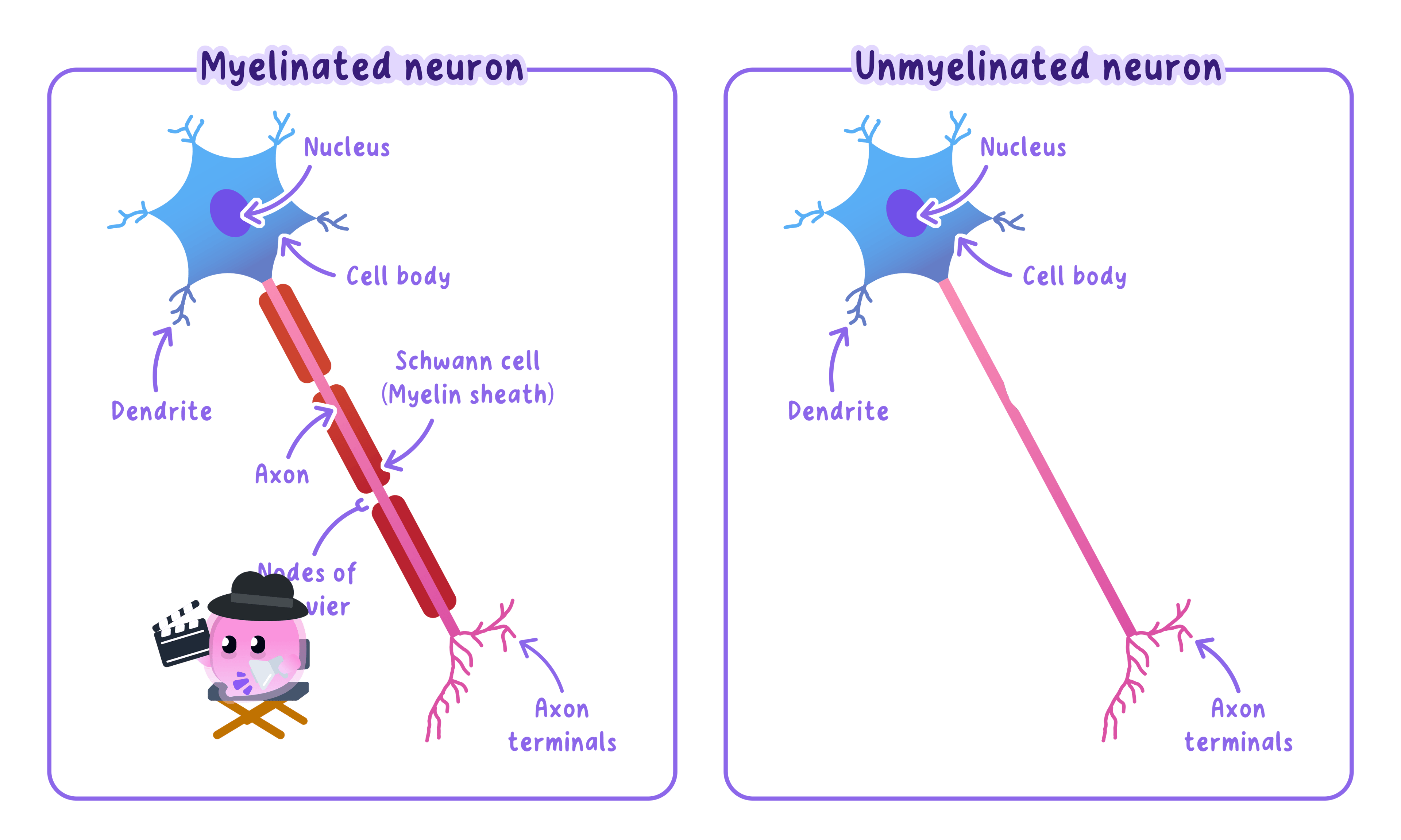
The longest axon in your body runs from the base of your spine to your big toe, about a metre long.
Muscle cells
- Muscle cells contract and relax to produce movement.
- They contain special proteins, actin and myosin, that slide over each other to make the cell contract.
- They have many mitochondria to release the energy needed for contraction.
- They can store glycogen, which is broken down in respiration to release that energy.
- Striated muscle is attached to bones and contracts to move the skeleton.
- Smooth muscle, such as in the gut and blood vessels, contracts to squeeze.
Sperm cells
- A sperm cell carries the father's genetic information to the egg for fertilisation.
- A long tail whips from side to side, and a streamlined head helps the sperm move towards the egg.
- The middle section is packed with mitochondria to release the energy the tail needs.
- The acrosome at the top of the head stores digestive enzymes that break down the egg's outer layers.
- The nucleus carries half the normal number of chromosomes, so a full set is restored at fertilisation.
To work out an unfamiliar cell's job, look at its shape and contents: a large surface area, many ribosomes for making proteins, or many mitochondria for energy.
Specialised Cells in Plants
Cell wall
A rigid outer layer around the cell membrane that supports and strengthens the cell; in plant and algal cells it is made of cellulose.
Root hair cells
- A root hair cell absorbs water by osmosis and mineral ions by active transport from the soil.
- A long, thin root hair gives a large surface area, so more water is absorbed by osmosis.
- Its wall is thinner than other plant cell walls, giving a shorter distance for water to cross.
- A large permanent vacuole helps draw water in from the soil.
- It has many mitochondria to release the energy needed for the active transport of mineral ions.
There is no light underground, so root hair cells contain no chloroplasts.
Xylem vessels
- Xylem carries water and dissolved mineral ions up from the roots to the shoots and leaves.
- As the cells form, lignin is deposited in their walls, which kills the cells.
- The dead cells become hollow and lose their end walls, joining into a continuous tube for water to move through.
- The lignin is laid down in spirals that strengthen the tube and help it withstand the pressure of the moving water.
Xylem is made of dead cells and carries water, so do not confuse it with phloem.
Phloem cells
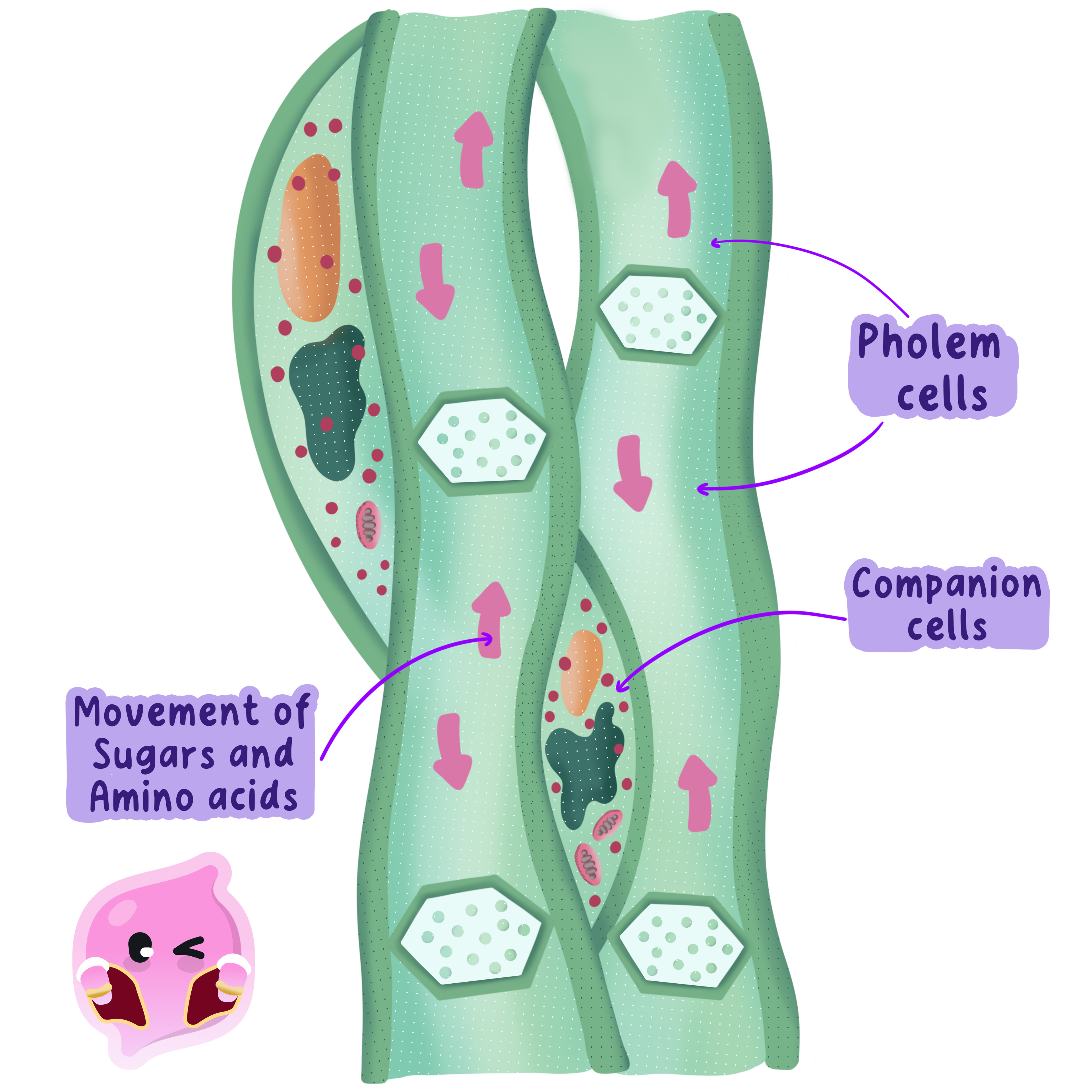
- Phloem carries dissolved food, such as sugars and amino acids made in photosynthesis, around the plant.
- The cells are living and joined end to end.
- Their end walls have holes, called sieve plates, that let the dissolved food flow through.
- Phloem cells lose most of their sub-cellular structures, so companion cells supply the energy that keeps them alive.
- What does it mean to say a cell is specialised?
- Give two ways a nerve cell is adapted to carry impulses quickly around the body.
- Name the two proteins that make a muscle cell contract.
- How is a root hair cell adapted to absorb water and mineral ions?
- State two differences between xylem and phloem.
