What you'll learn
- Why split-brain patients were so useful for studying regions of the brain.
- How Sperry tested the left and right hemispheres separately using divided visual-field tasks.
- The main findings: language is mainly in the left hemisphere, while the right hemisphere has important non-verbal abilities.
- How to evaluate Sperry (1968) for AO3 and compare it with the paired contemporary study, Casey et al. (2011).
The big idea: regions of the brain
Sperry (1968) is a classic core study in the Biological area for the theme Regions of the brain. It shows that different parts of the brain can have different functions.
Lateralisation of function
Lateralisation of function means that some mental or behavioural functions are more strongly associated with one side of the brain than the other. For example, in most people, speech and language are mainly controlled by the left hemisphere.
The brain has two large halves called hemispheres: the left hemisphere and the right hemisphere. In everyday life they usually work together, because they are connected by a thick bundle of nerve fibres.
Corpus callosum
The corpus callosum is the bundle of nerve fibres connecting the left and right cerebral hemispheres, allowing them to share information.
Background: why were the hemispheres separated?
Some people with very severe epilepsy — a neurological condition involving seizures — had surgery to cut the corpus callosum. This operation aimed to stop seizure activity spreading from one hemisphere to the other.
Split brain
A split-brain patient is someone whose corpus callosum has been surgically severed, so the two hemispheres cannot communicate normally.
Importantly, the surgery was done for medical reasons, not just for the research. Sperry and colleagues then studied these rare patients to understand what each hemisphere could do on its own.
Why split-brain patients matter
Split-brain patients created a natural opportunity to test the two hemispheres separately, because information sent to one hemisphere could not easily be passed to the other.
Prerequisite: visual fields are not the same as eyes
This is the part students often find tricky.
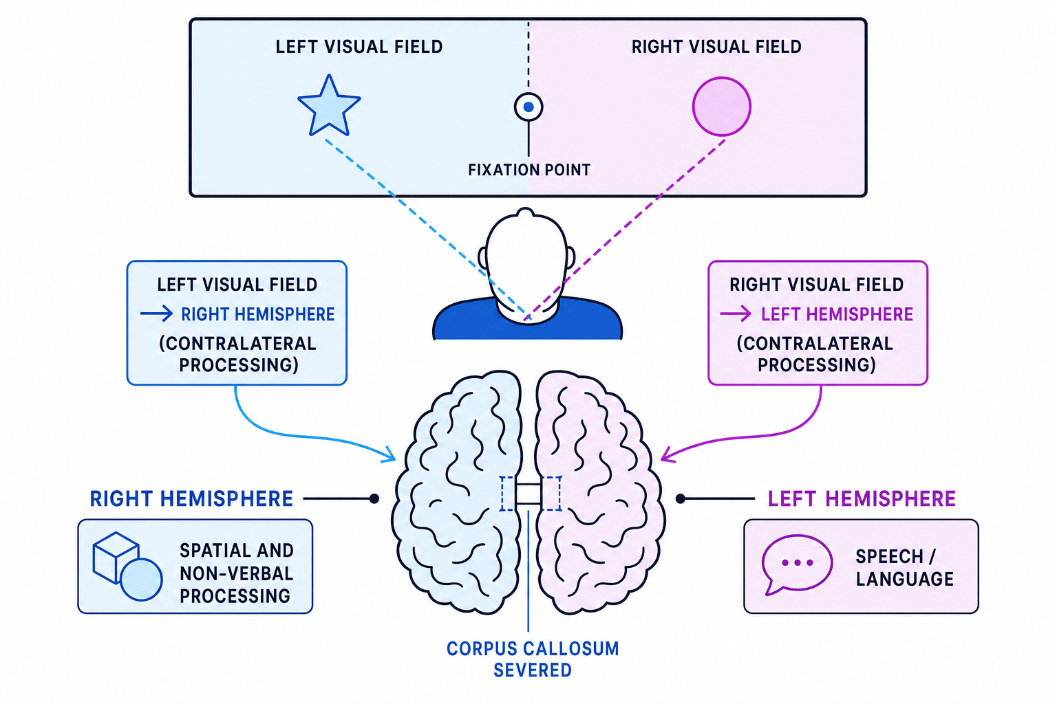
A visual field is the area of space you can see. The left visual field is everything to the left of where you are looking. The right visual field is everything to the right of where you are looking.
Because of the way the visual system is wired:
- Information from the left visual field goes to the right hemisphere.
- Information from the right visual field goes to the left hemisphere.
This crossing-over is called contralateral processing, meaning “opposite-side processing”.
Visual field, not eye
Do not write “the left eye sends information to the right hemisphere.” Each eye receives information from both visual fields. In Sperry’s task, the important issue is whether the stimulus is shown in the left visual field or right visual field.
Aim of the study
Sperry aimed to investigate the effects of hemisphere deconnection and to discover what each hemisphere could do when the two sides of the brain could not share information through the corpus callosum.
In simpler terms: what happens when each half of the brain receives information on its own?
Method
Design
Sperry’s study is best described as a quasi-experiment using highly controlled laboratory tasks.
Quasi-experiment
A quasi-experiment is a study where the researcher investigates an existing difference between participants, rather than randomly allocating them to conditions. Sperry could not randomly assign people to have split-brain surgery.
The study also has features of a case-study series, because the sample was very small and unusual, and the researchers collected detailed data from each participant.
Sample
The sample consisted of 11 split-brain patients who had undergone surgery to sever the corpus callosum, usually to treat severe epilepsy.
This is a very specialised sample. That is useful because the patients are rare, but it also limits how far the findings can be generalised to the wider population.
Materials and apparatus
Sperry used controlled apparatus to present information to one hemisphere at a time. Key materials included:
- A screen with a central fixation point, which the participant had to stare at.
- A device similar to a tachistoscope, which presents visual stimuli very briefly.
- Pictures, words or symbols flashed to the left or right visual field.
- Objects placed out of sight so participants could identify them by touch.
- Tasks requiring verbal responses, pointing, drawing or selecting objects with one hand.
Tachistoscope
A tachistoscope is a device that presents visual stimuli for a very short time. This prevents participants moving their eyes and accidentally sending the stimulus to both hemispheres.
Procedure
Participants focused on the centre of a screen. A word or picture was then flashed very briefly to either the left or right visual field.
If the stimulus appeared in the right visual field, it went to the left hemisphere, which usually controls speech. If it appeared in the left visual field, it went to the right hemisphere, which could not use the left hemisphere’s language system because the corpus callosum had been cut.
Participants were then asked to:
- Say what they had seen.
- Point to a matching object.
- Select an object using the left or right hand.
- Draw or copy objects.
- Identify objects by touch without seeing them.
Predicting a split-brain response
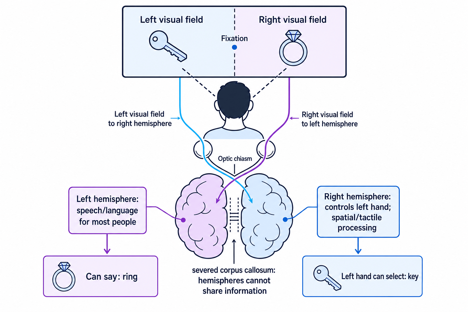
A split-brain participant fixates on the centre of a screen. The word “spoon” is flashed to the left visual field. They are then asked what they saw, and later asked to pick out the object using their left hand.
- The stimulus is in the left visual field, so it travels to the right hemisphere.
- In most people, speech is controlled mainly by the left hemisphere, but the corpus callosum has been severed, so the right hemisphere cannot pass the word to the speech system.
- The participant is therefore unlikely to say “spoon” and may report seeing nothing.
- The right hemisphere controls the left hand, so the participant may still pick out the spoon correctly using the left hand.
Results
Right visual field: left hemisphere
When information was presented to the right visual field, it went to the left hemisphere. Participants could usually name or describe what they had seen.
This suggested that, for these participants, the left hemisphere was specialised for speech and language.
Left visual field: right hemisphere
When information was presented to the left visual field, it went to the right hemisphere. Participants often said they had seen nothing, because the right hemisphere could not communicate with the speech centres in the left hemisphere.
However, they could often still respond non-verbally. For example, they could use their left hand to point to or select the correct object.
The right hemisphere was not unconscious
A participant saying “I saw nothing” did not mean the right hemisphere had not processed the stimulus. It meant the hemisphere with speech could not access that information.
Tactile tasks
In touch-based tasks, an object placed in the right hand could usually be named because the right hand is controlled by the left hemisphere.
An object placed in the left hand often could not be named, but the participant could still select a matching object by touch. Again, this showed that the right hemisphere could process information but struggled to produce speech.
Other abilities
Sperry found evidence that the right hemisphere was better at some non-verbal tasks, such as spatial processing, recognising patterns and drawing. The left hemisphere was stronger for language and speech.
Conclusions
Sperry concluded that the two hemispheres have different specialised functions.
- The left hemisphere is dominant for speech and language in most people.
- The right hemisphere can process visual and tactile information, especially spatial and non-verbal material.
- The corpus callosum normally allows the hemispheres to share information.
- When the corpus callosum is severed, the hemispheres can act almost like two separate systems.
This strongly supports the idea that particular regions of the brain are linked to particular psychological functions.
Evaluation: AO3
Strengths
One major strength is the high level of control. Stimuli were flashed very briefly, and participants had to fixate on the centre of the screen. This increased internal validity, meaning the researchers could be more confident that responses were due to which hemisphere received the information.
Internal validity
Internal validity is the extent to which a study measures what it intends to measure and can show a genuine cause-effect relationship within the study conditions.
Another strength is that Sperry used several different types of task: visual, tactile, verbal, pointing and drawing. This provided converging evidence for hemisphere specialisation.
The study was also highly useful. It contributed to understanding epilepsy surgery, brain organisation and the relationship between brain regions and behaviour.
Weaknesses
A key weakness is the small and unusual sample. The participants had severe epilepsy and had undergone major brain surgery. This lowers population validity, because their brains may not represent typical brains.
Population validity
Population validity is the extent to which findings from a sample can be generalised to the wider target population.
The study also has low ecological validity. Everyday perception does not usually involve staring at a fixation point while words are flashed for a fraction of a second.
Ecological validity
Ecological validity is the extent to which findings can be generalised to real-life settings and behaviours.
Reliability is mixed. The procedure was standardised and carefully controlled, which supports reliability. However, split-brain patients are rare, and individual differences in surgery, epilepsy history and brain organisation make exact replication difficult.
Overgeneralising the findings
Avoid writing that “everyone’s right hemisphere cannot understand language.” Sperry’s findings came from a tiny clinical sample, and the right hemisphere showed some understanding through non-verbal responses.
Ethics
Using modern BPS-style principles, Sperry’s study raises some ethical issues but is generally less problematic than studies involving deception or deliberate harm.
The surgery itself was not performed for the study; it was a treatment for severe epilepsy. That reduces ethical concern because researchers were not causing brain damage for experimental purposes.
However, the participants were a vulnerable clinical group. We would want clear evidence of informed consent, protection from stress or fatigue, confidentiality, and the right to withdraw.
Informed consent
Informed consent means participants understand what the study involves and voluntarily agree to take part.
Link to the Biological area and key theme
Sperry fits the Biological area because it explains behaviour through brain structures and nervous-system processes. It is scientific, controlled and focused on physical mechanisms.
It fits the key theme Regions of the brain because it shows that different hemispheres are linked with different functions. The study is also quite reductionist, because it explains complex behaviour, such as language and recognition, in terms of brain regions.
Reductionism
Reductionism means explaining complex behaviour by breaking it down into simpler components, such as brain structures, genes or neurotransmitters.
This can be a strength because it makes behaviour testable and scientific. It can also be a weakness if it ignores wider influences such as learning, context and individual experience.
Comparison with Casey et al. (2011)
The paired contemporary study for this theme is Casey et al. (2011), which investigated delay of gratification using brain imaging.
Both Sperry (1968) and Casey et al. (2011) are in the Biological area and link behaviour to brain regions. Sperry focused on hemisphere specialisation, while Casey focused on brain areas involved in self-control, such as the prefrontal cortex and ventral striatum.
There are also clear differences:
| Feature | Sperry (1968) | Casey et al. (2011) |
|---|---|---|
| Core theme | Regions of the brain | Regions of the brain |
| Main method | Split-brain laboratory tasks | Behavioural task plus brain imaging |
| Sample | 11 rare split-brain patients | Participants from a longitudinal delay-of-gratification study |
| Main focus | Hemispheric lateralisation | Neural correlates of self-control |
| Key issue | Very small clinical sample | More contemporary technology but still selective sample |
A good comparison point is that both studies support the usefulness of biological explanations, but both can be criticised for reducing complex behaviour to brain activity.
How to use Sperry in essays
For AO1, tell the story clearly: background, aim, sample, apparatus, procedure, results and conclusions.
For AO2, apply the logic of visual fields and contralateral processing. If a stimulus goes to the left visual field, think right hemisphere. If it goes to the right visual field, think left hemisphere.
For AO3, evaluate the research method rather than just saying “small sample” repeatedly. Link each point to the conclusion.
Simple AO2 rule
Use this chain: visual field → opposite hemisphere → available response. For example, left visual field → right hemisphere → no speech, but left-hand selection may be possible.
In the exam
- For AO1, include the specialist details: 11 split-brain patients, brief visual presentation, fixation point, left/right visual fields, and responses by speech or hand selection.
- For AO2 scenarios, work through the pathway carefully: left visual field to right hemisphere, or right visual field to left hemisphere.
- For AO3, balance strengths and weaknesses: high control and usefulness, but low population validity, low ecological validity, and ethical concerns around vulnerable clinical participants.
Check yourself
- Why could a split-brain patient say a word shown to the right visual field but not usually say one shown to the left visual field?
- What is the role of the corpus callosum in a typical brain?
- Give one strength and one weakness of Sperry’s study as evidence for localisation of function.