What you'll learn
- How key functions are localised in different parts of the brain.
- Why some functions, especially language, are lateralised to one hemisphere.
- What split-brain research tells us about the left and right hemispheres.
- How plasticity helps the brain recover after trauma.
Starting point: the brain is specialised and connected
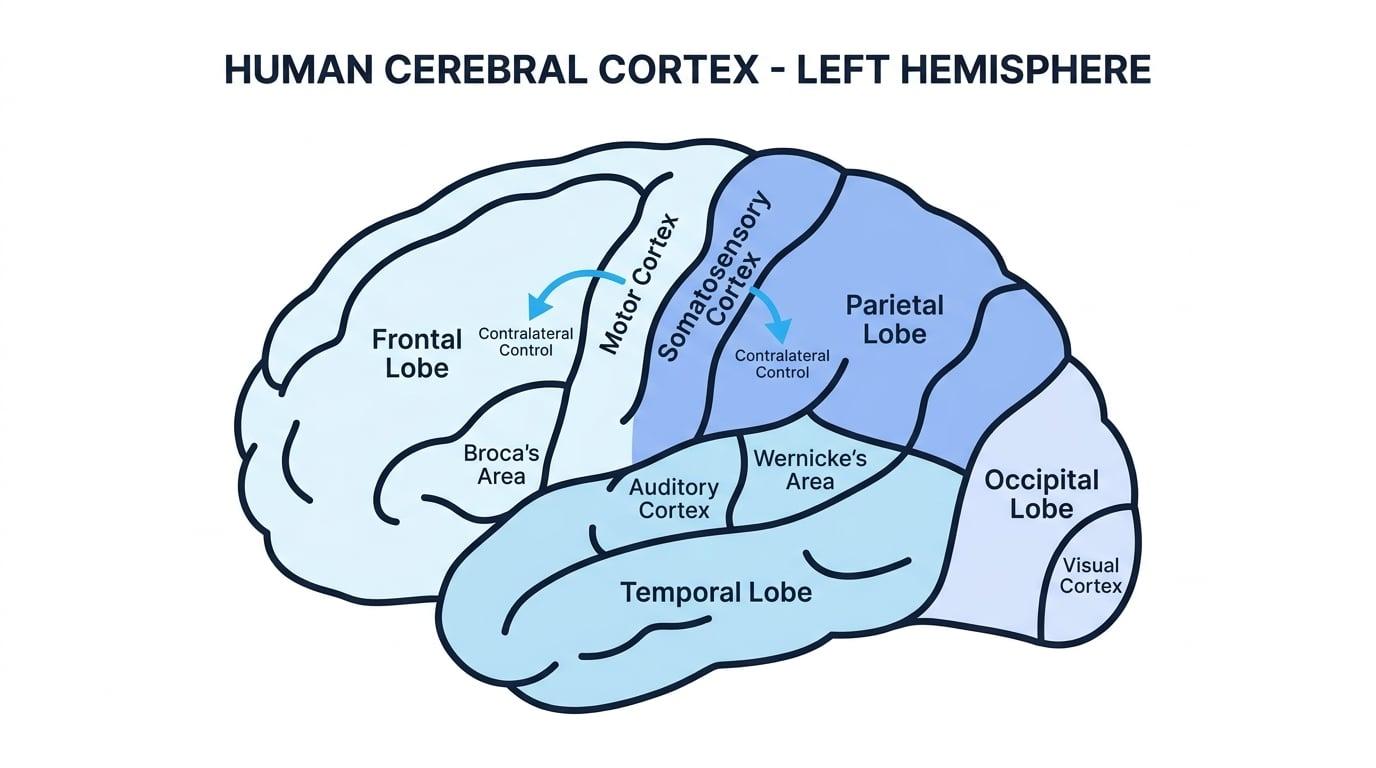
The cerebral cortex is the wrinkled outer layer of the brain. It is divided into two halves called hemispheres: the left hemisphere and the right hemisphere. These communicate through a bundle of nerve fibres called the corpus callosum.
Many brain functions are contralateral, meaning one side of the brain controls or receives information from the opposite side of the body. For example, the left motor cortex controls movement on the right side of the body.
Localisation and lateralisation
Localisation of function means that particular mental or physical functions are associated with specific areas of the brain. Hemispheric lateralisation means that some functions are mainly controlled by one hemisphere rather than being equally shared across both.
Localisation of function in the brain
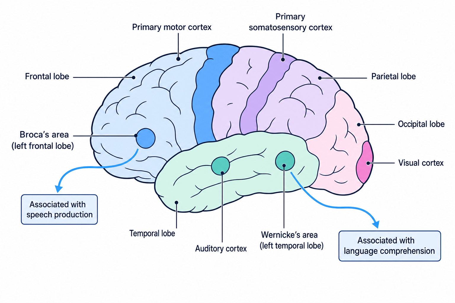
AQA expects you to know the main cortical areas: motor, somatosensory, visual, auditory, and language centres. The diagram below gives you the “map” to anchor the content.
Motor cortex
The motor cortex is in the frontal lobe. It controls voluntary movement, such as raising your hand or moving your lips to speak.
Each hemisphere controls the opposite side of the body, so damage to the left motor cortex may affect movement on the right side.
Somatosensory cortex
The somatosensory cortex is in the parietal lobe. It processes bodily sensations such as touch, pressure, temperature and pain.
More sensitive body areas, like the lips and fingertips, take up more cortical space than less sensitive areas.
Visual cortex
The visual cortex is in the occipital lobe at the back of the brain. It processes visual information from the eyes.
Importantly, the left visual field is processed by the right hemisphere, and the right visual field is processed by the left hemisphere.
Auditory cortex
The auditory cortex is in the temporal lobe. It processes sound information, including volume, pitch and rhythm.
Damage here can affect hearing or the interpretation of sounds, depending on the exact location and extent of damage.
Language centres: Broca’s and Wernicke’s areas
Broca’s area is usually found in the left frontal lobe. It is linked to speech production. Damage can lead to Broca’s aphasia, where speech becomes slow, effortful and grammatically simple, while comprehension is often relatively preserved.
Wernicke’s area is usually found in the left temporal lobe. It is linked to language comprehension. Damage can lead to Wernicke’s aphasia, where speech may be fluent but meaningless, and comprehension is poor.
Localisation is not the same as isolation
A brain area may be strongly associated with a function, but most behaviours still rely on networks of areas working together.
Linking symptoms to brain areas
- If a patient has weakness in their right hand, apply the contralateral rule: the problem is likely to involve the left hemisphere.
- Because voluntary movement is controlled by the motor cortex, right-hand weakness suggests damage to the left motor cortex.
- If the same patient also has slow, effortful speech but understands questions, this pattern fits Broca’s aphasia, so Broca’s area may also be affected.
- A strong answer would link both symptoms to localisation: movement is linked to the motor cortex, while speech production is linked to Broca’s area.
Evidence and evaluation of localisation
Early case studies support localisation. Broca (1861) studied a patient known as “Tan”, who had serious speech production difficulties and damage in the left frontal lobe. Wernicke (1874) identified patients with poor language comprehension and damage in the left temporal lobe.
Modern brain-scanning evidence also supports localisation because techniques such as fMRI can show different areas becoming active during different tasks.
However, localisation can be too simplistic. Lashley (1950) removed areas of rats’ cortex while they learned mazes and found that learning did not seem to depend on one single “memory centre”. This supports the idea that complex functions may be distributed across the brain.
Overstating localisation
Avoid writing that one brain area “does” a whole behaviour by itself. A better phrase is: “This area is strongly associated with…” or “This area plays an important role in…”
For AO3, case studies are useful because they provide rich detail, but they often involve unusual patients with complex damage, so generalisability is limited. Modern research must consider informed consent, confidentiality, protection from harm, the right to withdraw, minimal deception, and proper debriefing, especially when patients have brain injuries that may affect communication or decision-making.
Hemispheric lateralisation
Some functions are more dominant in one hemisphere. For most right-handed people, language is mainly lateralised to the left hemisphere. The right hemisphere is often more involved in visual-spatial tasks, face recognition and emotional tone, although this is not absolute.
The hemispheres usually share information through the corpus callosum. This is why everyday behaviour feels unified rather than split into two separate minds.
Split-brain research
Split-brain research studies people whose corpus callosum has been surgically cut. This operation, called a commissurotomy, was sometimes used to reduce severe epilepsy by preventing seizures spreading between hemispheres.
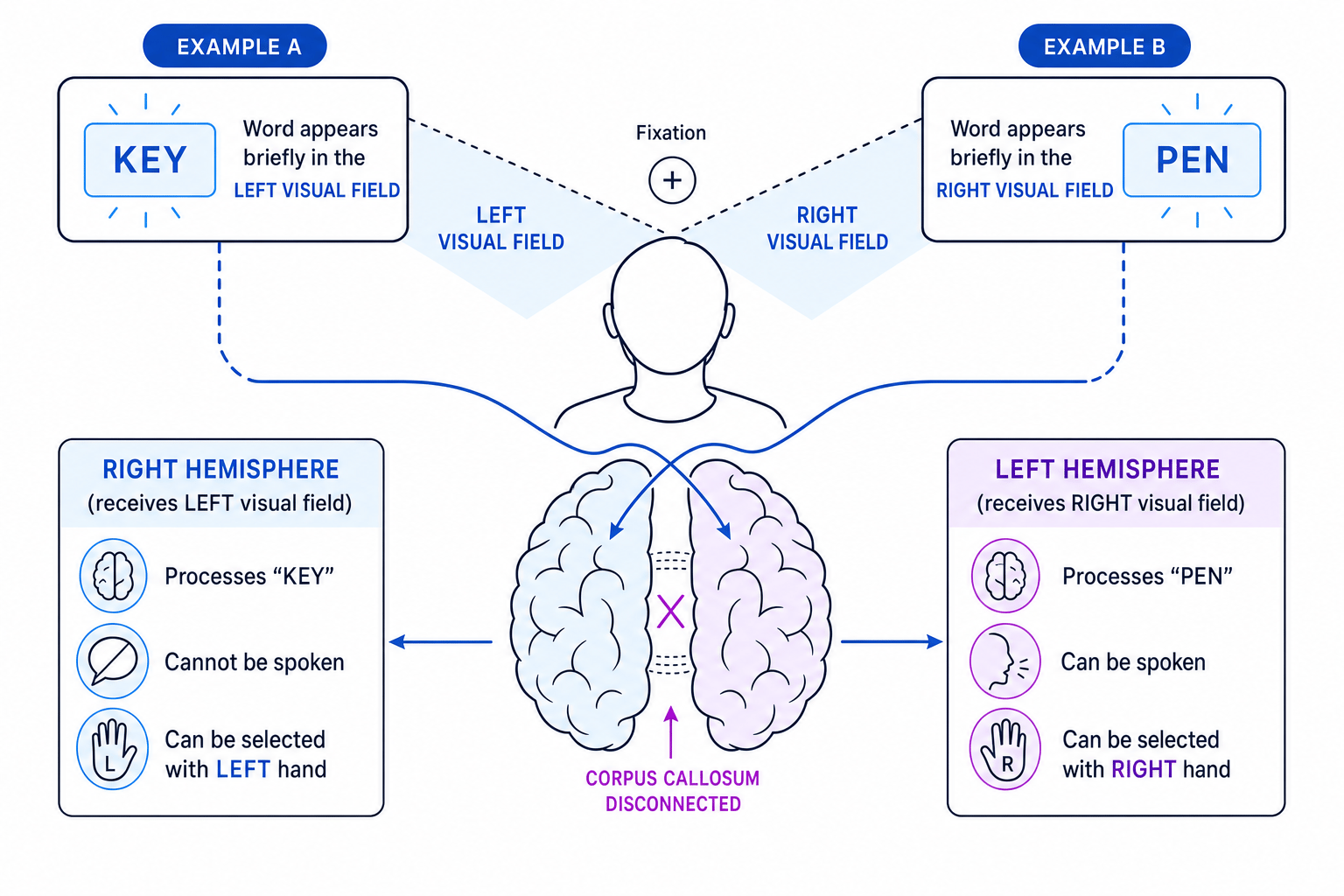
Sperry (1968) studied split-brain patients by presenting information to only one visual field at a time. Because the corpus callosum was cut, information could not be transferred easily between hemispheres.
The diagram below shows the key logic: information in the left visual field goes to the right hemisphere, while information in the right visual field goes to the left hemisphere.
Key findings
- If a word was shown to the right visual field, it went to the left hemisphere, where language is usually located, so the participant could say the word aloud.
- If a word was shown to the left visual field, it went to the right hemisphere, so the participant often could not say the word.
- However, the participant could often select or draw the object using the left hand, because the right hemisphere controls the left side of the body.
Predicting a split-brain response
- Suppose the word “key” is flashed to the left visual field. This information is processed by the right hemisphere.
- Speech production is usually controlled by the left hemisphere, but the corpus callosum is disconnected, so the information cannot be transferred for speech.
- The participant is therefore unlikely to say “key” aloud.
- Because the right hemisphere controls the left hand, the participant may still be able to pick out a key using the left hand.
Evaluation of split-brain research
A strength is that Sperry’s studies were highly controlled. Stimuli were presented briefly to one visual field, allowing researchers to test each hemisphere separately. This gives strong evidence for lateralisation.
However, the sample was very small and unusual. Split-brain patients had severe epilepsy, medication histories and major surgery, so findings may not generalise to typical brains. The tasks were also artificial, reducing ecological validity.
Ethically, the surgery was performed for medical reasons, not simply for research. Researchers still needed valid consent, protection from distress, confidentiality, the right to withdraw and debriefing, because participants were neurologically vulnerable.
Plasticity: the brain can change
Plasticity means the brain’s ability to change its structure and function in response to experience, learning or injury.
Functional recovery
Functional recovery is the transfer or rebuilding of functions after brain trauma, such as a stroke, tumour or head injury. Undamaged areas may take over functions previously carried out by damaged areas.
Plasticity can happen through several mechanisms:
- Axonal sprouting: surviving neurons grow new branches to connect with other neurons.
- Synaptic strengthening: frequently used connections become stronger.
- Recruitment of homologous areas: the opposite hemisphere may take over some functions.
- Neural unmasking: existing but inactive connections become active when usual pathways are damaged.
Recovery is often fastest in the first weeks and months after injury, but rehabilitation can support improvement for much longer.
Explaining recovery after a stroke
- If a stroke damages part of the left motor cortex, the patient may initially lose movement on the right side of the body.
- During recovery, nearby undamaged areas may form new connections through axonal sprouting and synaptic strengthening.
- Homologous areas in the right hemisphere may also help compensate, especially with rehabilitation and repeated practice.
- The improvement is therefore explained by functional recovery through plasticity, not by the damaged tissue simply “growing back” as normal.
Evidence and evaluation of plasticity
Research supports lifelong plasticity. Maguire et al. (2000) found that London taxi drivers had larger posterior hippocampi than controls, likely linked to years of spatial navigation experience. Draganski et al. (2004) found brain changes after participants learned to juggle, supporting experience-dependent plasticity.
Functional recovery is also supported by case evidence. For example, Danelli et al. (2013) reported language recovery after major left-hemisphere surgery in childhood, suggesting that other areas can take over language functions.
However, plasticity is not unlimited. Recovery depends on factors such as age, severity of damage, location of injury, rehabilitation and cognitive reserve. Schneider et al. (2014) found that people with more education were more likely to recover well after traumatic brain injury, suggesting individual differences matter.
There is also negative plasticity: the brain can change in unhelpful ways, such as chronic pain pathways becoming strengthened.
Strong AO3 balance
For essays, avoid saying “plasticity proves localisation is wrong”. A better argument is that localisation exists, but plasticity shows the brain is more flexible and interactive than a simple fixed map suggests.
Ethically, plasticity research using scans normally involves informed consent, confidentiality and protection from harm, such as checking participants are safe in MRI scanners. Trauma and rehabilitation studies must be especially careful not to pressure vulnerable patients and should not deprive them of normal treatment.
In the exam
- For localisation questions, name the area, state its function, then link damage to a specific symptom.
- For split-brain questions, track the pathway carefully: visual field → opposite hemisphere → speech or hand response.
- For evaluation, balance support from case studies and scans with limitations such as small samples, artificial tasks and brain plasticity.
Check yourself
- What symptom differences would you expect from damage to Broca’s area compared with Wernicke’s area?
- In a split-brain patient, why can a word in the left visual field be selected with the left hand but not spoken?
- How can plasticity help recovery after trauma, and why might recovery vary between individuals?