What you'll learn
- How fMRI, EEG, ERPs and post-mortem examinations are used to study the brain.
- The difference between spatial resolution and temporal resolution.
- How to evaluate each method using AO3: strengths, limitations, ethics and applications.
- How to apply the techniques to scenario questions in the exam.
Big picture: why study the brain?
Biopsychology assumes that thoughts, emotions and behaviour are linked to biological processes in the nervous system. To investigate this, psychologists need methods that show either:
- where activity or damage is located in the brain,
- when brain activity happens,
- or what the brain looked like after death.
No single technique is “best” for everything. A strong exam answer explains what the method measures and then evaluates whether that method is suitable for the research question.
Spatial and temporal resolution
- Spatial resolution means how accurately a technique can show where activity or damage is located in the brain.
- Temporal resolution means how accurately a technique can show when brain activity happens.
The central trade-off
Techniques with excellent timing, like EEG and ERPs, are often weaker at locating the exact brain area. Techniques with good localisation, like fMRI, are often slower at tracking rapid mental processes.
Functional magnetic resonance imaging: fMRI
What fMRI measures
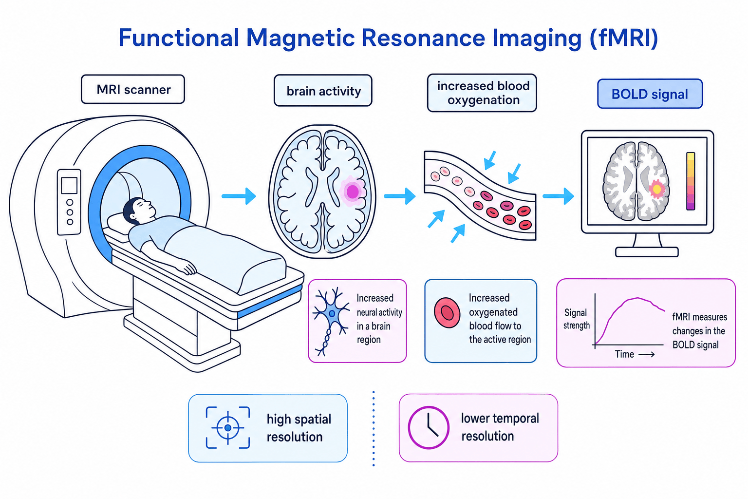
Functional magnetic resonance imaging, usually shortened to fMRI, is a brain-scanning technique that measures changes in blood oxygenation and blood flow while a person is alive and doing a task.
When neurons in a brain area become more active, they need more oxygen. The body responds by sending more oxygenated blood to that area. fMRI detects this change using the BOLD signal, which stands for blood oxygenation level-dependent signal.
BOLD signal
The BOLD signal is the change in oxygenated blood flow detected by fMRI. It is used as an indirect measure of neural activity.
In a typical fMRI study, a participant lies in a scanner and completes a task, such as recognising faces or listening to words. The researcher compares brain activity during the task with a control condition, then produces an image showing areas with increased blood oxygenation.
Strengths of fMRI
A major strength is high spatial resolution. fMRI can identify activity in specific brain areas more precisely than EEG, so it is useful for studying localisation of function — the idea that different brain areas specialise in different functions.
fMRI is also non-invasive. It does not require surgery or radioactive tracers, so it is safer than some other biological methods. This makes it useful in research with healthy volunteers and clinical groups.
It also has valuable real-world applications. For example, fMRI can help researchers understand language processing, memory, emotional responses and disorders such as depression or schizophrenia.
Limitations of fMRI
The biggest limitation is that fMRI is an indirect measure. It does not measure neurons firing directly; it measures blood oxygenation changes that are assumed to follow neural activity.
It also has poorer temporal resolution than EEG and ERPs because blood flow changes happen more slowly than electrical activity. This means fMRI is less suitable for studying very rapid cognitive processes.
Practical issues matter too. fMRI scanners are expensive, noisy and can be uncomfortable. Some participants may feel claustrophobic, and movement inside the scanner can distort the results.
Saying fMRI measures electrical activity
fMRI does not measure electrical activity directly. It measures changes in blood oxygenation, which are used as a proxy for neural activity.
Choosing fMRI for localisation
A researcher wants to find which brain area is most active when participants recognise emotional facial expressions.
- The key aim is to identify where activity occurs, so the researcher needs strong spatial resolution.
- fMRI is suitable because it can compare BOLD activity during emotional-face recognition with a control task, such as viewing neutral shapes.
- If the amygdala shows greater BOLD activity in the emotional-face condition, the researcher can say this area is associated with processing emotional faces.
- The researcher should not claim that fMRI proves the amygdala alone causes emotional recognition, because fMRI evidence is correlational and indirect.
Electroencephalogram: EEG
What EEG measures
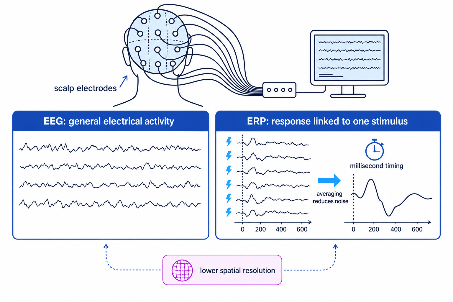
An electroencephalogram, or EEG, records electrical activity in the brain using electrodes placed on the scalp. These electrodes detect tiny electrical signals produced by many neurons firing together.
EEG does not produce a detailed picture of the brain like fMRI. Instead, it produces wave patterns showing general electrical activity over time.
EEG
An EEG is a recording of general brain electrical activity detected by electrodes placed on the scalp.
EEGs are often used in clinical settings. For example, they can help diagnose epilepsy, because epileptic seizures involve unusual electrical activity. They are also used in sleep research to identify different sleep stages.
Strengths of EEG
The main strength is excellent temporal resolution. EEG can detect changes in brain activity extremely quickly, so it is useful for studying the timing of neural responses.
EEG is also relatively cheap compared with fMRI and is non-invasive. Participants can often sit or sleep naturally while being recorded, which improves ecological validity for some types of research.
Limitations of EEG
EEG has poor spatial resolution. The electrical signals are detected at the scalp, but they may have come from several areas of the brain. Signals can also be distorted by the skull and scalp.
This means EEG is much better for saying when brain activity changes than for saying exactly where it started.
Movement, blinking and muscle tension can also create artefacts, which are unwanted signals that interfere with the recording.
Artefact
An artefact is unwanted noise in a biological recording, such as eye movement or muscle activity, which can make the brain signal harder to interpret.
Event-related potentials: ERPs
What ERPs measure
Event-related potentials, or ERPs, are derived from EEG recordings. Instead of looking at general brain activity, researchers present the same stimulus many times and average the EEG responses.
A stimulus is anything presented to the participant, such as a word, tone, image or face. If responses are time-locked to that stimulus, averaging helps remove unrelated background brain activity. What remains is a clearer waveform linked to the event.
ERP
An ERP is a small brain response, extracted from EEG data, that is time-locked to a specific stimulus or event.
Strengths of ERPs
ERPs are more specific than raw EEG because they isolate the brain’s response to a particular event. This makes them useful for studying cognitive processes such as attention, perception and language.
Like EEG, ERPs have excellent temporal resolution. This helps researchers investigate the order in which mental processes occur.
Limitations of ERPs
ERPs still have poor spatial resolution because they are based on scalp recordings. They tell us when a response happened more clearly than where it happened.
They also require many repeated trials and careful control. If the participant gets tired, distracted or moves too much, the data may become unreliable.
Another issue is interpretation. Researchers must decide which part of the waveform is meaningful, and different components can sometimes overlap.
Deciding between EEG and ERP
A researcher wants to study how quickly the brain responds to an unexpected tone in an attention task.
- The research question is about the brain’s response to a specific event: the unexpected tone.
- Raw EEG would show general electrical activity, but much of that activity would not be related to the tone.
- ERP is the better choice because repeated tone trials can be averaged to isolate the response time-locked to the stimulus.
- The researcher should still be cautious about locating the exact brain region involved, because ERPs have low spatial resolution.
Post-mortem examinations
What post-mortems involve
A post-mortem examination involves studying a person’s brain after death. Researchers may look for damage, abnormalities, lesions or differences in brain structure and then compare these findings with the person’s behaviour when they were alive.
Post-mortem examination
A post-mortem examination is the analysis of brain tissue after death, often to link brain abnormalities with symptoms or behaviour shown during life.
Post-mortems have been important in the history of brain research. For example, Paul Broca studied patients with speech problems and found damage in a region of the left frontal lobe after death. This contributed to the idea that Broca’s area is involved in speech production.
Strengths of post-mortem examinations
Post-mortems allow very detailed physical analysis of the brain. Researchers can examine tissue, lesions and abnormalities in ways that may not be possible with living participants.
They can also be useful when the person had a rare disorder or unusual symptoms. This can provide valuable insights into localisation of function and neurological illness.
Post-mortem evidence has helped support biological explanations of mental disorders by showing possible differences in brain structure or chemistry.
Limitations of post-mortem examinations
A key weakness is that they cannot show the living brain in action. Researchers are looking at the brain after death, so they cannot directly observe how the brain functioned during tasks.
Causality is also difficult. If a person had both brain damage and behavioural symptoms, we cannot be certain that the damage caused the symptoms. Other factors, such as illness, medication, ageing or substance use, may have affected the brain.
Samples are often small and unusual, so findings may not generalise to the wider population. Information about the person’s behaviour during life may also be incomplete or retrospective.
Ethical issues
Post-mortem research raises important ethical issues. Researchers need valid consent, either from the person before death or from relatives afterwards, depending on the situation. They must protect confidentiality and treat brain tissue respectfully.
For living techniques like fMRI, EEG and ERPs, researchers still need informed consent, the right to withdraw, protection from harm, confidentiality and debriefing. For example, fMRI participants should be warned about noise and confined spaces.
AO3 comparison shortcut
For evaluation, compare methods using three questions: What does it measure? How precise is it in space and time? What practical or ethical issues limit it?
Quick comparison
| Method | Measures | Best for | Main limitation |
|---|---|---|---|
| fMRI | Blood oxygenation changes | Locating active brain areas | Indirect and slower timing |
| EEG | General electrical activity | Tracking rapid brain activity | Poor localisation |
| ERP | Averaged response to a stimulus | Timing responses to specific events | Needs many trials and careful control |
| Post-mortem | Brain tissue after death | Detailed structural analysis | Cannot show living brain activity |
In the exam
- For AO1, state what the technique measures before evaluating it.
- For AO3, use comparison language: “high spatial resolution but lower temporal resolution” is stronger than simply saying “accurate”.
- In application questions, match the method to the research aim: fMRI for localisation, EEG or ERPs for timing, post-mortem for detailed tissue analysis after death.
Check yourself
- Why is fMRI described as an indirect measure of neural activity?
- What is the difference between EEG and ERP?
- Why is it difficult to infer causality from post-mortem evidence?
