Use of high-energy X-rays (A-level only)
Welcome to the physics of radiotherapy! You already know how X-rays are used to look inside the body (diagnostic imaging). In this topic, we will explore how we use them to treat the body.
What you'll learn
- The difference between diagnostic X-rays and therapeutic high-energy X-rays.
- Why treating tumours requires a delicate balancing act to protect healthy cells.
- The three main methods used to limit healthy cell exposure: overlapping beams, collimation, and fractionation.
1. Diagnostic vs. Therapeutic X-rays
In diagnostic radiography, the goal is to get a picture of the bones or tissues while doing as little damage to the patient as possible. We use X-rays with energies roughly between 20 keV and 150 keV.
However, sometimes we actually want to cause cellular damage.
Radiotherapy
Radiotherapy is the use of high-energy ionising radiation (like X-rays) to control or destroy malignant cancer cells by damaging their DNA, preventing them from multiplying.
To destroy cancer cells, we need much higher energies than diagnostic machines can provide. External beam radiotherapy uses high-energy X-rays in the range of 1 MeV to 20 MeV. These are usually produced by a linear accelerator (linac) rather than a standard X-ray tube.
The Goal of Radiotherapy
High-energy X-rays are highly ionising and will kill cells. The primary challenge in physics is designing a treatment that delivers a lethally high radiation dose to the tumour, while keeping the dose to surrounding healthy tissue as low as possible.
Worked Example 1: Energy comparison
Question: Explain why the X-rays used in external beam radiotherapy have much higher energies than those used for diagnostic imaging. (2 marks)
How to answer:
- State the purpose of diagnostic X-rays. They are used to image internal structures, so they need just enough energy to penetrate tissue but be absorbed by bone, minimising unnecessary cellular damage.
- State the purpose of therapeutic X-rays. They are specifically intended to destroy cancer cells by highly ionising the tissue and damaging DNA, which requires much higher energies (in the MeV range).
2. Limiting Exposure: Overlapping Beams
The biggest problem with treating a tumour deep inside the body is that the X-rays must pass through healthy tissue to get there, and then pass through more healthy tissue on the way out. If we just fired one massive, powerful beam straight at the tumour, we would destroy a "tunnel" of healthy cells along the entire path.
To prevent this, physicists use overlapping beams (often called the isocentric technique).
Instead of one high-intensity beam, the linac fires several lower-intensity beams from completely different angles. The machine is carefully calibrated so that all the beams cross over (intersect) at exactly one point: the tumour.
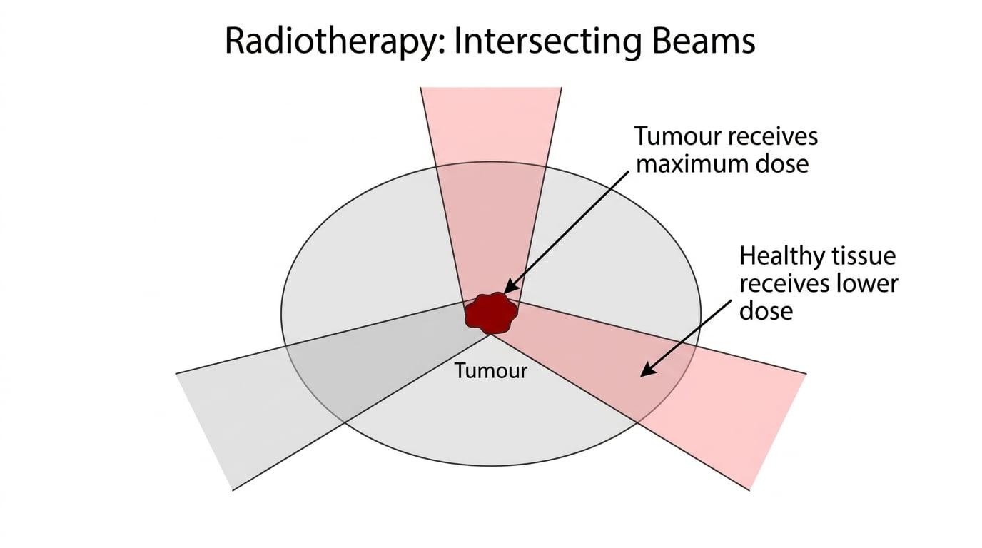
By doing this, the tumour sits at the "crossfire" and receives the combined total dose from all the beams. Meanwhile, any given patch of healthy tissue only sits in the path of a single beam, receiving a much smaller fraction of the dose. In modern machines, the linac often rotates in a continuous 360∘360^\circ360∘ arc around the patient while firing, spreading the "entry dose" over an entire ring of healthy tissue.
Worked Example 2: Dose ratio with intersecting beams
Question: A deep-tissue tumour requires a highly targeted total radiation dose of 60 Gy60 \text{ Gy}60 Gy. The treatment plan uses an isocentric linac that fires 4 equal-intensity beams from different angles, intersecting at the tumour. Assuming attenuation in the healthy tissue is negligible, determine the maximum dose received by a patch of healthy tissue in the path of one beam.
How to answer:
- Recognise that the tumour receives the sum of all overlapping beams.
- Since there are 4 equal-intensity beams, divide the total required dose by the number of beams to find the dose delivered by a single beam.
- Conclude the dose to the healthy tissue. Because any specific area of healthy tissue only intersects with one beam, it receives a maximum of 15 Gy15 \text{ Gy}15 Gy, which is exactly 14\frac{1}{4}41 of the dose delivered to the tumour.
3. Limiting Exposure: Beam Shaping (Collimation)
Tumours are rarely perfect spheres; they are irregular, jagged 3D shapes. If we fire a square X-ray beam at an irregularly shaped tumour, the corners of the square will hit healthy tissue. We need a way to shape the beam to match the exact profile of the tumour from the angle it is being fired.
Collimator
A collimator is a device made of dense radiation-absorbing material (usually lead or tungsten) used to narrow, block, and shape a beam of radiation.
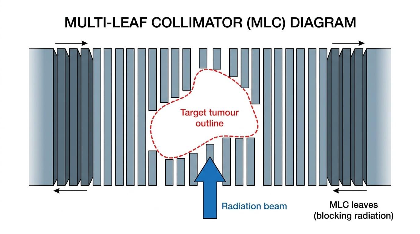
Modern radiotherapy machines use a brilliant piece of engineering called a Multi-Leaf Collimator (MLC). This consists of dozens of narrow, heavy metal plates (leaves) that can slide in and out independently. A computer controls the leaves, sliding them into position to create a custom jagged opening that perfectly matches the "silhouette" of the tumour.
As the linac rotates around the patient, the tumour's silhouette changes shape. The computer continuously moves the heavy metal leaves to dynamically reshape the beam in real-time, ensuring vital surrounding organs are always shielded in the shadow of the lead leaves.
Think of it like a shadow puppet
The multi-leaf collimator is essentially a highly advanced, automated shadow puppet. The X-ray source is the torch, the MLC leaves are the hands blocking the light, and the "shadow" protects the healthy organs while the "light" (the X-rays) only falls on the tumour.
4. Limiting Exposure: Fractionation
The final method for protecting healthy cells relies on a bit of biology. Even with overlapping beams and collimators, healthy cells still receive some radiation.
To help them survive, the total required radiation dose is not given in a single session. Instead, it is divided up into smaller doses (called fractions) delivered over several days or weeks. Healthy cells generally have better DNA-repair mechanisms than rapidly dividing cancer cells. By splitting up the dose, the healthy cells get time to recover and repair themselves between sessions, while the cancer cells progressively accumulate lethal damage and die off.
Forgetting the physics focus
While fractionation is a valid point in an exam question, remember that you are sitting a Physics exam, not a Biology exam. Always prioritise the physical methods (overlapping beams and multi-leaf collimators) as your primary answers when asked how healthy tissue is protected, unless the question specifically asks for a time-based or biological method.
Worked Example 3: Explaining the methods
Question: State and explain two physical methods used during external beam radiotherapy to limit the radiation exposure of healthy cells. (4 marks)
How to answer:
- Method 1: Use of overlapping beams / rotating the linac.
- Explanation 1: The machine fires beams from multiple different angles that intersect at the tumour. The tumour receives the maximum combined dose, while the surrounding healthy tissue only receives a small fraction of the dose from a single beam.
- Method 2: Using a multi-leaf collimator (MLC).
- Explanation 2: Heavy metal leaves (lead or tungsten) slide into position to shape the beam so its cross-section perfectly matches the shape of the tumour. This shields adjacent healthy tissue and vital organs from the primary beam.
Summary
When dealing with high-energy X-rays, controlling the dose is everything. By combining isocentric rotation (overlapping beams from multiple angles) with dynamic collimation (shaping the beam to the tumour) and fractionation (splitting the dose over time), physicists can safely deliver massive, targeted energy directly to cancer cells while keeping the patient safe.
In the exam
- Ensure you clearly state that therapeutic X-rays are "high energy" (usually MeV) compared to diagnostic X-rays.
- If asked to explain how healthy tissue is protected, your go-to answers should be intersecting beams and collimation.
- When describing collimation, use the key terms "lead/tungsten leaves", "shielding", and "shaping the beam to the tumour profile".
- When describing intersecting beams, ensure you mention that the beams cross at the tumour so the tumour gets the maximum/combined dose while healthy tissue gets a lower/single-beam dose.
Check yourself
- Can you explain why we cannot use an ordinary diagnostic X-ray tube for radiotherapy?
- Can you describe how an isocentric linac setup allows a high dose to be delivered to a deep tumour without destroying the skin above it?
- How does a multi-leaf collimator help protect a healthy organ that is located right next to a tumour?