Gamma camera (A-level only)
What you'll learn
- Why we need a gamma camera to image radioactive tracers inside the body.
- The step-by-step journey of a gamma photon through the camera's key components (collimator, scintillator, light guide, and photomultiplier tubes).
- How a photomultiplier tube (PMT) works to multiply a tiny light signal into a measurable electronic pulse.
In medical physics, we often inject patients with radioactive tracers (like Technetium-99m) that gather in specific organs and emit gamma radiation. But how do we "see" radiation that is entirely invisible to the human eye? This is where the gamma camera comes in. It sits outside the patient and detects the escaping gamma photons, building a 2D image of where the tracer is concentrated.
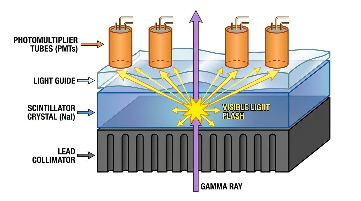
Let's break down the structure of a gamma camera, layer by layer, starting from the side closest to the patient.
1. The Collimator
Gamma photons are emitted in all random directions from the patient's body. If all of these photons hit our detector, we would just get a blurry mess of overlapping signals. We only want photons travelling in a straight line from a specific point in the patient to the corresponding point on our detector.
To achieve this, the first layer of the gamma camera is the collimator.
Collimator
A thick disk of heavy metal (usually lead) containing thousands of parallel, vertical holes. It acts as a strict filter for gamma photons.
Only gamma photons travelling vertically (parallel to the holes) can pass through the collimator. If a photon hits the collimator at an angle, it crashes into the lead walls between the holes and is absorbed.
Resolution over Intensity
The collimator dramatically reduces the number of gamma photons reaching the detector, but it is entirely necessary to create a sharp, high-resolution image. Without it, you wouldn't be able to tell where any photon came from!
AQA-Style Question: The Collimator
Explain the purpose of the collimator in a gamma camera and state the material it is typically made from. (3 marks)
- The collimator is made from lead.
- Its purpose is to absorb gamma photons that are travelling at an angle to the axis of the tubes.
- This ensures that only gamma photons travelling parallel to the holes reach the scintillator, which greatly improves the sharpness (spatial resolution) of the final image.
2. The Scintillator and Light Guide
Once a gamma photon makes it through a hole in the collimator, it needs to be detected. Gamma photons carry too much energy to be easily converted straight into an electrical signal. Instead, we use an intermediate step: we turn the gamma photon into visible light.
This happens in the scintillator.
Scintillator
A large single crystal, typically made of Sodium Iodide doped with Thallium — written as NaI(Tl) — which absorbs a single high-energy gamma photon and emits thousands of lower-energy visible light photons.
When the gamma photon interacts with the crystal, it transfers its energy to an electron in the crystal lattice. As this electron loses energy, it causes the crystal to emit a localized "flash" of visible light (scintillation).
Immediately behind the scintillator is the light guide. This is a solid piece of clear plastic or glass. It acts as a bridge, optically coupling the scintillator crystal to the array of photomultiplier tubes behind it, ensuring the flashes of light are guided smoothly into the tubes without reflecting backwards.
3. The Photomultiplier Tube (PMT)
Even though one gamma photon creates thousands of visible light photons in the scintillator, the resulting light flash is still incredibly faint. If we just placed a standard wire there, it wouldn't register a useful electrical signal.
We must amplify this faint light into a large electrical pulse. This is the job of the Photomultiplier Tube (PMT). A modern gamma camera contains an array of dozens of PMTs arranged in a hexagonal honeycomb pattern behind the light guide.
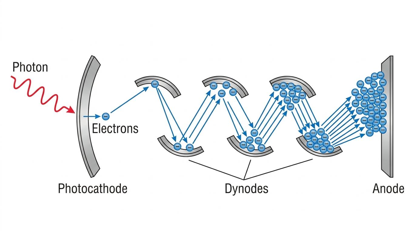
Here is the step-by-step workings of a single PMT:
The Photocathode
The visible light photons enter the front of the vacuum tube and hit the photocathode. Thanks to the photoelectric effect, each visible light photon that is absorbed causes the photocathode to emit an electron (called a photoelectron).
The Dynodes and Secondary Emission
These tiny numbers of electrons are then accelerated towards a series of positively charged electrodes called dynodes. Each dynode is kept at a progressively higher positive potential difference (voltage) than the last.
Because the electrons are accelerated through a vacuum, they gain significant kinetic energy. When a single fast-moving electron smashes into the first dynode, its impact is energetic enough to knock out multiple electrons from the dynode surface. This process is called secondary emission.
Secondary emission
The process where a single high-energy electron strikes a surface, causing several secondary electrons to be ejected.
These secondary electrons are then accelerated to the second dynode, where they each knock out multiple electrons again. This happens across roughly 10 to 12 dynodes, creating a massive avalanche of electrons.
The Anode
Finally, this huge swarm of electrons is collected by the anode at the back of the tube. What started as perhaps one electron at the photocathode has now multiplied into millions of electrons, creating a measurable electrical current pulse!
AQA-Style Calculation: PMT Amplification
In a photomultiplier tube, a single photoelectron is emitted from the photocathode. The PMT has 101010 dynodes. At each dynode, 444 secondary electrons are emitted for every 111 electron that strikes it. Calculate the total number of electrons that reach the anode for a single photoelectron.
- The initial number of electrons is 111.
- At the first dynode, the number of electrons becomes 1×4=41 \times 4 = 41×4=4.
- At the second dynode, those 444 electrons each produce 444 more, so 4×4=164 \times 4 = 164×4=16, which is 424^242.
- This multiplication happens 101010 times. Therefore, the final number of electrons at the anode is 4104^{10}410.
- Calculate the value: 410=1.05×10⁶ electrons4^{10} = 1.05 \times 10^6 \text{ electrons}410=1.05×10⁶ electrons.
Multiplication formula
If a PMT has nnn dynodes and the multiplication factor at each dynode is kkk, the total amplification factor is knk^nkn.
4. Forming the Final Image
So, a gamma photon has passed the collimator, flashed in the scintillator, and triggered a pulse in the PMTs. What next?
The electrical pulses from all the PMTs in the array are sent to a computer. The computer looks at the signals and figures out exactly where the original gamma flash occurred.
For example, if the flash happens directly under PMT number 5, then PMT 5 will output the strongest electrical pulse. But the neighboring PMTs (like PMT 4 and 6) will also detect a bit of the light and output smaller pulses. By comparing the exact strengths of the signals from all the different PMTs, the computer can triangulate the exact xxx and yyy coordinates of the original gamma photon hit.
Over several minutes, millions of these hits are recorded, and the computer builds up a 2D map showing the concentration of the radioactive tracer inside the patient!
Confusing X-rays and Gamma cameras
Students often confuse an X-ray machine with a Gamma camera. In X-ray imaging, the radiation source is outside the patient and shines through them to a detector. In Gamma camera imaging, the radiation source is the tracer inside the patient, and the camera simply acts as a passive detector catching the escaping rays.
In the exam
If asked to describe the full sequence of events in a gamma camera, use this checklist to ensure you don't drop sequence marks:
- Gamma photons are emitted from the tracer in the patient.
- The lead collimator absorbs angled photons, only letting parallel photons pass.
- The scintillator crystal (NaI) absorbs the gamma photon and emits many visible light photons.
- The photocathode in the PMT absorbs the visible light and emits photoelectrons.
- Secondary emission at the dynodes multiplies the electrons.
- The anode collects the electrons to form an electrical pulse.
- A computer processes the signals to locate the source and map the image.
Check yourself
- What material is the collimator made of, and why?
- What physical process causes the photocathode to emit electrons when struck by light?
- If a PMT has 8 dynodes and a multiplication factor of 5 per dynode, what is the total number of electrons reaching the anode per single initial photoelectron?
- Why do we use a scintillator rather than passing gamma rays directly into the PMT?