The Continuous Spectrum (Bremsstrahlung)
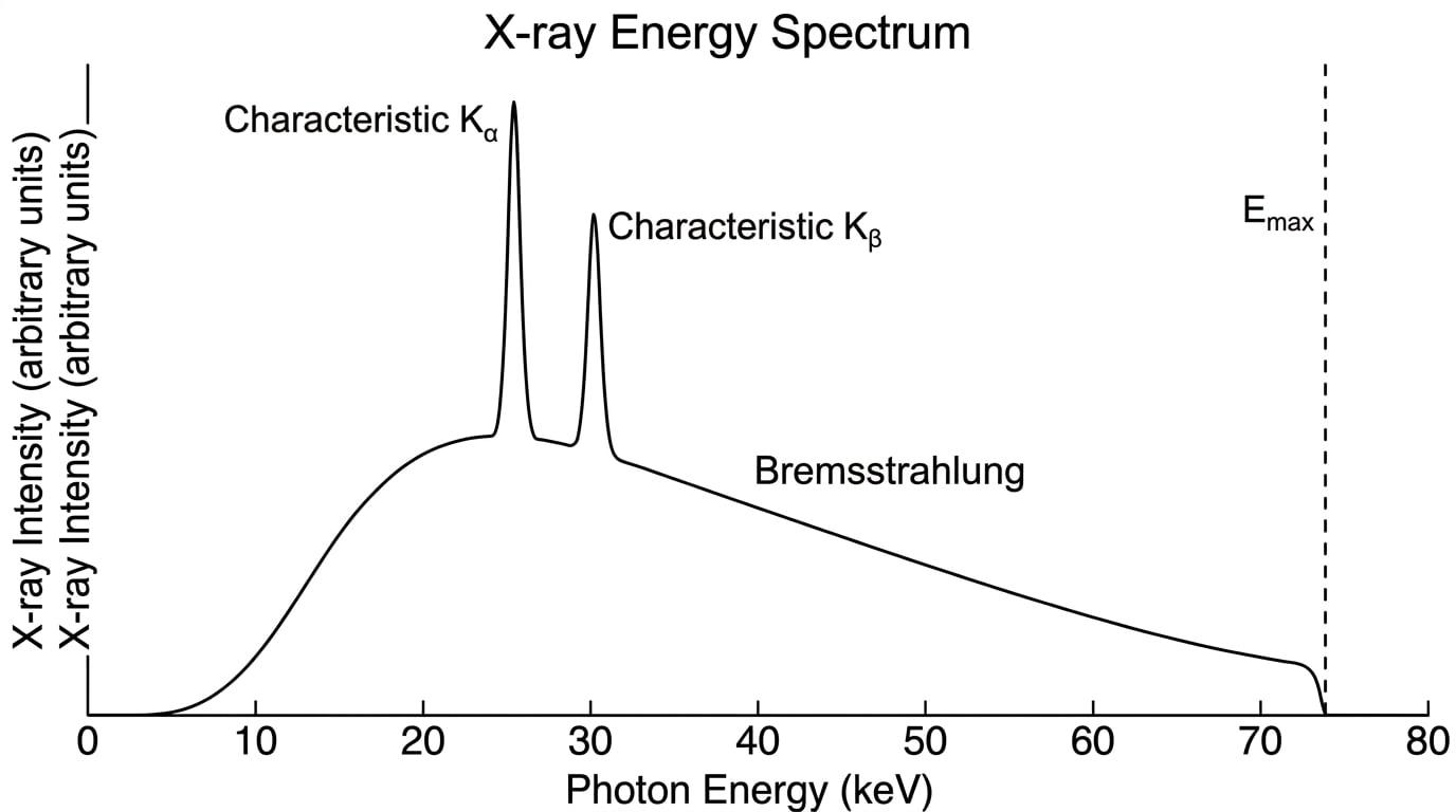
As the fast electrons plunge into the tungsten target, they are deflected and slowed down by the electric fields of the tungsten nuclei. When an electron decelerates, it loses kinetic energy, emitting it as an X-ray photon. This "braking radiation" is called Bremsstrahlung. Because electrons can be slowed down by varying amounts, they release photons of varying energies, creating the continuous "hump" on the graph.
The Characteristic Spectrum
The sharp spikes on the graph are characteristic X-rays. Occasionally, an incoming electron has enough energy to collide with and knock out an inner-shell electron from a tungsten atom. An outer-shell electron instantly drops down to fill the vacancy, emitting an X-ray photon. Because the atomic energy levels are quantized and unique to the target material, these photons have exact, specific energies—hence the sharp spikes.
Maximum Photon Energy
There is a hard limit to how much energy a single X-ray photon can have. The maximum possible photon energy (EmaxE_{\text{max}}Emax) occurs when a single incoming electron gives up all of its kinetic energy in one single braking interaction.
The kinetic energy of the electron is equal to the work done on it by the accelerating voltage, so:
Emax=eV \begin{aligned} E_{\text{max}} = eV \end{aligned} Emax=eVWhere eee is the elementary charge (1.60×10⁻¹⁹ C1.60 \times 10^{-19} \text{ C}1.60×10⁻¹⁹ C) and VVV is the accelerating potential difference.
Because photon energy is also given by E=hcλE = \frac{hc}{\lambda}E=λhc, we can find the minimum wavelength (λmin\lambda_{\text{min}}λmin) of the emitted X-rays:
hcλmin=eV \begin{aligned} \frac{hc}{\lambda_{\text{min}}} = eV \end{aligned} λminhc=eVMaximum Energy vs. Minimum Wavelength
The highest energy photon corresponds to the highest frequency and the shortest (minimum) wavelength. The continuous spectrum graph drops to exactly zero at EmaxE_{\text{max}}Emax (or λmin\lambda_{\text{min}}λmin).
Calculating the minimum wavelength
An X-ray tube operates at 70.0 kV. Calculate the minimum wavelength of the X-rays produced. (Planck constant h=6.63×10⁻³⁴ J sh = 6.63 \times 10^{-34} \text{ J s}h=6.63×10⁻³⁴ J s, speed of light c=3.00×10⁸ m s−1c = 3.00 \times 10^8 \text{ m s}^{-1}c=3.00×10⁸ m s−1)
- Equate the maximum photon energy to the work done on the electron:
- Rearrange the formula to make minimum wavelength the subject:
- Substitute the values into the equation (remembering to convert 70.0 kV to volts):
- Calculate the final answer:
3. Controlling the Beam
A radiographer must adjust the X-ray beam depending on what part of the body is being imaged. You need to understand the difference between controlling the intensity (quantity) and the photon energy (quality or "hardness").
Beam Intensity
Intensity refers to the number of X-ray photons produced per second.
- You control it by changing the tube current (the current flowing through the cathode filament).
- A higher current heats the filament more, causing more thermionic emission.
- More electrons cross the tube per second, meaning more collisions with the target, and therefore a greater number of X-ray photons are emitted per second. The overall shape of the spectrum gets taller, but EmaxE_{\text{max}}Emax doesn't change.
Photon Energy ("Hardness")
Photon energy refers to the penetrating power of the beam.
- You control it by changing the accelerating potential difference (VVV) across the tube.
- Increasing VVV gives the electrons more kinetic energy.
- This increases the maximum photon energy (EmaxE_{\text{max}}Emax) and shifts the peak of the continuous spectrum to the right (higher energy). The beam becomes "harder" and can penetrate denser tissue.
Confusing tube current with accelerating voltage
Students often mix these up. Remember:
- Filament current = How many X-rays (Intensity).
- Accelerating voltage = How energetic the X-rays are (Hardness / EmaxE_{\text{max}}Emax).
Explaining the effect of increasing tube voltage
Explain how and why the X-ray spectrum changes if the accelerating potential difference is increased, but the filament current is kept constant.
- State the effect on maximum energy: The maximum photon energy (EmaxE_{\text{max}}Emax) will increase.
- Explain why using physics principles: The electrons are accelerated through a greater potential difference, meaning they strike the target with more kinetic energy (Ek=eVE_k = eVEk=eV).
- State the effect on the spectrum's shape: The continuous spectrum shifts to the right (towards higher energies) and the overall intensity (area under the curve) increases slightly because the higher energy electrons are more efficient at producing X-rays.
- State what remains unchanged: The characteristic spikes remain at the same energy values, because these are fixed by the atomic energy levels of the target material.
4. Image Quality and Patient Dose
Taking an X-ray is a balancing act. We want a sharp, high-contrast image, but X-rays are ionizing radiation, so we must expose the patient to the absolute minimum dose necessary.
Sharpness
Image sharpness is limited by the physical size of the spot where the X-rays are generated (the focal spot). If the spot is too wide, the X-rays originate from slightly different angles, casting a blurry shadow (called a penumbra) on the detector. To improve sharpness, we need a small focal spot. However, a very small focal spot concentrates the heat and risks melting the anode. The rotating, bevelled edge of the anode elegantly solves this by providing a large actual focal area for cooling, but a small effective focal area when viewed from the perspective of the patient below.
Contrast
Contrast is the difference in blackening on the image between different tissues (e.g., bone vs. muscle). Bone absorbs X-rays much more readily than soft tissue because absorption is highly dependent on the atomic number (ZZZ) of the atoms in the tissue. To get high contrast between soft tissues, we use lower accelerating voltages (softer X-rays). If the voltage is too high, the X-rays just pass straight through everything, washing out the image.
Patient Dose
Soft (low-energy) X-rays are heavily absorbed by the patient's skin and outer tissue. They never make it to the detector to help form the image, meaning they just contribute to the patient's radiation dose without any diagnostic benefit. To reduce this unnecessary dose, we use filtration. An aluminium filter is placed across the X-ray window. It selectively absorbs the low-energy X-rays, letting the highly penetrating ("hard") X-rays through.
Safety measures checklist
Other ways to reduce dose include:
- Collimation: Using lead plates to narrow the beam so it only hits the exact area of interest.
- Intensifying screens / digital detectors: Using highly sensitive detectors so fewer X-rays are needed to form the image.
In the exam
- Be precise with your definitions. If asked about thermionic emission, mention both "heating" and the "release of electrons from the surface".
- Watch your prefixes. High voltage is almost always in kV, and wavelengths are incredibly short (10−1010^{-10}10−10 to 10−1210^{-12}10−12 m). Convert to standard SI units before putting numbers into E=eVE=eVE=eV or E=hcλE=\frac{hc}{\lambda}E=λhc.
- Link properties to controls. If a question asks how to increase the penetrating power of the beam, state that you must increase the accelerating potential difference, not the filament current.
- Use equations in explanations. If asked why the continuous spectrum ends abruptly, explicitly write down Emax=eVE_{\text{max}} = eVEmax=eV and state that it corresponds to an electron losing all its kinetic energy in a single collision.
Check yourself
- Can you describe the two mechanisms by which X-ray photons are produced when electrons hit the target?
- What happens to the minimum wavelength of the emitted X-rays if you double the accelerating voltage?
- Why does the rotating anode have an angled (bevelled) edge?
- Why is an aluminium filter placed in the path of the X-ray beam before it reaches the patient?