

What you'll learn
- How standard photographic X-ray detection is improved using intensifying screens.
- How image intensifiers allow for live, moving X-ray images (fluoroscopy).
- The inner workings of modern Flat Panel (FTP) detectors and why they have replaced photographic film.
- How and why we use contrast media, like barium meals, to see soft tissues clearly.
Traditional Photographic Detection
In the early days of radiography, X-ray images were captured directly onto photographic film. X-ray photons strike the film, causing a chemical change that turns the film black when developed. However, X-rays are highly penetrating—most of them pass straight through the film without interacting at all.
To get a clear image using bare film, you would need to use a very high intensity of X-rays, which means a dangerously high radiation dose for the patient.
The Intensifying Screen
To solve the dose problem, radiographers place the photographic film between two intensifying screens inside a light-tight cassette.
Intensifying Screen
A flat sheet coated with a fluorescent material (a scintillator) that absorbs high-energy X-ray photons and re-emits their energy as thousands of lower-energy visible light photons.
Here is the step-by-step process:
- X-rays pass through the patient and hit the intensifying screen.
- The scintillator material in the screen absorbs a single X-ray photon.
- It emits thousands of visible light photons in its place.
- It is this visible light that actually exposes the photographic film.
Why use intensifying screens?
Because one X-ray photon creates thousands of light photons, the film darkens much faster. This drastically reduces the exposure time and the radiation dose given to the patient, whilst maintaining a dark, high-contrast image.
Fluoroscopic Image Intensification
Sometimes doctors don't just want a static picture; they want a live, moving video of the body's internal workings (like watching swallowing or tracking a catheter). This is called fluoroscopy.
Historically, doctors just looked directly at a fluorescent screen, but the image was incredibly dim. If they turned up the X-ray intensity to make it brighter, the patient received a massive, unsafe dose. The modern solution is the image intensifier.
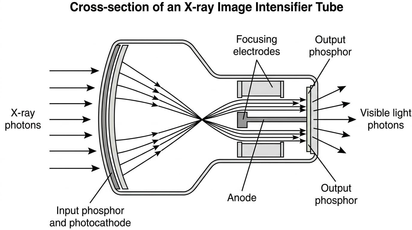
An image intensifier is a vacuum tube that converts a faint X-ray pattern into a brilliant visible image. It works like this:
- Input Phosphor: Converts X-rays into visible light photons.
- Photocathode: Absorbs this visible light and releases electrons via the photoelectric effect.
- Electron Acceleration: A high potential difference (around 25 kV25 \text{ kV}25 kV) accelerates these electrons down the tube. Focusing electrodes guide them so the image isn't lost.
- Output Phosphor: The high-speed electrons smash into a much smaller fluorescent screen at the end, converting their massive kinetic energy back into a very bright flash of visible light.
Worked Example: Explaining Image Intensification
Question: Explain how an image intensifier tube reduces the radiation dose required for dynamic X-ray imaging. (4 marks)
Answer:
- X-rays hit the input phosphor and are converted to visible light, which then ejects electrons from the photocathode.
- These electrons are accelerated by a high potential difference, gaining substantial kinetic energy.
- They hit a smaller output phosphor, converting this gained kinetic energy into a much larger number of visible light photons.
- Because the output image is both physically smaller (concentrating the light) and contains more light photons per initial X-ray, the image is vastly brighter. Therefore, a much lower initial X-ray intensity is needed to produce a visible image, reducing the patient dose.
The Modern Era: Flat Panel (FTP) Detectors
Photographic film is bulky, requires chemical processing, and degrades over time. Modern hospitals have overwhelmingly replaced film with digital systems, primarily Flat Panel (FTP) Detectors.
An FTP detector essentially takes the concept of the intensifying screen and pairs it with modern digital camera technology.
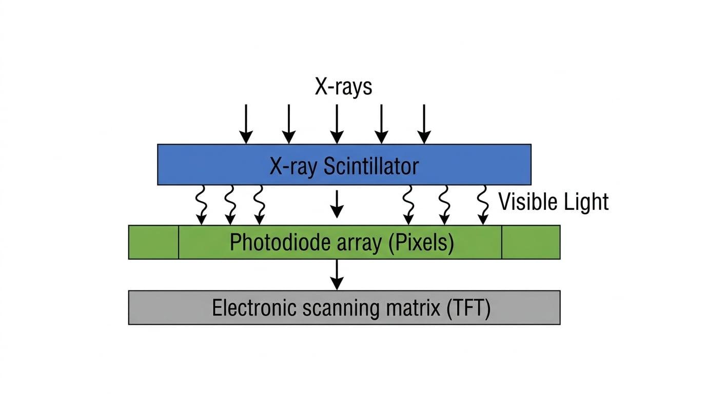
An FTP detector consists of three key layers:
- X-ray Scintillator: Usually made of caesium iodide. Just like in the intensifying screen, this layer absorbs X-rays and flashes visible light.
- Photodiode Pixels: A grid of microscopic photodiodes sits directly below the scintillator. They absorb the visible light and generate a small electrical charge proportional to the amount of light they received.
- Electronic Scanning (TFT Array): A matrix of Thin-Film Transistors (TFTs) acts like tiny switches. They are electronically scanned to read out the trapped charge from each photodiode pixel, one by one. This charge is sent to a computer to build a digital image.
Advantages of FTP detectors over photographic detection
- Lower dose: They are extremely sensitive, requiring even fewer X-rays than film with intensifying screens.
- Immediate digital image: No chemical processing time. The image pops up instantly on a monitor.
- Easier storage and sharing: Digital files can be stored securely and emailed to specialists anywhere in the world.
- Digital enhancement: The contrast and brightness can be adjusted by software after the image is taken, avoiding the need for a second X-ray if the first was too dark.
Worked Example: Comparing Detectors
Question: State two reasons why flat panel detectors have largely replaced photographic film in modern hospitals, referencing the physics of their operation. (4 marks)
Answer:
- FTP detectors use an electronic scanning matrix (TFT array) to read charge from photodiodes, creating a digital image. This eliminates the need for chemical processing, making images available instantly.
- Because the digital signal can be electronically amplified and mathematically processed by software, image contrast can be optimized post-exposure. This reduces the likelihood of needing a repeat scan, lowering the overall radiation dose to the patient.
Contrast Enhancement
X-rays are excellent at showing bone because bone has a high density and contains calcium (which has a relatively high atomic number). However, soft tissues—like the intestines, blood vessels, or muscles—have very similar densities and atomic numbers.
If you take a standard X-ray of the abdomen, the intestines barely show up. To solve this, we use an X-ray opaque material (a contrast medium).
Contrast Medium
A substance with a high atomic number introduced into the body to artificially increase the attenuation of X-rays in a specific organ or system, making it stand out as a bright white shape on the final image.
The two most common contrast materials are:
- Barium (Atomic number 565656): Used as a "barium meal" to image the digestive tract. The patient drinks a thick, chalky barium sulphate suspension, which coats the lining of the stomach and intestines.
- Iodine (Atomic number 535353): Injected into the bloodstream to visualize blood vessels (angiography) or the kidneys.
Why do high atomic number (ZZZ) materials work?
The probability of an X-ray photon being absorbed via the photoelectric effect is roughly proportional to the cube of the atomic number (Z3Z^3Z3).
Because soft tissue is mostly water (hydrogen Z=1Z=1Z=1, oxygen Z=8Z=8Z=8), the effective atomic number is low. Barium (Z=56Z=56Z=56) has a vastly higher atomic number.
The Z-cubed rule
If you compare oxygen (Z=8Z=8Z=8) to barium (Z=56Z=56Z=56), barium has an atomic number that is 777 times larger. Since absorption scales with Z3Z^3Z3, barium absorbs roughly 73=3437^3 = 34373=343 times more strongly per atom than oxygen via the photoelectric effect!
Worked Example: The Barium Meal
Question: A patient is experiencing stomach pain and requires an X-ray of their gastrointestinal tract. Explain why the patient is asked to swallow a barium meal prior to the X-ray, and how this affects the resulting image. (3 marks)
Answer:
- Soft tissues in the stomach have similar attenuation coefficients to the surrounding flesh, meaning they provide poor contrast on a standard X-ray.
- Barium has a large atomic number, meaning it is an X-ray opaque material that highly attenuates X-ray photons.
- Swallowing the barium meal coats the stomach lining, preventing X-rays from passing through that region. This creates a high-contrast (bright white) outline of the stomach on the final X-ray image.
Confusing Barium with Radiation
Students sometimes state that "barium emits X-rays". This is entirely wrong. Barium is stable (not radioactive) and its role is purely to absorb X-rays coming from the external X-ray tube. It acts as a shield, leaving a shadow on the detector.
In the exam
- Be precise with your verbs: Intensifying screens and FTP scintillators convert X-rays to visible light. Photodiodes convert light to electrical charge. Don't say they "bounce" or "reflect" X-rays.
- Remember the chain of events for FTP: X-ray →\to→ Scintillator →\to→ Visible Light →\to→ Photodiode →\to→ Charge →\to→ TFT Array. Knowing this sequence is an easy 3 or 4 marks on descriptive questions.
- Contrast enhancement keywords: Always use the phrases "soft tissue", "similar attenuation", "high atomic number", and "X-ray opaque" when discussing barium or iodine.
Check yourself
- Can you list the three main layers of a Flat Panel (FTP) detector?
- Why does a doctor use an intensifying screen rather than bare photographic film?
- In an image intensifier, what is the purpose of the high potential difference?
- Why is barium better than water at stopping X-rays?
